

Identification and anatomic description of the anterior choroidal artery by use of 3D-TOF source and 3D-CISS MR imaging
- PMID: 11156774
- PMCID: PMC7973945
Identification and anatomic description of the anterior choroidal artery by use of 3D-TOF source and 3D-CISS MR imaging
Abstract
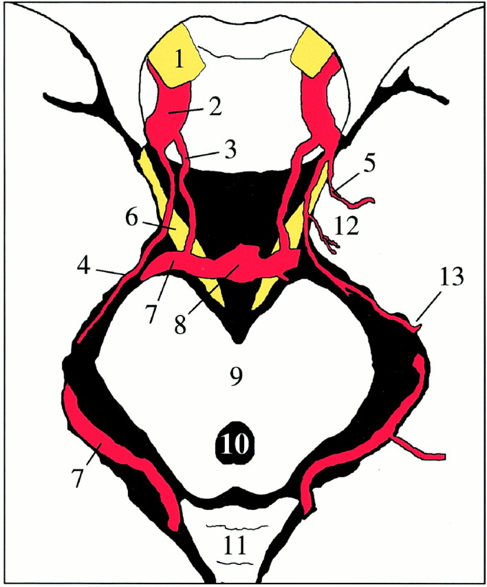
Background and purpose: The importance of the anterior choroidal artery (AChA) is related to its supply of crucial anatomic structures, such as the internal capsule. Angiographically, the AChA can be detected in 71% to 98% of patients, but as yet, its visibility on MR images has not been evaluated. Our goal was to assess the sensitivity of MR imaging in the identification of the AChA and its anatomic characteristics.
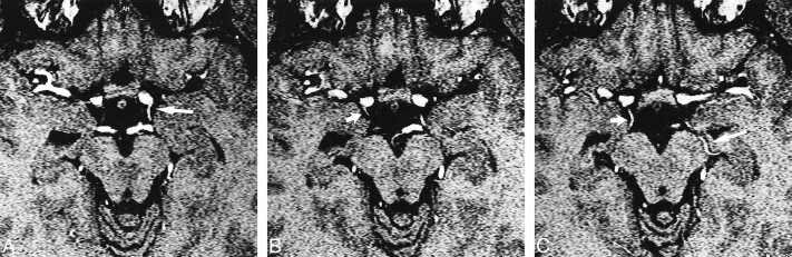
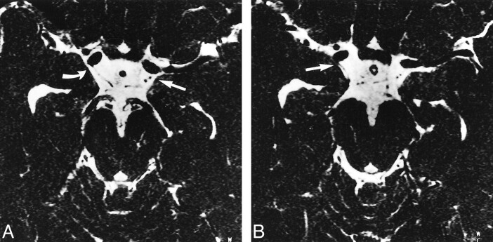
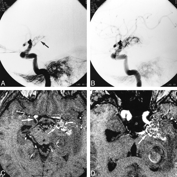
Methods: Twenty volunteers underwent MR imaging with a 3D time-of-flight (3D-TOF) sequence, 10 of them additionally with a 3D Fourier transformation constructive interference in steady state (3D-CISS) sequence. The MR angiographic source images and the 3D-CISS images were analyzed independently by two neuroradiologists, who evaluated the ability to identify the different segments of the AChA and the posterior communicating artery (PComA) according to a previously defined scoring system (0 = not identified, 1 = most probably identified, 2 = identified with certainty). Additionally, three patients were examined who had an arteriovenous malformation (AVM) supplied by the AChA.
Results: In the volunteers, the PComA was identified with certainty in 87.5% on 3D-TOF sequences and in 95% on 3D-CISS sequences; the AChA was identified with certainty in 92.5% on 3D-TOF sequences and in 90% on 3D-CISS sequences. 3D-CISS images showed additional anatomic information in six of 20 vessels. In the three patients, the enlarged AChA was identified with certainty on both imaging sequences.
Conclusion: The AChA can be reliably identified using both 3D-CISS sequences and the source images of the 3D-TOF sequence. MR imaging can be used to assess and follow-up AChA-related disorders, especially AVMs.
Figures
Similar articles
-
Trigeminal ganglion and its divisions: detailed anatomic MR imaging with contrast-enhanced 3D constructive interference in the steady state sequences.AJNR Am J Neuroradiol. 2005 May;26(5):1128-35. AJNR Am J Neuroradiol. 2005. PMID: 15891171 Free PMC article.
-
Preoperative Evaluation of Patients with Hemifacial Spasm by Three-dimensional Time-of-Flight (3D-TOF) and Three-dimensional Constructive Interference in Steady State (3D-CISS) Sequence.Clin Neuroradiol. 2016 Dec;26(4):431-438. doi: 10.1007/s00062-015-0382-2. Epub 2015 Mar 21. Clin Neuroradiol. 2016. PMID: 25795466
-
MR imaging of the cisternal segment of the posterior group of cranial nerves: neurovascular relationships and abnormal changes.Eur J Radiol. 2010 Jul;75(1):57-63. doi: 10.1016/j.ejrad.2009.03.036. Epub 2009 Apr 22. Eur J Radiol. 2010. PMID: 19395211
-
Role of the anterior choroidal artery in the endovascular treatment of brain arteriovenous malformations.Acta Neurol Belg. 2023 Feb;123(1):57-67. doi: 10.1007/s13760-022-01878-4. Epub 2022 Feb 11. Acta Neurol Belg. 2023. PMID: 35147868 Review.
-
MRI of hyperacute stroke in the AChA territory.Eur Radiol. 2004 Mar;14(3):417-24. doi: 10.1007/s00330-003-2220-1. Epub 2004 Jan 23. Eur Radiol. 2004. PMID: 14740166 Review.
Cited by
-
Perfusion Deficits and Association with Clinical Outcome in Patients with Anterior Choroidal Artery Stroke.J Stroke Cerebrovasc Dis. 2017 Aug;26(8):1755-1759. doi: 10.1016/j.jstrokecerebrovasdis.2017.04.001. Epub 2017 Apr 27. J Stroke Cerebrovasc Dis. 2017. PMID: 28457620 Free PMC article.
-
Choroid plexus perfusion in sickle cell disease and moyamoya vasculopathy: Implications for glymphatic flow.J Cereb Blood Flow Metab. 2021 Oct;41(10):2699-2711. doi: 10.1177/0271678X211010731. Epub 2021 Apr 28. J Cereb Blood Flow Metab. 2021. PMID: 33906512 Free PMC article.
-
Using a New Landmark of the Most External Point in the Embolization of Distal Anterior Choroidal Aneurysms: A Report of Two Cases.Front Neurol. 2020 Jul 31;11:693. doi: 10.3389/fneur.2020.00693. eCollection 2020. Front Neurol. 2020. PMID: 32849184 Free PMC article.
-
The Regulation of Cerebral Spinal Fluid Flow and Its Relevance to the Glymphatic System.Curr Neurol Neurosci Rep. 2020 Oct 19;20(12):58. doi: 10.1007/s11910-020-01077-9. Curr Neurol Neurosci Rep. 2020. PMID: 33074399 Free PMC article. Review.
-
The cisternal segment of the anterior choroidal artery: an anatomical study using magnetic resonance imaging.Childs Nerv Syst. 2017 Nov;33(11):2011-2016. doi: 10.1007/s00381-017-3525-8. Epub 2017 Jul 11. Childs Nerv Syst. 2017. PMID: 28698909
References
-
- Carpenter MR, Noback CR, Moss ML. The anterior choroidal artery: its origin, course distribution and variations. Arch Neurol Psychiatry 1954;71:714-722 - PubMed
-
- Saeki N, Rhoton AL. Microsurgical anatomy of the upper basilar artery and the posterior circle of Willis. Neurosurgery 1977;46:563-578 - PubMed
-
- Helgason CM. A new view of anterior choroidal artery territory infarction. J Neurol 1988;235:387-391 - PubMed
-
- Pullicino PM. The course and territories of cerebral small arteries. Adv Neurol 1993;62:11-39 - PubMed
-
- Foix C, Chavany JA, Hillemand P, Schiff-Wertheimer S. Oblitération de l'artère choroidienne antérieure: ramollissement de son territoire cérébral: hémiplégie, hémianesthésie, hémianopsie. Bull Soc Ophtalmol Paris 1925;37:221-223
Publication types
MeSH terms
LinkOut - more resources
Full Text Sources
Medical