

Case Reports
Endovascular management of a bleeding mandibular arteriovenous malformation by transfemoral venous embolization with NBCA
Affiliations
- PMID: 11156783
- PMCID: PMC7973946
Item in Clipboard
Case Reports
Endovascular management of a bleeding mandibular arteriovenous malformation by transfemoral venous embolization with NBCA
AJNR Am J Neuroradiol.
2001 Feb.
Abstract
A 13-year-old boy presented with an arteriovenous malformation (AVM) involving the left mandible that bled after intraoral biopsy. The AVM was treated on an emergency basis by primary intravenous delivery of n-butyl cyanoacrylate after transfemoral catheterization, resulting in complete anatomic and clinical cure.
Figures
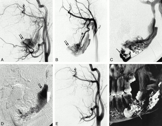
13-year-old boy with an AVM involving the left mandible that bled after intraoral biopsy. A and B, Initial arteriograms. A, Left common carotid arteriogram, lateral view, shows opacification of an arteriovenous shunting lesion with early filling of a large venous varix (double arrow) of the left mandibular vein draining into the retromandibular vein (single arrow). B, Left internal maxillary arteriogram, lateral view, shows the venous varix (arrows) is better filled and supplied mainly by the inferior dental artery (arrowheads), revealing the intraosseous location. C–F, Studies after embolization. C, Left mandibular phlebogram shows the tip of the microcatheter navigated into the very proximal part of the vein (arrow). The glue (arrowheads), already delivered by transarterial embolization, only partially occludes the mandibular vein. D, Roadmap image shows the glue cast (arrows) after the third intravenous injection filling the entire mandibular vein. E, Left common carotid arteriogram shows complete occlusion of the AVM by the end of the procedure. F, 3D reconstruction of the left mandible (rotational digital subtraction angiogram) shows the radiopaque embolic agent within the osseous cavity. The glue is positioned in the proximal and distal vein as well as in some of the small feeding vessels (arrows).
Similar articles
-
Embolization as the treatment for a life-threatening mandibular arteriovenous malformation.J Craniofac Surg. 2010 Mar;21(2):380-2. doi: 10.1097/SCS.0b013e3181cfa62a. J Craniofac Surg. 2010. PMID: 20186078
-
Treatment of mandibular arteriovenous malformation by transvenous embolization: A case report.Head Neck. 1999 Sep;21(6):574-7. doi: 10.1002/(sici)1097-0347(199909)21:6<574::aid-hed12>3.0.co;2-d. Head Neck. 1999. PMID: 10449675
-
Treatment of mandibular arteriovenous malformation by transvenous embolization through the mental foramen.J Oral Maxillofac Surg. 2008 Jan;66(1):139-43. doi: 10.1016/j.joms.2006.06.259. J Oral Maxillofac Surg. 2008. PMID: 18083429 No abstract available.
-
Transvenous embolisation of an arteriovenous malformation of the mandible via a femoral approach.Pediatr Radiol. 1997 Nov;27(11):855-7. doi: 10.1007/s002470050254. Pediatr Radiol. 1997. PMID: 9361043 Review.
-
A successful case of transcatheter arterial embolization with n-butyl-2-cyanoacrylate for pancreatic arteriovenous malformation.Intern Med. 2014;53(23):2683-7. doi: 10.2169/internalmedicine.53.3327. Epub 2014 Dec 1. Intern Med. 2014. PMID: 25447650 Review.
Cited by
-
Management of Late Post-traumatic Facial Artery Pseudoaneurysmal Cyst: Review of Literature.J Maxillofac Oral Surg. 2015 Jun;14(2):201-5. doi: 10.1007/s12663-014-0678-9. Epub 2014 Aug 13. J Maxillofac Oral Surg. 2015. PMID: 26028835 Free PMC article. Review.
-
Life-threatening arteriovenous malformation of the maxillomandibular region and treatment outcomes.Interv Neuroradiol. 2012 Mar;18(1):49-59. doi: 10.1177/159101991201800107. Epub 2012 Mar 16. Interv Neuroradiol. 2012. PMID: 22440601 Free PMC article.
-
Embolotherapy of Head and Neck Lesions: Basics and Clinical Tips.Interv Radiol (Higashimatsuyama). 2024 Oct 4;9(3):112-121. doi: 10.22575/interventionalradiology.2024-0017. eCollection 2024 Nov 1. Interv Radiol (Higashimatsuyama). 2024. PMID: 39559806 Free PMC article. Review.
-
Embolization in the head and neck.Semin Intervent Radiol. 2008 Sep;25(3):293-309. doi: 10.1055/s-0028-1085929. Semin Intervent Radiol. 2008. PMID: 21326519 Free PMC article.
-
Contralateral de novo intraosseous arteriovenous malformation in a child with arteriovenous malformation of mandible treated by endovascular embolotherapy. A case report.Interv Neuroradiol. 2012 Dec;18(4):484-9. doi: 10.1177/159101991201800415. Epub 2012 Dec 3. Interv Neuroradiol. 2012. PMID: 23217644 Free PMC article.
References
-
- Rodesch G, Soupre V, Vazquez MP, Alvarez H, Lasjaunias P. Arteriovenous malformations of the dental arcades: the place of endovascular therapy: results in 12 cases are presented. J Craniomaxillofac Surg 1998;26:306-313 - PubMed
-
- Engel JD, Supancic JS, Davis LF. Arteriovenous malformation of the mandible: life-threatening complications during tooth extraction [see comments]. J Am Dent Assoc 1995;126:237-242 - PubMed
-
- Kiyosue H, Mori H, Hori Y, Okahara M, Kawano K, Mizuki H. Treatment of mandibular arteriovenous malformation by transvenous embolization: a case report. Head Neck 1999;21:574-577 - PubMed
-
- Lamberg MA, Tasanen A, Jaaskelainen J. Fatality from central hemangioma of the mandible. J Oral Surg 1979;37:578-584 - PubMed
-
- Resnick SA, Russell EJ, Hanson DH, Pecaro BC. Embolization of a life-threatening mandibular vascular malformation by direct percutaneous transmandibular puncture. Head Neck 1992;14:372-379 - PubMed
Publication types
MeSH terms
Substances
LinkOut - more resources
Full Text Sources
Medical