. 2001 Feb 5;193(3):271-80.
doi: 10.1084/jem.193.3.271.

The relative importance of T cell subsets in immunity and immunopathology of airborne Mycobacterium tuberculosis infection in mice

Affiliations

The relative importance of T cell subsets in immunity and immunopathology of airborne Mycobacterium tuberculosis infection in mice

T Mogues et al. J Exp Med. .

Abstract

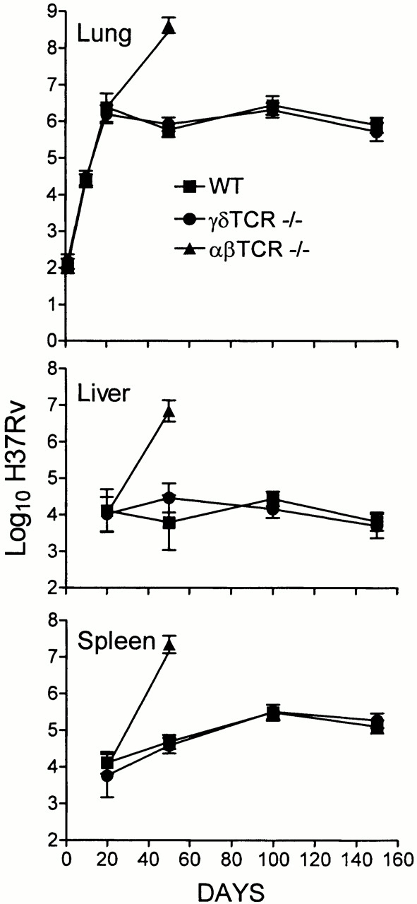
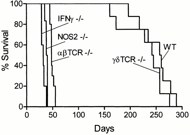
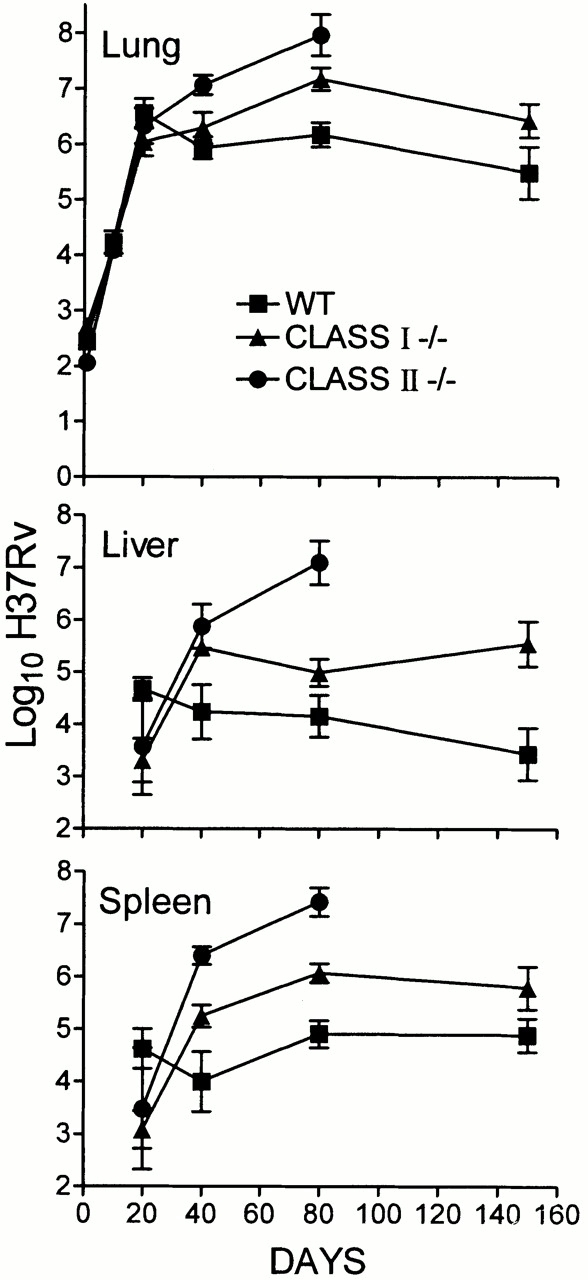
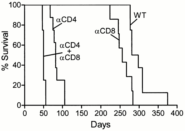
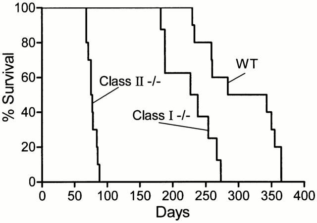
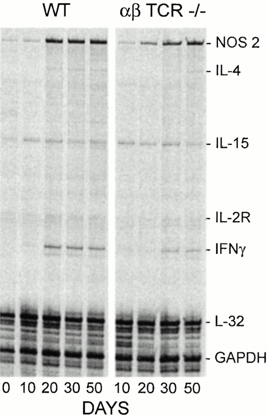
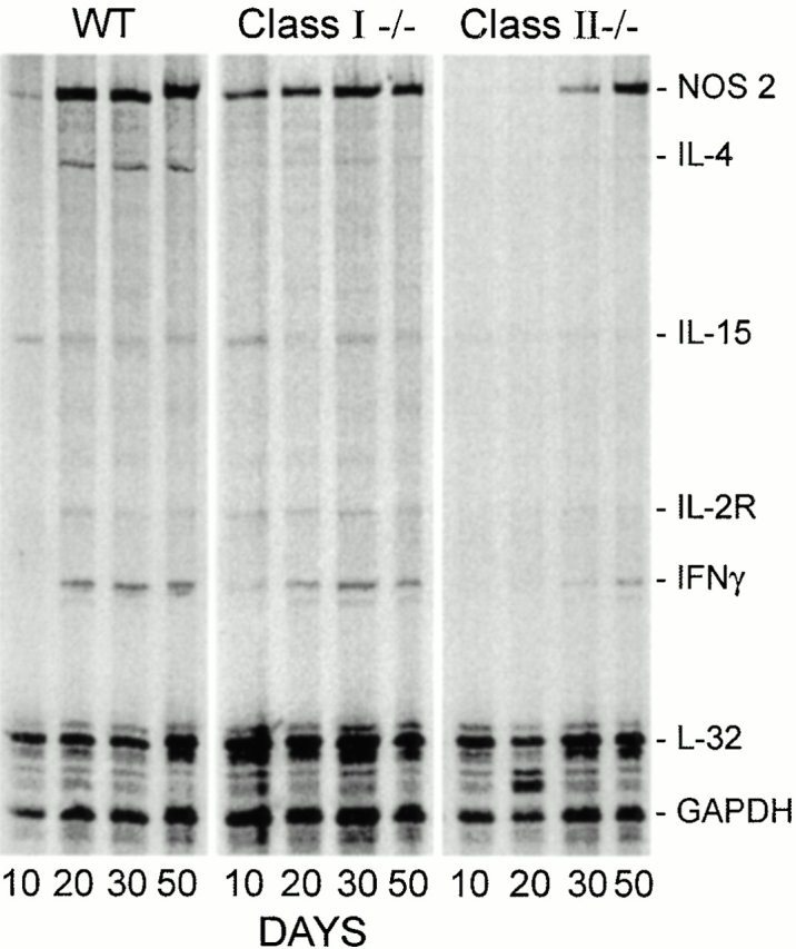
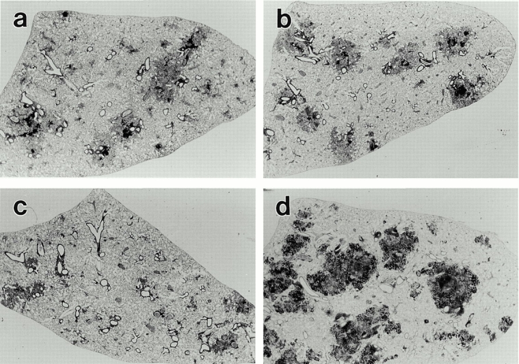
Wild-type (WT) and targeted-mutant mice incapable of making alphabeta T cells, gammadelta T cells, class I major histocompatibility complex (MHC), class II MHC, interferon (IFN)-gamma, or inducible nitric oxide synthase (NOS2), were infected with Mycobacterium tuberculosis (Mtb) by aerosol, and monitored over time for their ability to (a) control infection, (b) develop histopathology at sites of infection, and (c) survive. WT mice acquired the ability to control and to hold infection at a stationary level from day 20 on. This was associated with the development of a macrophage-dominated alveolitis at sites of infection, with increased synthesis of IFN-gamma and NOS2 mRNA, and with an median survival time (MST) of 258.5 d. In the absence of alphabeta T cells, Mtb grew progressively and rapidly to induce a necrotic, neutrophil-dominated lung pathology that killed mice with an MST of 48 d. In the absence of CD4-mediated immunity (class II(-/-) mice), progressive bacterial growth continued in the lungs and in other organs beyond day 20, resulting in an MST of 77 d. By contrast, in the absence of CD8 T cell-mediated immunity, lung infection was controlled at a 1 log higher stationary level that induced a similar histopathologic response to that of WT mice, and resulted in an MST of 232 d.


Figures

Figure 1
Figure 1
Growth of H37Rv in the lungs, livers, and spleens of WT, TCR-α/β−/−, and TCR-γ/δ−/− mice infected with 102 CFU via the respiratory route. WT and TCR-γ/δ−/− controlled infection and caused it to become stationary in all organs after day 20, whereas TCR-α/β−/− mice were unable to control infection in any organ. This is one of two experiments that gave essentially the same results. Means of five mice per time point per group ± SD.
Figure 3
Figure 3
Survival times of WT, TCR-α/β−/−, TCR-γ/δ−/−, IFN-γ−/−, and NOS2−/− mice (10 mice per group) infected with 102 H37Rv via the respiratory route. IFN-γ−/− mice died earlier (MST = 32 d) than NOS2−/− mice (MST = 39 d), and NOS2−/− mice died earlier than TCR-α/β−/− mice (MST = 48). TCR-γ/δ−/− mice died with an MST of 243.5 d, which was not significantly different from the 258.5-d MST of WT mice.
Figure 2
Figure 2
Growth of H37Rv in the lungs, livers, and spleens of WT, class I−/−, and class II−/− mice infected with 102 H37Rv via the respiratory route. Infection in class I−/− mice was stabilized in the lungs and other organs at a 1 log higher level than in WT mice. Class II−/− mice were unable to control infection in any organ. This is one of two experiments that gave essentially the same results. Means of five mice per time point per group ± SD.
Figure 5
Figure 5
The survival times of mice depleted of CD4, CD8, or both T cell subsets by treatment with mAbs were similar to survival times of class II−/−, class I−/−, and TCR-α/β−/− mice, respectively, as shown in Fig. 4. All mice (10 per group) were infected by aerosol with 102 H37Rv.
Figure 4
Figure 4
Survival times of WT (MST = 313.5 d), class I−/− (MST = 232 d), and class II−/− (MST = 77 d) mice (10 per group) infected with 102 H37Rv via the respiratory route.
Figure 6
Figure 6
Radioautograph of multiprobe RPA run using 35S-labeled probes to detect mRNA for IFN-γ, NOS2, IL-4, IL-13, IL-15, and IL-2Rα in the lungs of WT and TCR-α/β−/− mice on days 0, 10, 20, 30, and 50 of infection. The results clearly show upregulation of mRNA for NOS2 and IFN-γ from day 20 of infection in WT mice. Much less IFN-γ mRNA was present in the lungs of TCR-α/β−/− mice from day 30 on, and the production of NOS2 mRNA was delayed. IL-15 mRNA was constitutively produced in the lungs of both groups of mice, and its synthesis appeared to be little altered by infection. GAPDH, glyceraldehyde 3-phosphate dehydrogenase.
Figure 7
Figure 7
Changes in the quantity of mRNA for IFN-γ and NOS2 against time of infection in the lungs of WT, class I−/−, and class II−/− mice as determined by RPA. IFN-γ and NOS2 mRNA was upregulated from day 20 of infection on in WT mice. Likewise, IFN-γ and NOS2 mRNA was present in the lungs of class I−/− mice from days 20 on. In the lungs of class II−/− mice, in contrast, upregulation of IFN-γ mRNA was not evident until day 30, and relatively low levels were produced thereafter. Appreciable amounts of NOS2 mRNA were made in the lungs of these mice, but not until after day 30. GAPDH, glyceraldehyde 3-phosphate dehydrogenase.
Figure 8
Figure 8
Low power (×6.5) of 15-μm thick sections of the left lobe of lungs of a WT (a) class I−/− (b), class II−/− (c), and TCR-α/β−/− (d) mouse on day 50 of infection initiated with 102 H37Rv by aerosol. The individual lesions in WT and class I−/− mice are very similar in size and appearance, being made up of darkly stained (lymphoid) and lightly stained (macrophage) areas. The lung lesions in class II−/− mice are smaller and less well defined, whereas the lung lesions in TCR-α/β−/− mice are large, darkly stained, and granular in appearance.
Figure 9
Figure 9
Low power (×25) and high power (×550) micrographs of lung sections of a WT (a and b), class I−/− (c and d), class II−/− (e and f), and TCR-α/β−/− mouse (g and h) stained for NOS2 by immunocytochemistry. At low power, the immunocytochemical reaction product is seen to be located in cells in the lightly stained macrophage areas of the lesions of WT and class I−/− mice. In class II−/− and TCR-α/β−/− mice, the reaction product is scattered in smaller quantity in cells throughout the lesions. At high power, the reaction product is seen confined to macrophages in lesions of WT and class I−/− mice, with some of the macrophages containing acid-fast bacilli. The NOS2-positive macrophages of the class I−/− mouse contain more acid-fast bacilli than NOS2-positive macrophages in WT lesion. In the lesion of the class II−/− mouse, NOS2-positive macrophages are heavily infected with Mtb, although some heavily infected macrophages have little or no reaction product. In the lesion of the TCR-α/β−/− mouse, most of the NOS2 reaction product appears to be concentrated in scattered mononuclear cells.

References

    1. Boom W.H. The role of T cell subsets in Mycobacterium tuberculosis infection. Infect. Agents Dis. 1996;5:73–81. - PubMed
    1. Murray P.J. Defining the requirements for immunological control of mycobacterial infections. Trends Micribiol. 1999;7:366–372. - PubMed
    1. Stenger S., Modlin R.L. T cell-mediated immunity to Mycobacterium tuberculosis . Curr. Opin. Microbiol. 1999;2:89–93. - PubMed
    1. Cooper A.M., Saunders B.M., D'Souza C.D., Frank A.A., Orme I.M. Mycobacterium tuberculosis-driven processes in gene-disrupted mice. Bull. Inst. Pasteur. 1997;95:85–95.
    1. Orme I.M., Collins F.M. Adoptive protection of the Mycobacterium tuberculosis-infected lung. Cell. Immunol. 1984;84:113–120. - PubMed

Publication types

MeSH terms