. 2001 Feb 5;193(3):339-51.
doi: 10.1084/jem.193.3.339.

Interleukin 12 p40 production by barrier epithelial cells during airway inflammation

Affiliations

Interleukin 12 p40 production by barrier epithelial cells during airway inflammation

M J Walter et al. J Exp Med. .

Abstract

Human airway epithelial cells appear specially programmed for expression of immune response genes implicated in immunity and inflammation. To better determine how this epithelial system operates in vivo, we analyzed its behavior in mouse models that allow for in vitro versus in vivo comparison and genetic modification. Initial comparisons indicated that tumor necrosis factor alpha induction of epithelial intercellular adhesion molecule 1 required sequential induction of interleukin (IL)-12 (p70) and interferon gamma, and unexpectedly localized IL-12 production to airway epithelial cells. Epithelial IL-12 was also inducible during paramyxoviral bronchitis, but in this case, initial IL-12 p70 expression was followed by 75-fold greater expression of IL-12 p40 (as monomer and homodimer). Induction of IL-12 p40 was even further increased in IL-12 p35-deficient mice, and in this case, was associated with increased mortality and epithelial macrophage accumulation. The results placed epithelial cell overgeneration of IL-12 p40 as a key intermediate for virus-inducible inflammation and a candidate for epithelial immune response genes that are abnormally programmed in inflammatory disease. This possibility was further supported when we observed IL-12 p40 overexpression selectively in airway epithelial cells in subjects with asthma and concomitant increases in airway levels of IL-12 p40 (as homodimer) and airway macrophages. Taken together, these results suggest a novel role for epithelial-derived IL-12 p40 in modifying the level of airway inflammation during mucosal defense and disease.


Figures

Figure 1
Figure 1
TNF-α responsiveness of epithelial ICAM-1 gene expression is not found in vitro (A) but is found in vivo (B and C), where it depends on IL-12 and IFN-γ production. In A, airway epithelial (mTE) cells were treated with IFN-γ (0–1,000 U/ml for 24 h and 100 U/ml for 0–24 h) or with IFN-γ vs. TNF-α (100 U/ml for 24 h). For each condition, cell lysates were subjected to Western blotting with anti–ICAM-1 mAb detected by enhanced chemiluminescence. Equality of protein loading and specificity was demonstrated by reblotting against anti-Sp1 mAb (data not shown). For each condition, fold increase above control was determined using densitometry, and values represent mean ± SEM for three experiments. A significant increase from untreated control value (by ANOVA) is indicated by (*). In B, wild-type (WT), IFN-γ–deficient (−/−), and IL-12 p35 (−/−) mice (all in C57BL/6J background) underwent intratracheal injection of IFN-γ or TNF-α. At 12 h after treatment, tracheal sections were immunostained with anti–ICAM-1 mAb, biotinylated goat anti–hamster IgG, streptavidin-conjugated horseradish peroxidase, and 3,3′-diaminobenzidine, and then counterstained with hematoxylin. Wild-type, IFN-γ (−/−), and IL-12 p35 (−/−) mice injected with PBS vehicle exhibited levels of epithelial ICAM-1 similar to background (data not shown). Immunostaining with control nonimmune IgG also gave no signal above background (data not shown). Bar, 20 μm. In C, tracheal sections from conditions in B underwent quantification of epithelial ICAM-1 immunostaining relative to a cartilage reference set at a value of 100. For each condition, values represent mean ± SEM for three experiments, and a significant increase from PBS-treated wild-type cohort is indicated by (*).
Figure 1
Figure 1
TNF-α responsiveness of epithelial ICAM-1 gene expression is not found in vitro (A) but is found in vivo (B and C), where it depends on IL-12 and IFN-γ production. In A, airway epithelial (mTE) cells were treated with IFN-γ (0–1,000 U/ml for 24 h and 100 U/ml for 0–24 h) or with IFN-γ vs. TNF-α (100 U/ml for 24 h). For each condition, cell lysates were subjected to Western blotting with anti–ICAM-1 mAb detected by enhanced chemiluminescence. Equality of protein loading and specificity was demonstrated by reblotting against anti-Sp1 mAb (data not shown). For each condition, fold increase above control was determined using densitometry, and values represent mean ± SEM for three experiments. A significant increase from untreated control value (by ANOVA) is indicated by (*). In B, wild-type (WT), IFN-γ–deficient (−/−), and IL-12 p35 (−/−) mice (all in C57BL/6J background) underwent intratracheal injection of IFN-γ or TNF-α. At 12 h after treatment, tracheal sections were immunostained with anti–ICAM-1 mAb, biotinylated goat anti–hamster IgG, streptavidin-conjugated horseradish peroxidase, and 3,3′-diaminobenzidine, and then counterstained with hematoxylin. Wild-type, IFN-γ (−/−), and IL-12 p35 (−/−) mice injected with PBS vehicle exhibited levels of epithelial ICAM-1 similar to background (data not shown). Immunostaining with control nonimmune IgG also gave no signal above background (data not shown). Bar, 20 μm. In C, tracheal sections from conditions in B underwent quantification of epithelial ICAM-1 immunostaining relative to a cartilage reference set at a value of 100. For each condition, values represent mean ± SEM for three experiments, and a significant increase from PBS-treated wild-type cohort is indicated by (*).
Figure 2
Figure 2
TNF-α induction of IL-12 expression drives downstream production of IFN-γ. Wild-type (WT), IL-12 p35 (−/−), and IFN-γ (−/−) C57BL/6J mice were treated with vehicle alone (PBS) or TNF-α as described in the legend to Fig. 1, followed 12 h later by BAL. The BAL fluid was concentrated 10-fold and used for duplicate measurements of IL-12 and IFN-γ levels by ELISA. Values represent mean ± SEM (n = 4). Levels of IL-12 were undetectable in IL-12 p35 (−/−) mice. A significant increase from PBS-treated wild-type cohort (by ANOVA) is indicated by (*).
Figure 4
Figure 4
Viral induction of IL-12 p40 without a change in constitutive p35 expression in airway epithelial cells. Wild-type C57BL/6J mice underwent intranasal inoculation with SdV (5,000 EID50 in 30 μl of PBS), and lungs were removed and fixed in formalin on day 1, 3, 5, and 8 after inoculation. In each case, tissue was immunostained for IL-12 P-40 and p35 as described in the legend to Fig. 3 and similarly for viral protein (labeled SdV) using rat anti-SdV pAb. In mice inoculated with UV-inactivated Sdv, IL-12 p40 immunostaining was not detected and IL-12 p35 constitutive immunostaining was unchanged from untreated control mice (not shown). Control rat, goat, or rabbit nonimmune IgG gave no signal above background (data not shown). Bar, 20 μm.
Figure 3
Figure 3
TNF-α induction of IL-12 p40 without a change in constitutive p35 expression in airway epithelial cells. In A, wild-type (WT) and same-strain IFN-γ (−/−) C57BL/6J mice were treated with PBS vehicle or TNF-α as described in the legend to Fig. 1. At 12 h after treatment, tracheal (rows 1 and 3) or bronchial (rows 2 and 4) tissue was fixed in formalin, blocked with nonimmune goat or rabbit serum, and then incubated with anti–IL-12 p40 or p35 Ab. For p35 immunostaining, tissues were also subjected to antigen retrieval (using 10 mM Citra solution for 10 min at 98°C). Primary Ab binding was detected by incubation with biotinylated rabbit anti–goat or goat anti–rabbit IgG, streptavidin-conjugated alkaline phosphatase complex, and a red chromogenic substrate, and tissues were counterstained with hematoxylin. Control goat nonimmune IgG gave no signal above background (data not shown). In B, wild-type C57BL/6J mice were treated with PBS as described in the legend Fig. 1. Tracheal tissue sections were incubated with control rabbit nonimmune IgG, anti–IL-12 p35 pAb, or anti–IL-12 p35 pAb in the presence of recombinant murine IL-12 p40 or IL-12 followed by detection of primary Ab binding and hematoxylin counterstaining as described in A. Bars, 20 μm.
Figure 6
Figure 6
Decreased survival from viral bronchopneumonia in IL-12 p35–deficient mice. In A, wild-type (WT), and IL-12 p35 (−/−) and p40 (−/−) C57BL/6J mice were inoculated with SdV (50,000 EID50) and monitored for survival by Kaplan-Meier analysis (n = 29, 19, and 27 in each group, respectively). A significant decrease in survival (by Wilcoxon rank-sum test) is indicated by (*). In B, the same cohorts were inoculated with SdV (50,000 EID50), and lungs were removed at days 7, 9, and 10 after inoculation for hematoxylin/eosin staining and photomicrography. Bar, 50 μm.
Figure 5
Figure 5
Early induction of IL-12 p70 followed by predominant release of IL-12 p40 into the airway during viral bronchitis. Wild-type mice were inoculated with SdV or UV-inactivated SdV as described in the legend to Fig. 4. In A, at the indicated times after inoculation, BAL fluid was obtained for duplicate measurements of IL-12 and IL-12 p40 levels by ELISA. All values represent mean ± SEM (n = 4). A significant increase from PBS-treated cohort or UV-activated SdV cohort (by ANOVA) is indicated by (*). In B, BAL fluid from day 5 after inoculation was subjected to Western blotting against anti–IL-12 p40 mAb under nonreducing conditions or control anti–mouse IgG Ab under reducing conditions with detection by enhanced chemiluminescence. Bands corresponding to IL-12 p40 homodimer (p80), IL-12 (p70), IL-12 p40 monomer (p40), and mouse IgG are indicated by arrows.
Figure 8
Figure 8
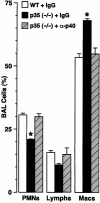
Inhibition of macrophage accumulation by anti–IL-12 p40 Ab treatment. Wild-type (WT) and IL-12 p35 (−/−) mice were inoculated with SdV (50,000 EID50) and treated with control rat IgG or rat anti–IL-12 p40 IgG (1 mg given intraperitoneally on postinoculation days 2 and 6), and differential cell counts were determined in BAL fluid obtained at day 7 after inoculation. Values represent mean ± SEM (n = 4), and a significant difference from the wild-type cohort treated with control IgG is indicated by (*).
Figure 7
Figure 7
Decreased survival from viral bronchopneumonia driven by overexpression of IL-12 p40 homodimer and macrophage accumulation. In A and B, wild-type (WT) and IL-12 p35 (−/−) and p40 (−/−) C57BL/6J mice were inoculated with PBS vehicle, UV-inactivated SdV, or SdV (50,000 EID50), and IFN-γ, TNF-α, and IL-12 p40 levels were determined in BAL fluid obtained at day 7 after inoculation. In C, wild-type and IL-12 p35 (−/−) and p40 (−/−) mice were inoculated with SdV (50,000 EID50) and differential cell counts were determined in BAL fluid obtained at day 7 after inoculation. In A–C, values represent mean ± SEM (n = 4), and a significant difference from the wild-type cohort is indicated by (*). In D, wild-type, IL-12 p35 (−/−), and IL-12 p40 (−/−) mice were inoculated with SdV (50,000 EID50) and lungs at day 7 were removed and immunostained with anti-Mac3 mAb and counterstained with hematoxylin. Control nonimmune IgG gave no signal above background (not shown). Bar, 20 μm. Quantitation of macrophages per mm in length of basement membrane are provided as mean ± SEM (n = 5) for each condition.
Figure 9
Figure 9
Selective induction of epithelial IL-12 p40 expression in asthmatic subjects. In A, endobronchial biopsies from normal control, asthmatic, and chronic bronchitis subjects were immunostained with control nonimmune IgG or with anti–IL-12 p40 or p35 Ab as described in the legend to Fig. 3. Bar, 20 μm. Representative photomicrographs of biopsies from seven control, seven asthmatic, and eight chronic bronchitis subjects are shown. In B, endobronchial biopsy sections from conditions in A underwent quantification of epithelial IL-12 p40 immunostaining relative to a subepithelial reference set at a value of 100. For each condition, values represent mean ± SEM for each cohort, and a significant increase from immunostaining with control nonimmune IgG is indicated by (*).
Figure 9
Figure 9
Selective induction of epithelial IL-12 p40 expression in asthmatic subjects. In A, endobronchial biopsies from normal control, asthmatic, and chronic bronchitis subjects were immunostained with control nonimmune IgG or with anti–IL-12 p40 or p35 Ab as described in the legend to Fig. 3. Bar, 20 μm. Representative photomicrographs of biopsies from seven control, seven asthmatic, and eight chronic bronchitis subjects are shown. In B, endobronchial biopsy sections from conditions in A underwent quantification of epithelial IL-12 p40 immunostaining relative to a subepithelial reference set at a value of 100. For each condition, values represent mean ± SEM for each cohort, and a significant increase from immunostaining with control nonimmune IgG is indicated by (*).
Figure 10
Figure 10
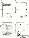
Selective IL-12 p40 release in asthmatic subjects is independent of glucocorticoid (GC) treatment and associated with macrophage accumulation. In A and B, BAL fluid from 10 normal, 4 asthmatic (without glucocorticoid treatment,) and 7 asthmatic subjects (with glucocorticoid treatment) was concentrated 35-fold and used for duplicate measurements of IL-12 p70 and p40 levels by ELISA. Mean value for IL-12 p40 is represented by bold line and P values are indicated. In C, concentrated BAL fluid from normal and asthmatic subjects was subjected to immunoprecipitation and Western blotting against anti–IL-12 p40 Ab using nonreducing (top blot) or reducing (bottom blot) conditions and enhanced chemiluminescence detection. Bands corresponding to IL-12 (p70) and IL-12 P-40 monomer (p40) were identified using recombinant protein standards (Std) and are indicated by arrows. Immunoprecipitation with control nonimmune IgG gave no signal above background (data not shown). In D, BAL fluid levels of macrophages and IL-12 p40 were determined for each sample obtained from each asthmatic subject and then subjected to correlation analysis (r, correlation coefficient).

Similar articles

Cited by

References

    1. Medzhitov R., Janeway C.A., Jr. An ancient system of host defense. Curr. Opin. Immunol. 1998;10:12–15. - PubMed
    1. Holtzman M.J., Zhang V., Hussain H., Roswit W.T., Wilson J.D. Prostaglandin H synthase and lipoxygenase gene families in the epithelial cell barrier. In: Goetzl E.J., Lewis R.A., Rola-Pleszczynski M., editors. Cellular Generation, Transport and Effects of EicosanoidsBiological Roles and Pharmacological Intervention. Ann. NY. Acad. Sci; New York: 1995. pp. 58–77. - PubMed
    1. Holtzman M.J., Castro M., Look D.C., O'Sullivan M., Walter M.J. Regulation of epithelial-leukocyte interaction and epithelial immune-response genes. In: Busse W., Holgate S., editors. Asthma and Rhinitis. Blackwell Scientific; Cambridge, MA: 2000. pp. 784–800.
    1. Look D.C., Keller B.T., Rapp S.R., Holtzman M.J. Selective induction of intercellular adhesion molecule-1 by interferon-γ in human airway epithelial cells. Am. J. Physiol. 1992;263:L79–L87. - PubMed
    1. Nakajima S., Look D.C., Roswit W.T., Bragdon M.J., Holtzman M.J. Selective differences in vascular endothelial- vs. airway epithelial-T cell adhesion mechanisms. Am. J. Physiol. 1994;267:L422–L432. - PubMed

Publication types

MeSH terms