. 2001 Feb 5;193(3):365-74.
doi: 10.1084/jem.193.3.365.

The importation of hematogenous precursors by the thymus is a gated phenomenon in normal adult mice

Affiliations

The importation of hematogenous precursors by the thymus is a gated phenomenon in normal adult mice

D L Foss et al. J Exp Med. .

Abstract

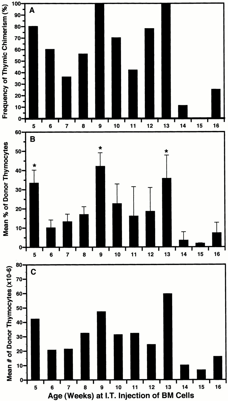
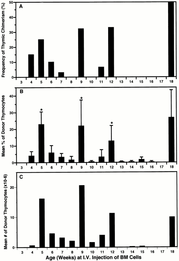
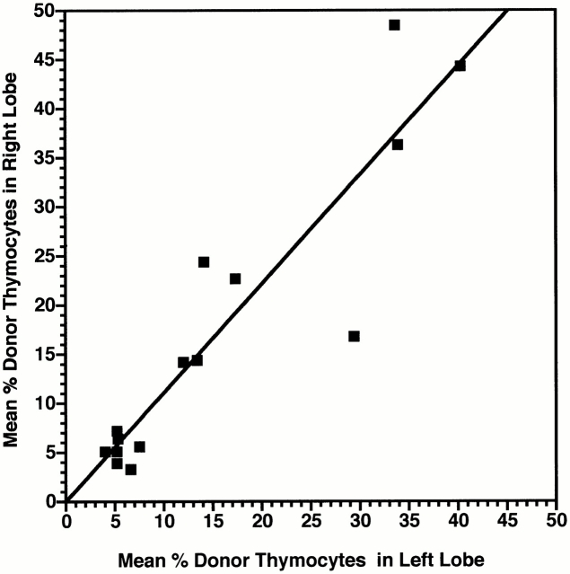
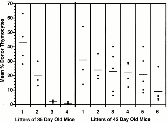
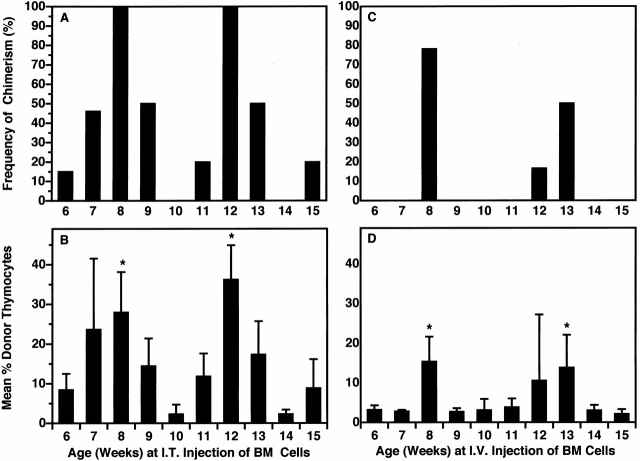
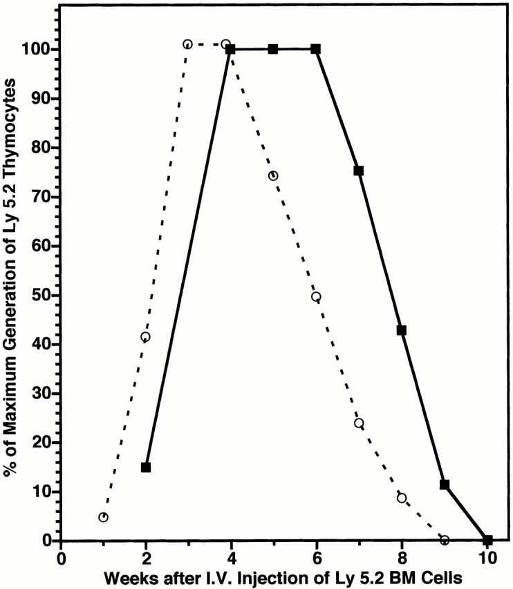
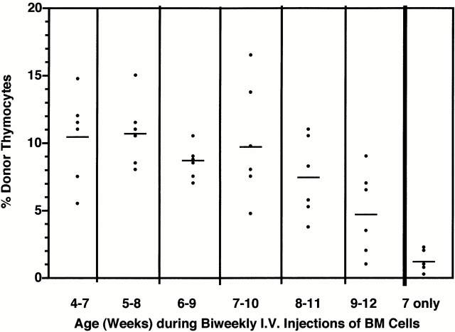
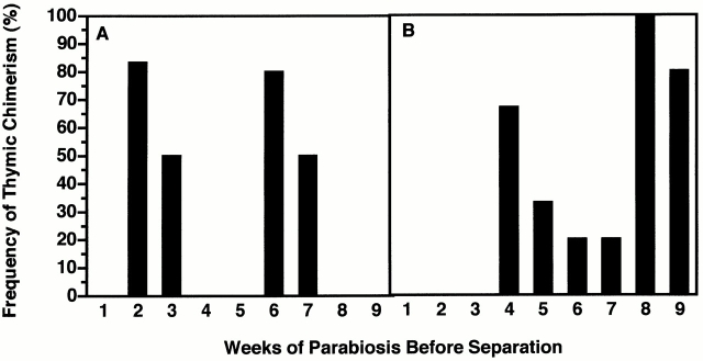
Hematogenous precursors repopulate the thymus of normal adult mice, but it is not known whether this process is continuous or intermittent. Here, two approaches were used to demonstrate that the importation of prothymocytes in adult life is a gated phenomenon. In the first, age-dependent receptivity to thymic chimerism was studied in nonirradiated Ly 5 congenic mice by quantitative intrathymic and intravenous bone marrow (BM) adoptive transfer assays. In the second, the kinetics of importation of blood-borne prothymocytes was determined by timed separation of parabiotic mice. The results showed that >60% of 3-18-wk-old mice developed thymic chimerism after intrathymic injection of BM cells, and that the levels of chimerism (range, 5-90% donor-origin cells) varied cyclically (periodicity, 3 to 5 wk). In contrast, only 11-14% of intravenously injected recipients became chimeric, and chimerism occurred intermittently (receptive period approximately 1 wk; refractory period approximately 3 wk). In the intravenously injected mice, chimerism occurred simultaneously in both thymic lobes; gate opening occurred only after most intrathymic niches for prothymocytes had emptied; and the ensuing wave of thymocytopoiesis encompassed two periods of gating. These kinetics were confirmed in parabiotic mice, and in cohorts of mice in whom gating was synchronized by an initial intrathymic injection of BM cells. In addition, a protocol was developed by which sequential intravenous injections of BM cells over a 3 to 4 wk period routinely induces thymic chimerism in the apparent absence of stem cell chimerism. Hence, the results not only provide a new paradigm for the regulation of prothymocyte importation during adult life, but may also have applied implications for the selective induction of thymocytopoiesis in nonmyeloablated hosts.


Figures

Figure 1
Figure 1
Induction of thymic chimerism in normal adult mice by intravenous and intrathymic injection of BM cells. Groups of normal adult Ly 5.1 mice, obtained from multiple cohorts, were randomized for age (7–12 wk) and sex, and injected intravenously (black bars) or intrathymically (hatched bars) with suspensions of sex-matched Ly 5.2 BM cells (20 × 106 intravenously or 2 × 10⁶ intrathymically) from 6-wk-old donors. The frequency and levels of thymic chimerism attained 28 d later were determined by FCM analysis. Data for male and female recipients are pooled, as no differences were observed. Percentage of chimeric mice (≥5% donor-origin cells): intravenous injection = 12.7% (21 of 165); intrathymic injection = 85.9% (122 of 142).
Figure 2
Figure 2
The induction of thymic chimerism in adult mice is cyclical after intrathymic (I.T.) injection of BM cells. A cohort of 5-wk-old (± 3 d) Ly 5.1 mice was divided into 12 groups (9–12 mice each) and, at weekly intervals, a different group was injected intrathymically with a saturating dose (2 × 10⁶) of Ly 5.2 BM cells. The percentage of Ly 5.2 thymocytes present 28 d later was determined by FCM analysis. Results for total mice in each group (5–16 wk of age) are presented as: (A) frequency of thymic chimerism (≥5% donor-origin cells); (B) mean percentage of donor-origin thymocytes (± SD); and (C) mean number of donor-origin thymocytes. Percentage of chimeric mice = 62.5%. Maximum level of thymic chimerism = 88% (120 × 106 donor-origin cells). *P < 0.05 between highest and lowest values in each cycle. This experiment was repeated in part on two occasions using cohorts of mice varying in age from 5–8, 7–12, and 12–16 wk. Similar results were obtained, with peaks and valleys shifting by no more than 1 wk from those illustrated.
Figure 3
Figure 3
The induction of thymic chimerism in adult mice is periodic after intravenous (I.V.) injection of BM cells. A cohort of 3-wk-old (± 3 d) Ly 5.1 mice was divided into 16 groups (15–20 mice each) and, at weekly intervals, a different group was injected intravenously with 20 × 106 Ly 5.2 BM cells. The percentage of Ly 5.2 thymocytes present 28 d later was determined by FCM analysis. Results for total mice in each group (3–18 wk of age) are presented as: (A) frequency of thymic chimerism (≥5% donor-origin cells); (B) mean percentage of donor-origin thymocytes (± SD); and (C) mean number of donor-origin thymocytes. Percentage of chimeric mice = 10.9% (30 of 275). Maximum level of thymic chimerism = 78% (98 × 106 donor-origin cells). *P < 0.05 between highest and lowest values in sequential periods of receptivity and refractivity. This experiment was repeated in part on two occasions using cohorts of mice varying in age from 4–13, 7–12, and 12–18 wk. Similar results were obtained (± 1 wk).
Figure 4
Figure 4
The induction of thymic chimerism in adult mice occurs in both lobes after intravenous injection of BM cells. Three cohorts of 5-wk-old (± 3 d) Ly 5.1 mice (total 30) were injected intravenously with a saturating dose of Ly 5.2 BM cells. The percentage of Ly 5.2 thymocytes present 28 d later in the left and right lobes of each thymus was determined by FCM analysis. Squares represent paired data for each animal in which at least one thymic lobe had ≥5% Ly 5.2+ cells. Line of best fit was determined by regression analysis (r 2 = 0.84; slope = 1.11; x and y intercepts = 0). Mean levels of thymic chimerism: left lobe = 16 ± 13%; right lobe = 17 ± 15%; P > 0.1.
Figure 5
Figure 5
Thymic chimerism is heterogeneous between age-matched litters of mice injected intrathymically with BM cells. Litters of age-matched male Ly 5.1 mice from timed matings were injected intrathymically with 2 × 10⁶ Ly 5.2 BM cells on days 35 or 42. The percentage of Ly 5.2 thymocytes present 28 d later was determined by FCM analysis. Dots represent results for individual animals. Bars indicate mean level of thymic chimerism for each litter.
Figure 6
Figure 6
Intrathymic gating is synchronized by initial intrathymic (I.T.) injection of BM cells. A cohort of 5-wk-old (± 3 d) Ly 5.1 mice was injected intrathymically with a saturating dose of Ly 5.1 BM cells, and at weekly intervals thereafter a separate group of 9–12 of these mice was reinjected with saturating doses of Ly 5.2 BM cells (A and B) intrathymically or (C and D) intravenously. The percentage of Ly 5.2 thymocytes present 28 d later was determined by FCM analysis. The frequencies (A and C) and mean levels (B and D) of thymic chimerism within each group were plotted as a function of age. Percentage of chimeric mice (≥5% donor-origin cells) after intrathymic injection = 62. Percentage of chimeric mice after intravenous (I.V.) injection = 13.8.
Figure 7
Figure 7
A single wave of thymocytopoiesis is induced by intravenous (I.V.) injection of BM cells into normal or synchronized adult mice. A cohort of 6-wk-old Ly 5.1 mice was injected intravenously (▪) with a saturating dose of Ly 5.2 BM cells. In addition, a cohort of 4-wk-old Ly 5.1 mice was synchronized by intrathymic injection of Ly 5.1 BM cells and reinjected intravenously 3 wk later with Ly 5.2 BM cells (○). Mean numbers of Ly 5.2 thymocytes present in groups of 5–10 mice were determined at weekly intervals thereafter. Results at each time point are expressed as percentage of maximal numbers of Ly 5.2 thymocytes generated to allow for differences in peak levels of chimerism.
Figure 9
Figure 9
Sequential intravenous (I.V.) injections of BM cells routinely induce thymic chimerism in cohorts of 4 to 9-wk-old mice. A cohort of 4-wk-old (± 3 d) Ly 5.1 mice was divided into seven groups (six mice each) and, at weekly intervals, a different group was enrolled in a course of seven biweekly intravenous injections (3 wk) of Ly 5.2 BM cells. Levels of thymic chimerism for each group were determined 2 wk after the final injection. Dots indicate results for individual animals. Bars indicate mean chimerism for each group (identified by elapsed ages during injections). The group designated “7 only” received two intravenous injections of BM cells during week 7 only and was analyzed for thymic chimerism at week 12 (i.e., at the same time as the 7–10 wk group).
Figure 8
Figure 8
Sequential intravenous (I.V.) injections of BM cells routinely induce thymic chimerism in cohorts of 7-wk-old mice. Groups of 30 7-wk-old (± 3 d) Ly 5.1 mice from a single cohort were sequentially injected intravenously with saturating doses of Ly 5.2 BM cells (A) weekly on four occasions or (B) biweekly on eight occasions. Sets of 10 mice from each group were killed 1, 2, and 3 wk after the final injection, and levels of thymic chimerism were determined by FCM analysis. Dots represent results for individual animals. Bars indicate mean levels of thymic chimerism.
Figure 11
Figure 11
Integrated scheme of the kinetics of prothymocyte gating, occupation of microenvironmental niches, and generation of thymocytes in normal mice. Clusters of vertical arrows represent receptive periods (open gate) and horizontal black bars represent refractory periods (closed gate) for importation of hematogenous prothymocytes. Shaded triangles represent filling/equilibration (up slope) and emptying (down slope) phases of occupation of a finite number of intrathymic niches by prothymocytes and their immediate descendants. Dashed, dotted, and mixed symbol curves represent sequential waves of thymocytopoiesis, each generated by the gated importation of a saturating wave of prothymocytes. The lag period of thymocytopoiesis corresponds roughly to the filling/equilibration phase of occupation of the intrathymic niches. The duration of each wave of thymocytopoiesis exceeds the periodicity of gate-opening by twofold, so as to maintain relatively constant levels of total thymocytes. Gate closing appears to be initiated by occupation of intrathymic niches. Gate opening appears to be regulated in a threshold-dependent (all-or-none) manner by emptying of intrathymic niches, and to be synchronized between thymus lobes. The onset of thymic involution occurs at about week 12 and is partly related to a decrease in the number of available intrathymic niches for prothymocytes (unpublished observations). Although drawn as discrete curves, each idealized wave of thymocytopoiesis may actually consist of a series of partially overlapping waves (reference 9). Similarly, the prothymocyte “gate” may actually be a series of individual microvascular gates. The idealized time scale (weeks) approximates, but is not necessarily identical to, chronological age. The receptivity of normal neonatal mice and rats (week 0) to the induction of thymic chimerism by intravenously injected BM cells has been established in earlier studies (references 23 and 24).
Figure 10
Figure 10
The induction of thymic chimerism is periodic in parabiotic mice. Ly 5.1 and Ly 5.2 congenic mice were parabiosed at 5 wk of age, and groups of 4–6 parabiotic pairs were surgically separated at weekly intervals over a 9-wk period. The frequency of thymic chimerism (≥5% donor-origin cells) in the separated (A) Ly 5.2 and (B) Ly 5.1 parabiotic partners was determined 28 d later.

Similar articles

Cited by

References

    1. Jotereau F.V., LeDouarin N.M. Demonstration of a cyclic renewal of the lymphocyte precursor cells in the quail thymus during embryonic and perinatal life. J. Immunol. 1982;129:1869–1877. - PubMed
    1. Jotereau F.V., Heuze F., Salmon-vie V., Gascan H. Cell kinetics in the fetal mouse thymusprecursor cell input, proliferation, and emigration. J. Immunol. 1987;138:1026–1030. - PubMed
    1. Bechtold T.E., Smith P.B., Turpen J.B. Differential stem cell contributions to thymocyte succession during development of Xenopus laevis . J. Immunol. 1992;148:2975–2982. - PubMed
    1. Shortman K., Wu L. Early T lymphocyte progenitors. Annu. Rev. Immunol. 1996;14:29–47. - PubMed
    1. Dunon D., Courtois D., Vainio O., Six A., Chen C.H., Cooper M.D., Dangy J.P., Imhof B.A. Ontogeny of the immune systemγ/δ and α/β T cells migrate from thymus to periphery in alternating waves. J. Exp. Med. 1997;186:977–988. - PMC - PubMed

Publication types