. 2001 Feb 1;21(3):823-33.
doi: 10.1523/JNEUROSCI.21-03-00823.2001.

Ultrastructural localization of the CB1 cannabinoid receptor in mu-opioid receptor patches of the rat Caudate putamen nucleus

Affiliations

Ultrastructural localization of the CB1 cannabinoid receptor in mu-opioid receptor patches of the rat Caudate putamen nucleus

J J Rodriguez et al. J Neurosci. .

Abstract

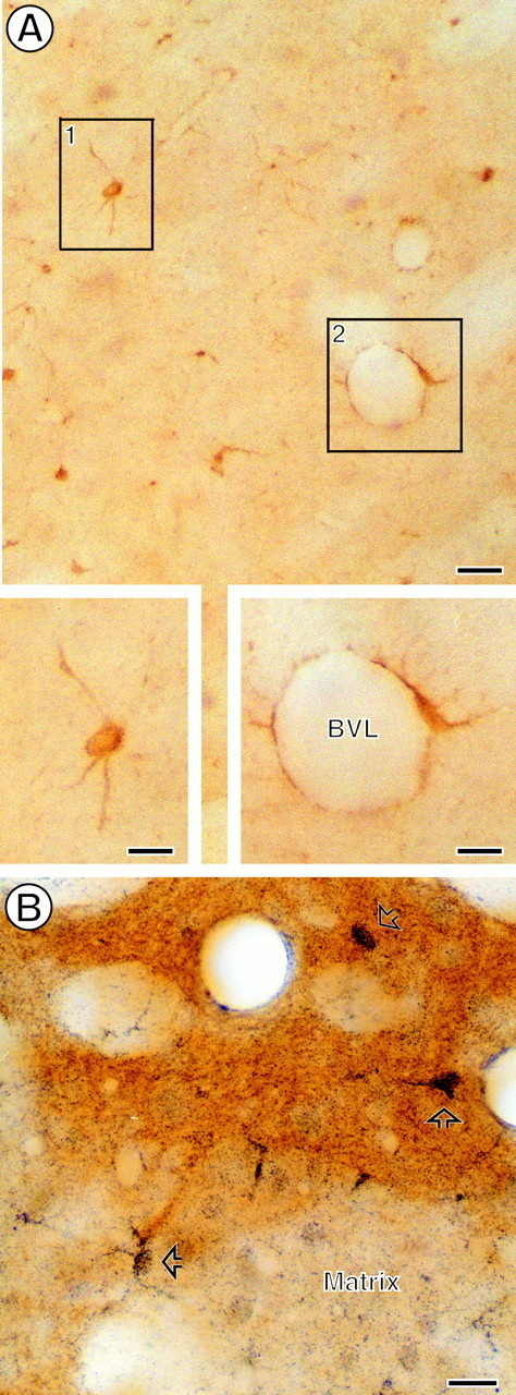
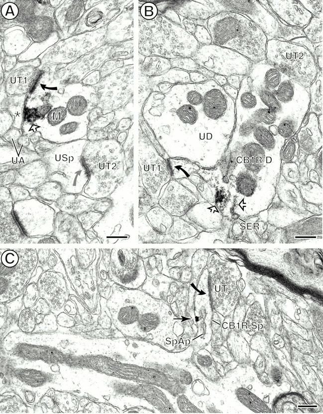
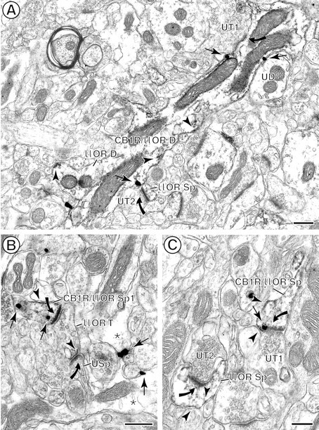
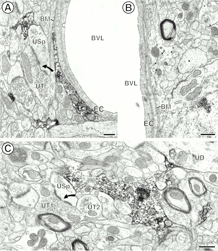
Cannabinoids and opioids are widely consumed drugs of abuse that produce motor depression, in part via respective activation of the cannabinoid subtype 1 receptor (CB1R) and the mu-opioid receptor (muOR), in the striatal circuitry originating in the caudate putamen nucleus (CPN). Thus, the CB1R and muOR may show similar targeting in the CPN. To test this hypothesis, we examined the electron microscopic immunocytochemical labeling of CB1R and muOR in CPN patches of rat brain. Of the CB1R-labeled profiles, 34% (588) were dendrites, presumably arising from spiny as well as aspiny-type somata, which also contained CB1R immunoreactivity. In dendrites, CB1R often was localized to nonsynaptic and synaptic plasma membranes, particularly near asymmetric excitatory-type junctions. Almost one-half of the CB1R-labeled dendrites contained muOR immunoreactivity, whereas only 20% of all muOR-labeled dendrites expressed CB1R. Axons and axon terminals as well as abundant glial processes also showed plasmalemmal CB1R and were mainly without muOR immunoreactivity. Many CB1R-labeled axon terminals were small and without recognizable synaptic junctions, but a few also formed asymmetric, or more rarely symmetric, synapses. The CB1R-labeled glial processes were often perivascular or perisynaptic, surrounding asymmetric excitatory-type axospinous synapses. Our results show that in CPN patches CB1R and muOR are targeted strategically to some of the same postsynaptic neurons, which may account for certain similarities in motor function. Furthermore, they also provide evidence that CB1R may play a major role in the modulation of presynaptic transmitter release and glial functions that are unaffected in large part by opioids active at muOR in CPN.


Figures

Fig. 1.
Fig. 1.
Photomicrographs showing striatal CB1R and μOR.A, Immunoperoxidase labeling for CB1R in a medium-sized spiny neuron (inset, boxed region 1) and in a perivascular glial cell (inset, boxed region 2). B, Immunogold labeling (open arrows) for CB1R within a striatal patch, which shows brown peroxidase reaction product identifying μOR immunoreactivity. Gold CB1R labeling also is seen in numerous cells and processes within the matrix compartment, which is without μOR peroxidase reaction product. Scale bars: A, 50 μm; insets inA, B, 25 μm.
Fig. 2.
Fig. 2.
Bar graphs showing the proportions of different profiles containing CB1R and μOR immunoreactivity in the CPN patch. These profiles include dendrites (DEN), dendritic spines (SP), axon terminals (TER), small axons (AX), and glial processes (GLIA). A, Percentage of each type of profile that contains either CB1R or CB1R and μOR. B, Percentage of each type of profile that contains either μOR or μOR and CB1R. Data were collected from nine vibratome sections through CPN patch regions of three animals, for a total analyzed surface of 8113.90 μm2. All sections were processed by using immunogold for CB1R and immunoperoxidase for μOR.
Fig. 3.
Fig. 3.
Electron micrographs from the CPN showing immunoperoxidase localization of CB1R within neuronal perikarya.A, Peroxidase reaction product (open arrows) on Golgi lamellae (G) close to the nucleus (Nu) and on a segment of the plasma membrane (PM) apposed to small unmyelinated axons (UA). Saccules of presumed smooth endoplasmic reticulum near a mitochondrion (M) also show peroxidase reaction product, whereas the rough endoplasmic reticulum (ER) is without immunoreactivity. B, Immunoperoxidase labeling within an endosome-like organelle (END). The END is located in a portion of the cytoplasm near a contact from an unlabeled terminal (UT).M, Mitochondrion, Nu, nucleus. Scale bars, 0.4 μm.
Fig. 4.
Fig. 4.
Bar graph showing the percentage of cytoplasmic versus plasmalemmal distribution of immunogold silver particles for CB1R in neuronal [dendrites (DEN), dendritic spines (SP), axon terminals (TER), small axons (AX)] and glial (GLIA) processes in the CPN patch. Data were collected from nine vibratome sections of three animals, for a total analyzed surface of 8113.90 μm2.
Fig. 5.
Fig. 5.
Dendrites and dendritic spines containing immunoperoxidase reaction product (A, B, open arrows) and immunogold (C, arrows) labeling for CB1R.A, Peroxidase reaction product on the plasma membrane and aggregated within the adjacent cytoplasm, separating the plasma membrane from a nearby mitochondrion (M). The labeling is near an asymmetric synapse (black curved arrow) established by an unlabeled terminal (UT1). The postsynaptic specialization in this dendrite also appears more electron dense than the one established (gray curved arrow) by a nearby unlabeled terminal (UT2) with an unlabeled spine (USp). The labeled dendritic plasma membrane is apposed by an unlabeled glial process (asterisk) located near small unmyelinated axons (UA). B, Peroxidase reaction product associated with smooth endoplasmic reticulum (SER) and with the plasma membrane at the base of a dendritic spine. The reaction product also is distributed throughout the spine neck and head, which receives an asymmetric synapse (curved arrow) from an unlabeled terminal (UT1). The CB1R-labeled dendrite (CB1R D) is apposed by another unlabeled terminal (UT2) and by an unlabeled dendrite (UD). C, Immunogold labeling on the plasma membrane close to the spinous apparatus (SpAp) in a dendritic spine neck. The labeled spine (CB1R Sp) receives an asymmetric synapse (curved arrow) from an unlabeled terminal (UT). Scale bars: A, B, 0.25 μm; C, 0.3 μm.
Fig. 6.
Fig. 6.
Colocalization of immunoperoxidase reaction product for μOR (arrowheads) and immunogold particles for CB1R (arrows) in spiny dendrites in the CPN patch.A, A longitudinal section of a dually labeled dendrite (CB1R/μOR D) containing CB1R and μOR. Both peroxidase labeling and gold labeling are shown prominently on the plasma membrane. Diffuse μOR-reaction product also is seen throughout the cytoplasm. CB1R/μOR D is contacted by two unlabeled terminals (UT1, UT2), but only UT1 forms an asymmetric synapse (curved arrow) with the dually labeled dendrite. UT2 forms an asymmetric synapse (curved arrow) with a μOR-labeled spine (μOR Sp). This dendrite also is apposed by a μOR-labeled dendrite (μOR D) and an unlabeled dendrite (UD). B, Dually labeled dendritic spine (CB1R/μOR Sp). Cytoplasmic CB1R immunogold labeling and μOR peroxidase labeling are seen on the plasma membrane. The peroxidase μOR also is distributed more diffusely throughout the spine. The spine receives an asymmetric synapse (curved arrow) from a μOR-labeled terminal (μOR T). C, Perisynaptic CB1R and μOR distributions near an asymmetric axospinous synapse from an unlabeled terminal (UT1). The peroxidase reaction product for μOR also is expressed, however, on other portions of the plasma membrane and cytoplasm in this spine as well as in a second spine (μOR Sp). This spine receives an asymmetric synapse from an unlabeled terminal (UT2). Scale bars: A, B, 0.4 μm;C, 0.25 μm.
Fig. 7.
Fig. 7.
Localization of CB1R in axon terminals, one of which contains μOR. A, Immunoperoxidase CB1R labeling (open arrow) on the plasma membrane of an axon terminal (CB1R T). The reaction product also is distributed diffusely on membranes of nearby small synaptic vesicles (SSVs). The CB1R-labeled terminal establishes an asymmetric synapse (curved arrow) with a CB1R-labeled dendritic spine (CB1R Sp). B, Cytoplasmic CB1R reaction product (open arrow) associated with tubulovesicles near a mitochondrion (M) in an axon terminal (CB1R T) without any recognizable synaptic specialization. The terminal also contains many small synaptic vesicles (SSVs). C, Plasmalemmal (open arrows) and cytoplasmic CB1R peroxidase labeling within an axon terminal (CB1R T) forming an asymmetric synapse (curved arrow) with an unlabeled spine (USp). One plasmalemmal aggregate of reaction product is seen on the presynaptic membrane of the asymmetric junction and is apposed by an unlabeled terminal (UT). The spine contains a spine apparatus (SpAp) in the neck region.D, Immunogold CB1R (arrow) labeling on the presynaptic plasma membrane of a terminal (CB1R T) forming asymmetric synapses (curved arrows) with two unlabeled dendritic spines (USp1, USp2). The location of the presynaptic gold particle is almost identical to that of the presynaptic plasmalemmal peroxidase reaction in C. The terminal and the spine are apposed by an unlabeled terminal (UT). E, Diffuse cytoplasmic immunoperoxidase reaction product for μOR is seen in an axon terminal (CB1R/μOR T) that contains gold particles (arrows) for CB1R. The terminal is apposed to an unlabeled dendrite (UD1) and establishes a synapse (solid thick arrow) with an unlabeled small dendrite (UD2). Scale bars: A, B, 0.2 μm; C, D, 0.25 μm; E, 0.4 μm.
Fig. 8.
Fig. 8.
Immunoperoxidase and immunogold CB1R labeling of glial processes (asterisks). A, Peroxidase reaction product within the cytoplasm of glial processes (asterisks) apposed to the basal membrane (BM) of endothelial cells (EC) lining the blood vessel lumen (BVL). Another CB1R-labeled glial process is apposed to an unlabeled spine (USp) that receives a perforated asymmetric synapse (curved arrow) from an unlabeled terminal (UT). B, Immunogold-labeled perivascular glial process. Abbreviations are the same as inA. C, Peroxidase labeling for CB1R within the cytoplasm and along discrete segments of the plasma membrane of a perisynaptic glial profile. The glial process apposes an unlabeled dendrite (UD) and an unlabeled spine (USp). The spine receives a perforated asymmetric synapse (curved arrow) from an unlabeled terminal (UT1) and is apposed to another unlabeled terminal (UT2). Scale bars: A, 0.3 μm; B, C, 0.5 μm.

Similar articles

Cited by

References

    1. Alkayed NJ, Birks EK, Narayanan J, Petrie KA, Kohler-Cabot AE, Harder DR. Role of P-450 arachidonic acid epoxygenase in the response of cerebral blood flow to glutamate in rats. Stroke. 1997;28:1066–1072. - PubMed
    1. Arvidsson U, Riedl M, Chakrabarti S, Lee J, Nakano AH, Dado RJ, Loh HH, Law P, Wessendorf MW, Elde R. Distribution and targeting of a μ-opioid receptor (MOR1) in brain and spinal cord. J Neurosci. 1995;15:3328–3341. - PMC - PubMed
    1. Auclair N, Otani S, Soubrié P, Crépel F. Cannabinoids modulate synaptic strength and plasticity at glutamatergic synapses of rat prefrontal cortex pyramidal neurons. J Neurophysiol. 2000;83:3287–3293. - PubMed
    1. Bouaboula M, Bourrié B, Rinaldi-Carmona M, Shire D, Le Fur G, Casellas P. Stimulation of cannabinoid receptor CB1 induces krox-24 expression in human astrocytoma cells. J Biol Chem. 2000;270:13973–13980. - PubMed
    1. Boudin H, Pelaprat D, Rostene W, Pickel VM, Beaudet A. Correlative ultrastructural distribution of neurotensin receptor proteins and binding sites in the rat substantia nigra. J Neurosci. 1998;18:8473–8484. - PMC - PubMed

Publication types

MeSH terms