. 2001 Feb 1;21(3):884-96.
doi: 10.1523/JNEUROSCI.21-03-00884.2001.

Excessive activation of serotonin (5-HT) 1B receptors disrupts the formation of sensory maps in monoamine oxidase a and 5-ht transporter knock-out mice

Affiliations

Excessive activation of serotonin (5-HT) 1B receptors disrupts the formation of sensory maps in monoamine oxidase a and 5-ht transporter knock-out mice

N Salichon et al. J Neurosci. .

Abstract

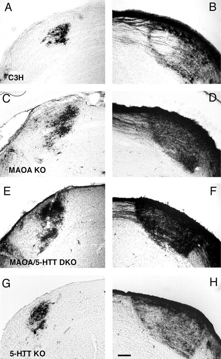
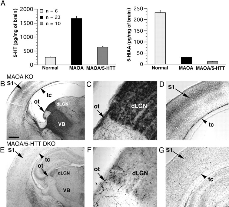
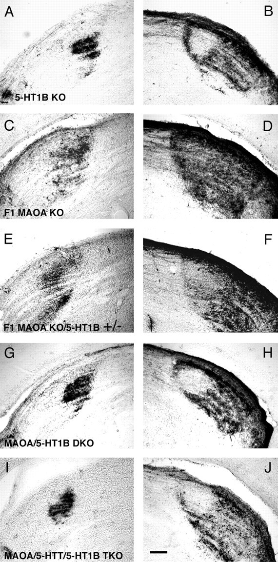
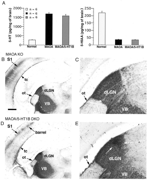
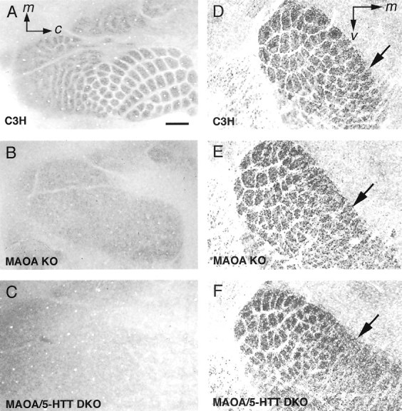
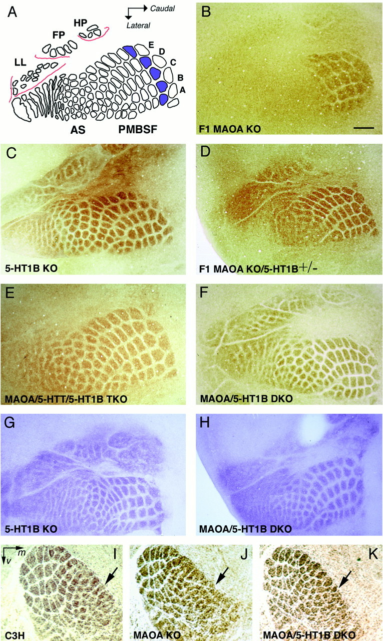
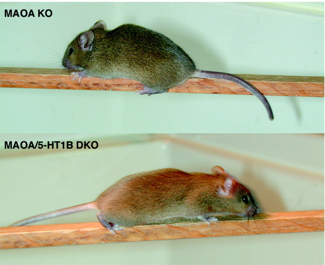
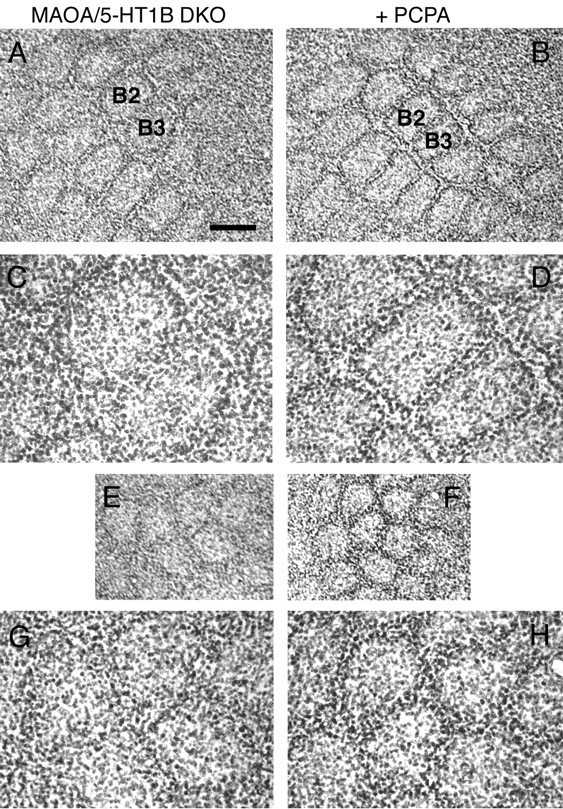
Deficiency in the monoamine degradation enzyme monoamine oxidase A (MAOA) or prenatal exposure to the monoamine uptake inhibitor cocaine alters behavior in humans and rodents, but the mechanisms are unclear. In MAOA knock-out mice, inhibiting serotonin synthesis during development can prevent abnormal segregation of axons in the retinogeniculate and somatosensory thalamocortical systems. To investigate this effect, we crossed MAOA knock-outs with mice lacking the serotonin transporter 5-HTT or the 5-HT1B receptor, two molecules present in developing sensory projections. Segregation was abnormal in 5-HTT knock-outs and MAOA/5-HTT double knock-outs but was normalized in MAOA/5-HT1B double knock-outs and MAOA/5-HTT/5-HT1B triple knock-outs. This demonstrates that the 5-HT1B receptor is a key factor in abnormal segregation of sensory projections and suggests that serotonergic drugs represent a risk for the development of these projections. We also found that the 5-HT1B receptor has an adverse developmental impact on beam-walking behavior in MAOA knock-outs. Finally, because the 5-HT1B receptor inhibits glutamate release, our results suggest that visual and somatosensory projections must release glutamate for proper segregation.


Figures

Fig. 1.
Fig. 1.
Lack of segregation of retinal afferents in MAOA KO mice, 5-HTT KO mice, and MAOA/5-HTT DKO mice. HRP was injected into the left eye in adult mice (P30 or more). Ipsilateral projections to the dLGN are shown in the left-hand column(A, C, E,G), and contralateral projections are shown in theright-hand column (B, D,F, H). The sections illustrated were chosen at similar rostrocaudal levels. A, B, In wild-type C3H mice, the eye-specific segregation is clear with tightly packed ipsilateral retinal axons (A) and a clear gap contralaterally (B). Within this gap, the only contralateral fibers correspond to retinal axon bundles traversing the nucleus. C, D, In MAOA KO mice (mixed C3H-129 genetic background), the ipsilateral projections are diffuse in the dLGN (C), and no gap is visible contralaterally (D). This aspect is identical to that of the MAOA KO mice having an inbred strain background (C3H, 129, or C57BL).E, F, In MAOA/5-HTT DKO mice (mixed 129-C3H-C57BL-CD1 background), there is no segregation of the ipsilateral and contralateral retinal projections. G, H, In 5-HTT KO mice (C3H-backcrossed), the contralateral projection does not retract completely from the normal ipsilateral territory. Scale bar, 0.1 mm.
Fig. 2.
Fig. 2.
Comparison of 5-HT brain levels and 5-HT localization in the MAOA KO and MAOA/5-HTT DKO mice. A, 5-HT and 5-HIAA brain levels were measured in normal and mutant mice aged P2. In normal mice, the ratio 5-HT/5-HIAA is low: 1.2 (5-HT concentration, 278 ± 14 pg/mg; 5-HIAA concentration, 232 ± 15 pg/mg; mean ± SEM, n = 6). In MAOA KO mice, the ratio 5-HT/5-HIAA is very high: 53.9 (5-HT, 1672 ± 52 pg/mg; 5-HIAA, 31 ± 2 pg/mg; n = 23). In MAOA/5-HTT DKO mice, the ratio 5-HT/5-HIAA is similarly high: 53.5 (5-HT, 642 ± 24 pg/mg; 5-HIAA, 12 ± 2 pg/mg;n = 10). B–D, 5-HT immunostaining in the MAOA KO mice (aged P4) shows an excessive accumulation of the amine in the VB and dLGN neurons (B), as well as in the somatosensory thalamocortical fibers (tc) in the somatosensory cortex (S1) (B,D). 5-HT is also visible in the retinal axons that course superficially to the dLGN in the optic tract (ot) (C).E–G, In MAOA/5-HTT DKO mice (P4), 5-HT immunostaining is strongly reduced in the dLGN (E,F), the VB (E), and S1 (E,G). Retinal axons in the optic tract contain no detectable 5-HT (F). Scale bar:B, E, 0.4 mm; C,F, 0.025 mm; D, G, 0.2 mm.
Fig. 3.
Fig. 3.
Deficient segregation of retinogeniculate axons is mediated by 5-HT1B receptors. HRP was injected into the left eye in adult mice (P30 or more). Ipsilateral projections to the dLGN are shown on the left (A, C,E, G, I), and contralateral projections on the right (B,D, F, H,J). The sections illustrated were chosen at similar rostrocaudal levels. A, B, Normal segregation in 5-HT1B KO mice (C3H-backcrossed). C, D, F1 control MAOA KO mice [F1 hybrid males from crosses between MAOA KO (C3H) females and normal 129 males] have identical abnormalities as the other MAOA KO strains (Fig. 1C,D). E, F, F1 MAOA KO mice that are heterozygous knock-outs for the 5-HT1B gene [F1 hybrid males from crosses between MAOA KO (C3H) females and 5-HT1B KO (129) males] display a partial correction of the abnormalities with the outline of a gap contralaterally (F), whereas the ipsilateral projections are still exuberant (E). G, H, In MAOA/5-HT1B DKO mice (C3H-backcrossed), the eye-specific segregation is normal and comparable with that of wild-type control mice (Fig.1A,B). I, J, In MAOA/5-HTT/5-HT1B TKO mice, the eye-specific segregation of retinogeniculate projections is also normal. Scale bar, 0.1 mm.
Fig. 4.
Fig. 4.
Comparison of 5-HT brain levels and 5-HT localization in MAOA KO and MAOA/5-HT1B DKO mice. A, In P6 MAOA KO (5-HT concentration, 1694 ± 38 pg/mg; mean ± SEM, n = 6) and MAOA/5-HT1B DKO mice (5-HT, 1592 ± 34 pg/mg; n = 6), 5-HT brain levels are similarly increased compared with the normals (5-HT, 278 ± 9 pg/mg; n = 6). In both mutants, the 5-HIAA brain levels are very low (5-HIAA in MAOA KO, 38 ± 2 pg/mg; 5-HIAA in MAOA/5-HT1B DKO, 36 ± 2 pg/mg; n = 6) compared with those of normal mice (5-HIAA, 220 ± 8 pg/mg;n = 6). B, C, 5-HT immunostaining in MAOA KO (P6) shows accumulation in the dLGN and VB (B), as well as in retinal fibers (C) and somatosensory thalamocortical fibers (B). D, E, Excess 5-HT accumulation in the thalamus and thalamocortical fibers is identical in MAOA/5-HT1B DKO mice and MAOA KO mice. Furthermore, 5-HT immunostaining reveals the ipsilateral patch of retinal terminals in MAOA/5-HT1B DKO mice (E, white arrows), whereas this is not visible in MAOA KO mice (C). Abbreviations are as in Figure 2. Scale bar: B, D, 0.4 mm; C, E, 0.2 mm.
Fig. 5.
Fig. 5.
Similar alterations in the somatosensory systems of MAOA KO mice and MAOA/5-HTT DKO mice. A–C, Normal barrel field in (P8) C3H mice revealed by CO-staining on a tangential section through layer IV (A). Barrel-less phenotype of (P8) MAOA KO mice (B) and (P10) MAOA/5-HTT DKO mice (C). Genetic backgrounds are as indicated in the legend to Figure 1. D–F, Normal barreloids in P7 C3H mice (D). Large barreloids of the VB appear to be close to normal in P7 MAOA KO mice (E) and P7 MAOA/5-HTT DKO mice (F). However, the small medioventral barreloids corresponding to the AS vibrissae are not delineated (arrow). Scale bar: A–C, 0.5 mm;D–F, 0.125 mm.
Fig. 6.
Fig. 6.
Effects of removing the 5-HT1B receptors on the cortical and thalamic somatosensory maps in normal mice, MAOA KO mice, and MAOA/5-HTT DKO mice. A, Scheme of a normal barrel field showing the different subfields: the posteromedial barrel subfield (PMBSF), anterior snout (AS), forepaw (FP), lower lip (LL), and hindpaw (HP). The first barrels of the five rows (A–E) of large whiskers are colored. B–F, CO histochemistry on tangential 40 μm sections from P8–P12 flattened hemispheres. B, In the barrel field of (P9) F1 MAOA KO mice, some patterning is visible in the PMBSF, particularly in row B. However, these barrels have a blurred aspect and are visible on a single section instead of two or three sections per hemisphere in normal mice. C, Normal barrel field in (P8) 5-HT1B KO mice (129 genetic background).D, F1 MAOA KO/5-HT1B+/− mice display normal barrels in the PMBSF, whereas small barrels in the AS are missing (P9).E, Normal barrel field in (P12) MAOA/5-HTT/5-HT1B TKO mice. F, Normal barrel field in P8 MAOA/5-HT1B DKO mice.G, H, 5-HTT immunostaining shows that thalamocortical patterning is normal in P6 5-HT1B KO mice (G) and P6 MAOA/5-HT1B DKO mice (H).I–K, Normal patterning in the thalamus of MAOA/5-HT1B DKO mice. I, Normal barreloids in P5 C3H mice.J, Abnormal smallest barreloids in P5 MAOA KO mice.K, Normal barreloids in P5 MAOA/5-HT1B DKO mice. Scale bar: B–H, 0.5 mm; I–K, 0.19 mm.
Fig. 7.
Fig. 7.
Removal of the 5-HT1B receptor restores normal beam-walking ability in MAOA KO mice. When placed on a narrow beam, MAOA KO mice with a C3H background crawl rather than walk, with hindlimbs grasping the beam, whereas MAOA/5HT1B DKO mice walk as C3H control mice (MAOA KO mice walk normally on slightly larger beams). The different coat color of the DKO mouse is an artifact.
Fig. 8.
Fig. 8.
Incomplete segregation of the barrel cytoarchitecture in P16 MAOA/5-HT1B DKO mice. A–H, Lack of clear segregation of cortical cells in the PMBSF and AS of P16 MAOA/5-HT1B DKO mice compared with MAOA/5-HT1B DKO mice (from the same litter) treated with PCPA for P0–P8, to reduce 5-HT brain levels.A–D, PCPA-treated MAOA/5-HT1B DKO mice show a sharp pattern of cortical cells in the PMBSF barrels (B) compared with untreated littermates (A). The B2 and B3 barrels (A,B) have been enlarged (C,D) to show the normal distribution of cells in the treated mouse. E–H, Improved cortical cytoarchitecture in the AS of PCPA-treated MAOA/5-HT1B DKO mice (F,H), compared with untreated littermates (E,G). Nevertheless, the sides of neighboring barrels are not clearly separated by septa (H). An absence of septa in this region was similarly observed in wild-type 129 mice and 5-HT1B KO mice (data not shown). Scale bar: A, B,E, F, 0.3 mm; C,D, G, H, 0.075 mm.

Similar articles

Cited by

References

    1. Adams JC. Stabilizing and rapid thionin staining of TMB-based HRP reaction product. Neurosci Lett. 1980;17:7–9. - PubMed
    1. Agmon A, Yang LT, O'Dowd DK, Jones EG. Organized growth of thalamocortical axons from the deep tier of terminations into layer IV of developing mouse barrel cortex. J Neurosci. 1993;13:5365–5382. - PMC - PubMed
    1. Bengel D, Murphy DL, Andrews AM, Wichems CH, Feltner D, Heils A, Mössner R, Westphal H, Lesch KP. Altered brain serotonin homeostasis and locomotor insensitivity to 3,4-methylenedioxymethamp-hetamine (“Ecstasy”) in serotonin transporter-deficient mice. Mol Pharmacol. 1998;53:649–655. - PubMed
    1. Bennett-Clarke CA, Leslie MJ, Chiaia NL, Rhoades RW. Serotonin 1B receptors in the developing somatosensory and visual cortices are located on thalamocortical axons. Proc Natl Acad Sci USA. 1993;90:153–157. - PMC - PubMed
    1. Bennett-Clarke CA, Chiaia NL, Rhoades RW. Thalamocortical afferents in rat transiently express high-affinity serotonin uptake sites. Brain Res. 1996;733:301–306. - PubMed

Publication types

MeSH terms