. 2001 Feb 1;21(3):897-910.
doi: 10.1523/JNEUROSCI.21-03-00897.2001.

GABAb receptors regulate chick retinal calcium waves

Affiliations

GABAb receptors regulate chick retinal calcium waves

M Catsicas et al. J Neurosci. .

Abstract

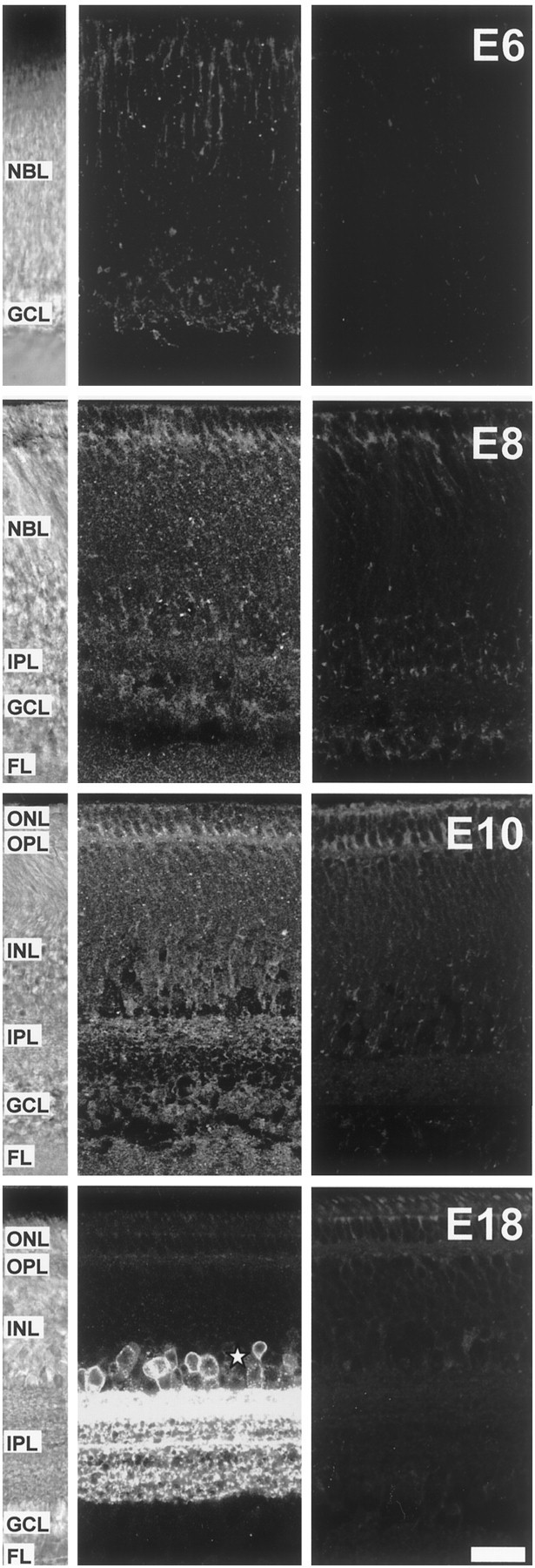
Correlated spiking activity and associated Ca(2+) waves in the developing retina are important in determining the connectivity of the visual system. Here, we show that GABA, via GABA(B) receptors, regulates the temporal characteristics of Ca(2+) waves occurring before synapse formation in the embryonic chick retina. Blocking ionotropic GABA receptors did no affect these Ca(2+) transients. However, when these receptors were blocked, GABA abolished the transients, as did the GABA(B) agonist baclofen. The action of baclofen was prevented by the GABA(B) antagonist p-3-aminopropyl-p-diethoxymethyl phosphoric acid (CGP35348). CGP35348 alone increased the duration of the transients, showing that GABA(B) receptors are tonically activated by endogenous GABA. Blocking the GABA transporter GAT-1 with 1-(4,4-diphenyl-3-butenyl)-3-piperidine carboxylic acid (SKF89976A) reduced the frequency of the transients. This reduction was prevented by CGP35348 and thus resulted from activation of GABA(B) receptors by an increase in external [GABA]. The effect of GABA(B) receptor activation persisted in the presence of activators and blockers of the cAMP-PKA pathway. Immunocytochemistry showed GABA(B) receptors and GAT-1 transporters on ganglion and amacrine cells from the earliest times when Ca(2+) waves occur (embryonic day 8). Patch-clamp recordings showed that K(+) channels on ganglion cell layer neurons are not modulated by GABA(B) receptors, whereas Ca(2+) channels are; however, Ca(2+) channel blockade with omega-conotoxin-GVIA or nimodipine did not prevent Ca(2+) waves. Thus, the regulation of Ca(2+) waves by GABA(B) receptors occurs independently of N- and L-type Ca(2+) channels and does not involve K(+) channels of the ganglion cell layer. GABA(B) receptors are likely to be of key importance in regulating retinal development.


Figures

Fig. 1.
Fig. 1.
Ionotropic GABA receptors are expressed and depolarize retinal cells when Ca2+ waves can first be detected. A, Spontaneous Ca2+transients from an E9 retina in control situation. The levels of fluorescence averaged over the field of view were normalized to the basal level at the beginning of the recording session.B, Traces plotting the changes in fluorescence in an E6 (top) and an E12 (bottom) retina. Application of GABA (100 μm) elicited a transient increase in [Ca2+]i at both ages (left traces), which was largely prevented by coapplication of the GABAA receptor antagonist bicuculline (50 μm;middle traces). The response to GABA recovered after 15 min washout of bicuculline (right traces).C, Developmental profile of the response to GABA. The amplitude of the responses is plotted as a percentage of the maximum response, observed at E8. All responses observed were increases in [Ca2+]i. GABA responses were present at E4 and peaked at E8. By E14, GABA no longer evoked increases in [Ca2+]i(see Results). n = 3–6 retinas for each age. The experiments in B and C were done in HEPES-buffered Ringer's solution and performed at room temperature to prevent spontaneous Ca2+ transients.
Fig. 2.
Fig. 2.
Blocking ionotropic GABA receptors does not affect spontaneous Ca2+ transients under control conditions. A, Ca2+ transients recorded from an E9 retina upon successive application of drugs, as indicated. The activity recovered in the presence of bicuculline and picrotoxin upon washout of GABA; some transients had an increased duration under these conditions. B, Histogram of the results pooled from four retinas at E9. The frequency (gray bars) and duration (black bars) of Ca2+transients in the presence of drugs is shown as a percentage change compared with the control condition. Application of the ionotropic GABA receptor antagonists bicuculline (bicu; 50 μm) and PTX (100 μm) did not affect the frequency of the transients. In contrast, application of GABA (50 μm) in the presence of bicuculline and PTX abolished the transients. After a 15 min wash of the drugs, the transients recovered with a frequency initially higher than in control (189 ± 29%;p = 0.09) and a duration comparable with that in control.
Fig. 3.
Fig. 3.
GABAB receptors modulate spontaneous Ca2+ transients. A, B,D, Activating GABAB receptors with the agonist baclofen (100 μm) reversibly abolished the transients. The effect of baclofen was dose-dependent (D) and prevented by application of the GABAB receptor antagonist CGP35348 (CGP, 100 μm; B, D,n = 3–4 retinas per condition). C,E, Blocking GABAB receptors prolongs spontaneous Ca2+ transients. Spontaneous Ca2+ transients in an E9 retina under control conditions (top trace), in the presence of CGP35348 (100 μm; middle trace), and after 10 min washout of CGP35348 (bottom trace). E, Pooled data taken from four retinas (comprising 4–10 transients for each condition) showing the duration of Ca2+transients as measured at half-peak.
Fig. 4.
Fig. 4.
Blocking neuronal GABA transport reduces the frequency of spontaneous Ca2+ transients.A, The frequency of transients was reversibly reduced with application of the GAT-1 transporter blocker SKF89976A (SKF; 100 μm; p < 0.001; n = 7). B, The effect of SKF was prevented by coapplication of CGP35348 (CGP; 100 μm). In the presence of SKF89976A and CGP35348, the frequency of Ca2+ transients increased approximately threefold compared with control levels (p < 0.01). This overshoot in the frequency was prevented if bicuculline (bicu; 50 μm) and PTX (100 μm) were applied together with SKF89976A and CGP35348 (n = 5 retinas). The duration of the transients in the presence of SKF89976A and CGP35348 was similar to that in control (data not shown).
Fig. 5.
Fig. 5.
GABAB receptors act in push–pull with a cAMP-dependent pathway. A, Spontaneous Ca2+ transients recorded from an E9 retina upon sequential application of forskolin and baclofen, as indicated.B, Pooled data taken from six retinas of the Ca2+ transient frequency normalized to control in each condition. Activating adenylate cyclase with forskolin (1 μm) increased the frequency of transients. This effect could be reversibly prevented by coapplication of baclofen (50 μm). The effect of both drugs reversed after 20 min wash.C, Bath application of 8-bromo-cAMP (500 μm, 30 min) mimicked the effect of forskolin (n = 2) and was antagonized by coapplication of baclofen (50 μm). Blocking protein kinase A with bath application of Rp-cAMPS (500 μm, 20–30 min) reduced the frequency of transients, which could be further decreased by baclofen (50 μm). The activity recovered only partially (D). D, Pooled data from three retinas showing the frequency of Ca2+ transients normalized to control.
Fig. 6.
Fig. 6.
Expression of GABAB receptors in the developing chick retina. Confocal microscope sections of GABAB receptor immunostaining of transverse vibratome slices from E6, E8, E10, and E18 chick retina. Left panels, White-light micrographs from the slices in themiddle panels showing the structure of the retina for each age. Middle panels, Sections immunoreacted with the anti-GABAB receptor antibody. Right panels, Negative controls in which the primary antibody was omitted. At E6, GABABreceptor-immunoreactive cells, presumptive ganglion cells, were present at the inner margin of the NBL in which the GCL forms. Punctate immunostaining was also present through the thickness of the NBL. At E8, the GCL is separated from the NBL by a layer of processes, the IPL. GABAB receptor immunoreactivity was prominent in cells of the GCL. Cells at the inner margin of the NBL, presumptive amacrine cells, were also positive, as well as fibers of the IPL. Immunoreactivity was still present in the rest of the NBL, in particular surrounding distal cell bodies of the NBL outlining the prospective OPL (presumptive horizontal cells). At E10, the pattern of staining was essentially the same as at E8, except there were more immunoreactive plaques in the outer retina at later times. By E18, the retina has reached its mature morphology. GABAB receptor immunoreactivity was present in cells of the GCL (ganglion and displaced amacrine cells), of the inner part of the INL (amacrine and displaced ganglion cells), and bordering the OPL. The pattern of staining showed a clear lamina distribution in both OPL and IPL, and immunoreactivity was present in the FL. Some autofluorescence is apparent in the outer retina at the level of the outer nuclear layer (ONL) and OPL (see negative controls). Scale bar, 25 μm.
Fig. 7.
Fig. 7.
Expression of GAT-1 transporters in the developing chick retina. Confocal microscope sections of GAT-1 transporter immunostaining of transverse vibratome slices from E6, E8, E10, and E18 chick retina. Left panels, White-light micrographs from the slices in the middle panels showing the structure of the retina for each age. Middle panels, Sections immunoreacted with the anti-GAT-1 antibody. Right panels, Negative controls in which the primary antibody was omitted. At E6, GAT-1 immunostaining was punctate and restricted to the outer NBL and the GCL. At E8, immunoreactive puncta were distributed throughout the retina. By E10, GAT-1 lamina-specific staining was apparent in the IPL, with laminas closer to the INL more strongly labeled. At E18, GAT-1 staining is restricted to the IPL, in which it has a clear laminar distribution, and to somata in the proximal INL, presumptive amacrine cells (asterisk). Some autofluorescence is apparent in the outer retina at the level of the ONL and OPL (see negative controls). Scale bar, 20 μm.
Fig. 8.
Fig. 8.
GABAB receptors do not modulate K+ currents in ganglion cell layer neurons.A, Currents evoked in an E9 neuron by 200 msec voltage steps to the indicated potentials from a holding potential of −90 mV in control solution (left) and in the presence of 100 μm baclofen (right). B, Current–voltage relationship from six neurons evoked by voltage steps in 10 mV increments from −139 to +4 mV from a holding potential of −90 mV in the absence (control, filledcircles) and presence of 100 μm baclofen (open circles). These data have been corrected for any voltage error produced by current flow through the access resistance in the steady state.
Fig. 9.
Fig. 9.
Effect of GABAB receptors on the Ca2+ current in ganglion cell layer neurons.A, Current–voltage relationship from an E11 cell in response to depolarizing steps in 10 mV increments from a holding potential of −90 to +60 mV in control solution. B, Current traces in response to a depolarizing step to 0 mV from a holding potential of −90 mV in control solution and during the sequential application of the L-type Ca2+ channel blocker nimodipine (10 μm) and of the peptide N-type Ca2+ channel blocker cgtxGVIA (5 μm).wash indicates washout of nimodipine before application of cgtxGVIA. The effect of cgtxGVIA was irreversible. C, Current traces in response to a depolarizing step to 0 mV from a holding potential of −90 mV in control solution (5 mmBa2+ and 1 μm TTX;left) in the presence of baclofen (100 μm;middle) and after wash to control (right). The histogram shows pooled data from 15 cells between E9 and E12 in which the amplitude of the currents was normalized to the control current and plotted as percentage change from control. Baclofen reversibly decreased the current by ∼50% (p < 0.001). D, Current traces in response to a depolarizing step to 0 mV from a holding potential of −90 mV in control solution (left), in the presence of CGP35348 (100 μm; CGP;middle) and after wash (right). The histogram shows pooled data from six cells at E10–E11 in which the amplitude of the currents was normalized to the control current and plotted as percentage change from control. CGP35348 reversibly increased the current by ∼10% (p < 0.01). E, Example of a Lucifer yellow-filled ganglion cell in an E10 retina photographed after whole-cell patch-clamp recording in situ. The patch pipette, still attached to the cell, is visible to the left. The image is an overlay of four photographs taken at different focal planes to reveal the extent of the dendritic tree. An asterisk marks the axon, running more superficial than the dendrites in the fiber layer. Scale bar, 20 μm.
Fig. 10.
Fig. 10.
GABAB receptors modulate Ca2+ currents in ganglion cell layer neurons in a cAMP–PKA-independent manner. A, B, Current traces in response to a depolarizing step to 0 mV from a holding potential of −90 mV in the presence of either cAMP (2 mm; A) or the PKA blocker Rp-cAMPS (500 μm; B, left traces) in the patch pipette, upon application of baclofen (100 μm;middle traces) and after wash of baclofen (right traces). The histograms show pooled data from 10 (A) or six (B) cells at E9–E11 in which the amplitude of the currents in baclofen was normalized to the currents in the presence of cAMP or Rp-cAMPS, respectively, and plotted as percentage change.
Fig. 11.
Fig. 11.
Blocking N- or L-type Ca2+channels does not block spontaneous Ca2+ transients. Application of cgtxGVIA (5 μm; A) or nimodipine (10 μm; B) to block N- and L-type Ca2+ channels, respectively, did not affect the frequency of transients (n = 4 retinas). Coapplication of baclofen (10 μm) reversibly abolished the transients in both cases.
Fig. 12.
Fig. 12.
Mechanisms regulating calcium waves during early development (E8–E12). Before synaptogenesis, the frequency of the calcium waves that propagate between ganglion and amacrine cells (GC and AC, respectively) is regulated by a number of the transmitter molecules released by amacrine cells, including ACh acting at nicotinic receptors, glycine (Gly), adenosine (data not shown), and GABA acting at GABAB receptors. The GABA transporter GAT-1 acts to keep GABA in the extracellular space at low levels. Glutamate receptors appear not to be involved in the regulation of calcium waves until after E12, at which time glutamate antagonists block wave activity probably through an action at the newly formed synapses between bipolar and ganglion cells (not shown). At both early and late times, gap junctions (GJ) are important for wave propagation because agents that block the junctions inhibit the waves, whereas agents known to influence coupling through modulation of intracellular levels of cAMP (forskolin, dopamine, and adenosine) increase wave frequency (see Discussion). Positive and negative signs indicate wave-promoting and wave-inhibiting signals, respectively. Shaded figures and pathways indicate that they are present but not instrumental in wave activity.

Similar articles

Cited by

References

    1. Amico C, Marchetti C, Nobile M, Usai C. Pharmacological types of calcium channels and their modulation by baclofen in cerebellar granules. J Neurosci. 1995;15:2839–2848. - PMC - PubMed
    1. Bettler B, Kaupmann K, Bowery N. GABAB receptors: drugs meet clones. Curr Opin Neurobiol. 1998;8:345–350. - PubMed
    1. Blazynski C. Displaced cholinergic, GABAergic amacrine cells in the rabbit retina also contain adenosine. Vis Neurosci. 1989;3:425–431. - PubMed
    1. Bodnarenko SR, Jeyarasasingam G, Chalupa LM. Development and regulation of dendritic stratification in retinal ganglion cells by glutamate-mediated afferent activity. J Neurosci. 1995;15:7037–7045. - PMC - PubMed
    1. Bonness V, Catsicas M, Cunningham J, Hutson P, Murray F, Neal M, Mobbs P. Depolarization-evoked transmitter release and agonist-evoked increases in [Ca2+]i in the early development of the chick retina. J Physiol (Lond) 1996;494:79.P.

Publication types

MeSH terms

Substances

LinkOut - more resources