

Expression and localization of endothelin receptors: implications for the involvement of peripheral glia in nociception
- PMID: 11157085
- PMCID: PMC6762331
- DOI: 10.1523/JNEUROSCI.21-03-00999.2001
Expression and localization of endothelin receptors: implications for the involvement of peripheral glia in nociception
Abstract
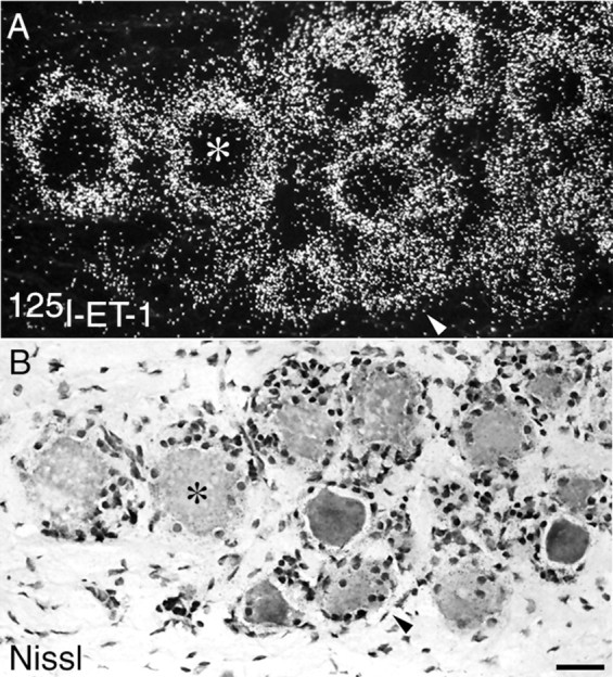
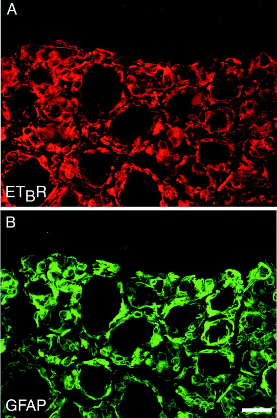
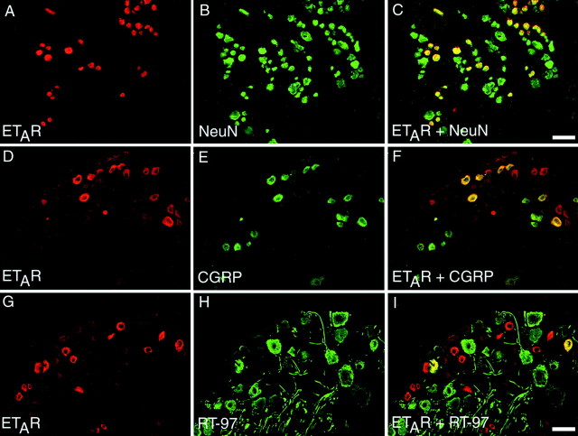
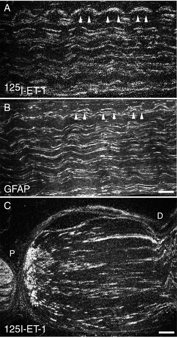
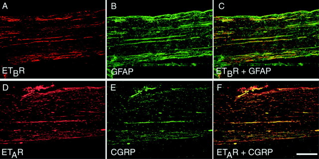
The endothelins (ETs) are peptides that have a diverse array of functions mediated by two receptor subtypes, the endothelin A receptor (ET(A)R) and the endothelin B receptor (ET(B)R). Pharmacological studies have suggested that in peripheral tissues, ET(A)R expression may play a role in signaling acute or neuropathic pain, whereas ET(B)R expression may be involved in the transmission of chronic inflammatory pain. To begin to define the mechanisms by which ET can drive nociceptive signaling, autoradiography and immunohistochemistry were used to examine the distribution of ET(A)R and ET(B)R in dorsal root ganglia (DRG) and peripheral nerve of the rat, rabbit, and monkey. In DRG and peripheral nerve, ET(A)R-immunoreactivity was present in a subset of small-sized peptidergic and nonpeptidergic sensory neurons and their axons and to a lesser extent in a subset of medium-sized sensory neurons. However, ET(B)R-immunoreactivity was not seen in DRG neurons or axons but rather in DRG satellite cells and nonmyelinating ensheathing Schwann cells. Thus, when ETs are released in peripheral tissues, they could act directly on ET(A)R-expressing sensory neurons and on ET(B)R-expressing DRG satellite cells or nonmyelinating Schwann cells. These data indicate that ETs can have direct, nociceptive effects on the peripheral sensory nervous system and that peripheral glia may be directly involved in signaling nociceptive events in peripheral tissues.
Figures
Similar articles
-
Distribution of the adhesion molecules N-CAM and L1 on peripheral neurons and glia in adult rats.J Neurocytol. 1986 Dec;15(6):799-815. doi: 10.1007/BF01625196. J Neurocytol. 1986. PMID: 3819781
-
Expression of endothelin-B receptors by glia in vivo is increased after CNS injury in rats, rabbits, and humans.Exp Neurol. 1997 May;145(1):180-95. doi: 10.1006/exnr.1997.6468. Exp Neurol. 1997. PMID: 9184120
-
S100beta and fibroblast growth factor-2 are present in cultured Schwann cells and may exert paracrine actions on the peripheral nerve injury.Acta Cir Bras. 2008 Nov-Dec;23(6):555-60. doi: 10.1590/s0102-86502008000600014. Acta Cir Bras. 2008. PMID: 19030756
-
Ultrastructure of dorsal root ganglia.Cell Tissue Res. 2023 Jul;393(1):17-36. doi: 10.1007/s00441-023-03770-w. Epub 2023 Apr 20. Cell Tissue Res. 2023. PMID: 37079097 Free PMC article. Review.
-
Estrogen receptor immunoreactivity in Schwann-like brain macroglia.J Neurobiol. 1999 Sep 15;40(4):458-70. doi: 10.1002/(sici)1097-4695(19990915)40:4<458::aid-neu4>3.0.co;2-9. J Neurobiol. 1999. PMID: 10453049 Review.
Cited by
-
Mechanism of cancer pain.Mol Interv. 2010 Jun;10(3):164-78. doi: 10.1124/mi.10.3.7. Mol Interv. 2010. PMID: 20539035 Free PMC article. Review.
-
Thrombin inhibits NMDA-mediated nociceptive activity in the mouse: possible mediation by endothelin.J Physiol. 2003 Jun 15;549(Pt 3):903-17. doi: 10.1113/jphysiol.2002.036384. Epub 2003 Apr 25. J Physiol. 2003. PMID: 12717003 Free PMC article.
-
The Involvement of Endothelin Pathway in Chronic Psychological Stress-Induced Bladder Hyperalgesia Through Capsaicin-Sensitive C-Fiber Afferents.J Inflamm Res. 2022 Feb 22;15:1209-1226. doi: 10.2147/JIR.S346855. eCollection 2022. J Inflamm Res. 2022. PMID: 35228812 Free PMC article.
-
Updated mechanisms underlying sickle cell disease-associated pain.Neurosci Lett. 2019 Nov 1;712:134471. doi: 10.1016/j.neulet.2019.134471. Epub 2019 Sep 7. Neurosci Lett. 2019. PMID: 31505241 Free PMC article. Review.
-
Endothelin-1 and endothelin receptors in the basilar artery of the capybara.J Mol Histol. 2005 Feb;36(1-2):25-34. doi: 10.1007/s10735-004-2912-0. J Mol Histol. 2005. PMID: 15703996
References
-
- Aramori I, Nakanishi S. Coupling of two endothelin receptor subtypes to differing signal transduction in transfected Chinese hamster ovary cells. J Biol Chem. 1992;267:12468–12474. - PubMed
-
- Baba A. Role of endothelin B receptor signals in reactive astrocytes. Life Sci. 1998;62:1711–1715. - PubMed
-
- Basbaum AI. Spinal mechanisms of acute and persistent pain. Reg Anesth Pain Med. 1999;24:59–67. - PubMed
-
- Berger UV, Hediger MA. Distribution of the glutamate transporters GLAST and GLT-1 in rat circumventricular organs, meninges, and dorsal root ganglia. J Comp Neurol. 2000;421:385–399. - PubMed
-
- Bozic CR, Lu B, Hopken UE, Gerard C, Gerard NP. Neurogenic amplification of immune complex inflammation. Science. 1996;273:1722–1725. - PubMed
Publication types
MeSH terms
Substances
Grants and funding
LinkOut - more resources
Full Text Sources
Medical