

Heterogeneity in the basic membrane properties of postnatal gonadotropin-releasing hormone neurons in the mouse
- PMID: 11157093
- PMCID: PMC6762336
- DOI: 10.1523/JNEUROSCI.21-03-01067.2001
Heterogeneity in the basic membrane properties of postnatal gonadotropin-releasing hormone neurons in the mouse
Abstract
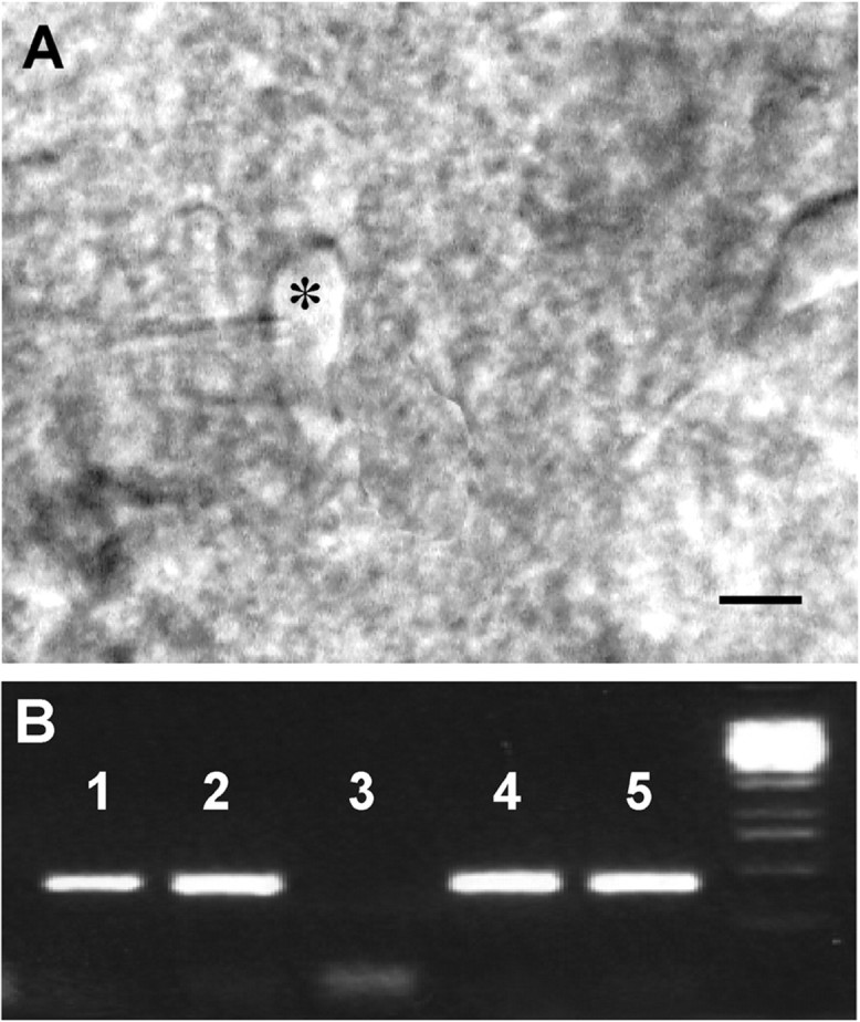
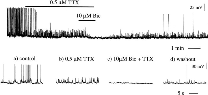
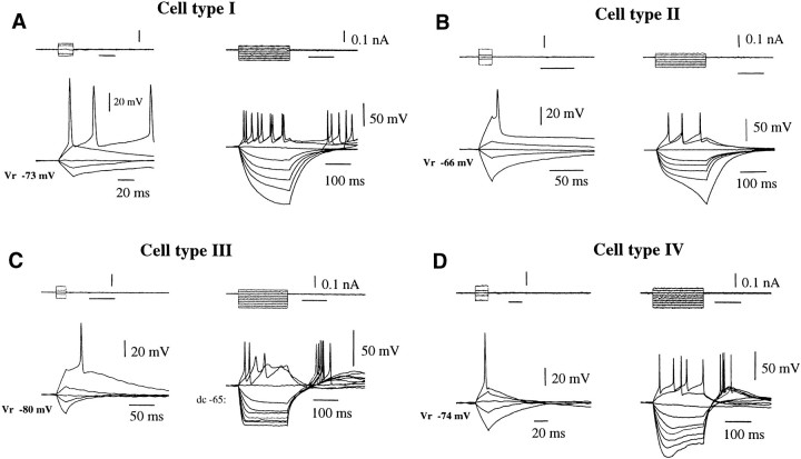
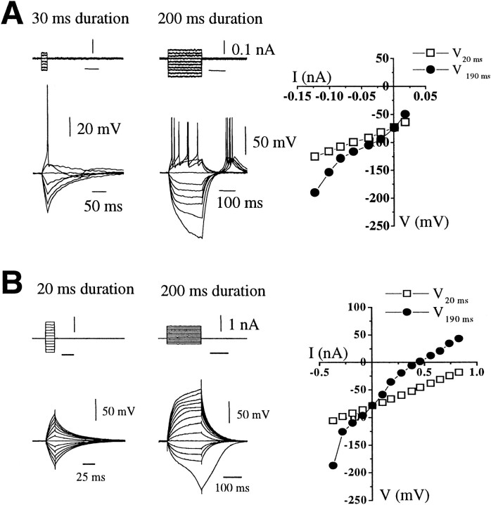
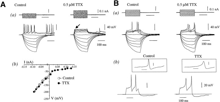
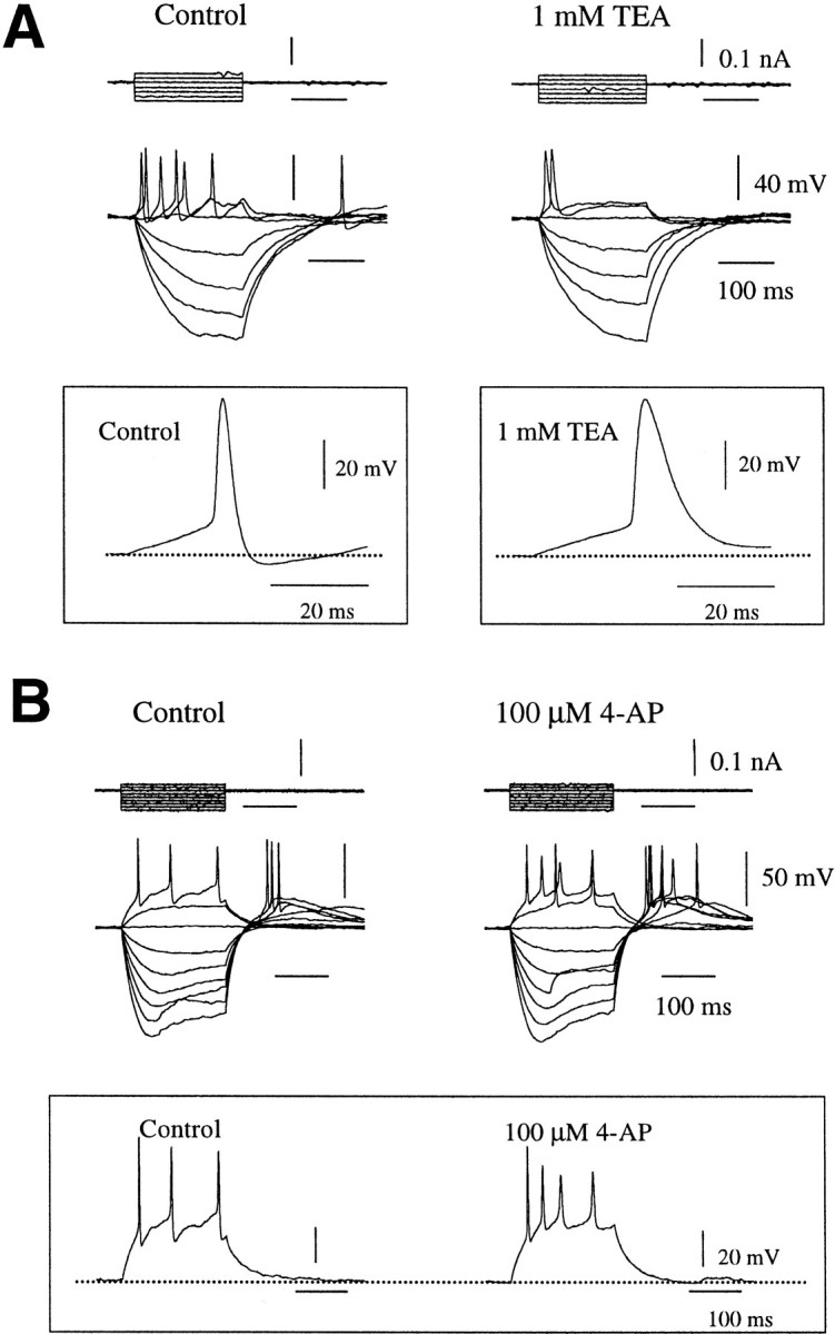
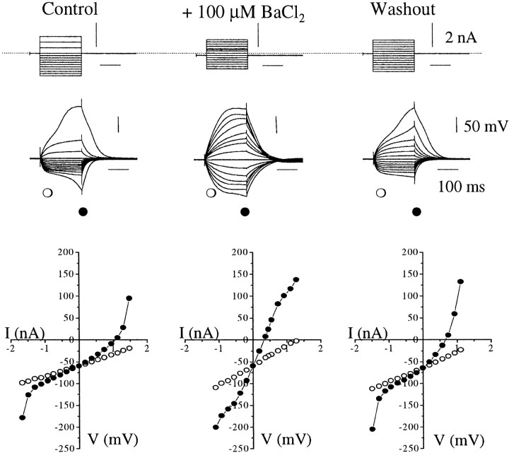
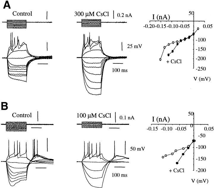
The electrophysiological characteristics of unmodified, postnatal gonadotropin-releasing hormone (GnRH) neurons in the female mouse were studied using whole-cell recordings and single-cell RT-PCR methodology. The GnRH neurons of adult animals fired action potentials and exhibited distinguishable voltage-current relationships in response to hyperpolarizing and depolarizing current injections. On the basis of their patterns of inward rectification, rebound depolarization, and ability to fire repetitively, GnRH neurons in intact adult females were categorized into four cell types (type I, 48%; type II, 36%; type III, 11%; type IV, 5%). The GnRH neurons of juvenile animals (15-22 d) exhibited passive membrane properties similar to those of adult GnRH neurons, although only type I (61%) and type II (7%) cells were encountered, in addition to a group of "silent-type" GnRH neurons (32%) that were unable to fire action potentials. A massive, action potential-independent tonic GABA input, signaling through the GABA(A) receptor, was present at all ages. Afterdepolarization and afterhyperpolarization potentials (AHPs) were observed after single action potentials in subpopulations of each GnRH neuron type. Tetrodotoxin (TTX)-independent calcium spikes, as well as AHPs, were encountered more frequently in juvenile GnRH neurons compared with adults. These observations demonstrate the existence of multiple layers of functional heterogeneity in the firing properties of GnRH neurons. Together with pharmacological experiments, these findings suggest that potassium and calcium channels are expressed in a differential manner within the GnRH phenotype. This heterogeneity occurs in a development-specific manner and may underlie the functional maturation and diversity of this unique neuronal phenotype.
Figures
Similar articles
-
Physiologic regulation of a tetrodotoxin-sensitive sodium influx that mediates a slow afterdepolarization potential in gonadotropin-releasing hormone neurons: possible implications for the central regulation of fertility.J Neurosci. 2006 Nov 15;26(46):11961-73. doi: 10.1523/JNEUROSCI.3171-06.2006. J Neurosci. 2006. PMID: 17108170 Free PMC article.
-
Late postnatal reorganization of GABA(A) receptor signalling in native GnRH neurons.Eur J Neurosci. 2000 Oct;12(10):3497-504. doi: 10.1046/j.1460-9568.2000.00261.x. Eur J Neurosci. 2000. PMID: 11029619
-
Profiling gamma-aminobutyric acid (GABA(A)) receptor subunit mRNA expression in postnatal gonadotropin-releasing hormone (GnRH) neurons of the male mouse with single cell RT-PCR.Neuroendocrinology. 2001 Nov;74(5):300-8. doi: 10.1159/000054697. Neuroendocrinology. 2001. PMID: 11694762
-
Understanding calcium homeostasis in postnatal gonadotropin-releasing hormone neurons using cell-specific Pericam transgenics.Cell Calcium. 2012 Mar-Apr;51(3-4):267-76. doi: 10.1016/j.ceca.2011.11.005. Epub 2011 Dec 15. Cell Calcium. 2012. PMID: 22177387 Review.
-
Autocrine regulation of calcium influx and gonadotropin-releasing hormone secretion in hypothalamic neurons.Biochem Cell Biol. 2000;78(3):359-70. Biochem Cell Biol. 2000. PMID: 10949086 Review.
Cited by
-
Epilepsy-associated increase in gonadotropin-releasing hormone neuron firing in diestrous female mice is independent of chronic seizure burden severity.Epilepsy Res. 2022 Aug;184:106948. doi: 10.1016/j.eplepsyres.2022.106948. Epub 2022 May 20. Epilepsy Res. 2022. PMID: 35690025 Free PMC article.
-
Kisspeptin can stimulate gonadotropin-releasing hormone (GnRH) release by a direct action at GnRH nerve terminals.Endocrinology. 2008 Aug;149(8):3926-32. doi: 10.1210/en.2007-1487. Epub 2008 May 1. Endocrinology. 2008. PMID: 18450966 Free PMC article.
-
Simulated GABA synaptic input and L-type calcium channels form functional microdomains in hypothalamic gonadotropin-releasing hormone neurons.J Neurosci. 2012 Jun 27;32(26):8756-66. doi: 10.1523/JNEUROSCI.4188-11.2012. J Neurosci. 2012. PMID: 22745478 Free PMC article.
-
Estrogen receptor beta mediates rapid estrogen actions on gonadotropin-releasing hormone neurons in vivo.J Neurosci. 2003 Jul 2;23(13):5771-7. doi: 10.1523/JNEUROSCI.23-13-05771.2003. J Neurosci. 2003. PMID: 12843281 Free PMC article.
-
The initiation and maintenance of gonadotropin-releasing hormone neuron identity in congenital hypogonadotropic hypogonadism.Front Endocrinol (Lausanne). 2023 Apr 27;14:1166132. doi: 10.3389/fendo.2023.1166132. eCollection 2023. Front Endocrinol (Lausanne). 2023. PMID: 37181038 Free PMC article. Review.
References
-
- Bean BP, McDonough SI. Two for T. Neuron. 1998;20:825–828. - PubMed
-
- Bordey A, Sontheimer H. Ion channel expression by astrocytes in situ: comparison of different CNS regions. Glia. 2000;30:27–38. - PubMed
-
- Bosma MM. Ionic channel properties and episodic activity in isolated immortalized gonadotropin-releasing hormone (GnRH) neurons. J Membr Biol. 1993;136:85–96. - PubMed
Publication types
MeSH terms
Substances
LinkOut - more resources
Full Text Sources