. 2001 Feb;67(2):575-85.
doi: 10.1128/AEM.67.2.575-585.2001.

gfp-based N-acyl homoserine-lactone sensor systems for detection of bacterial communication

Affiliations

gfp-based N-acyl homoserine-lactone sensor systems for detection of bacterial communication

J B Andersen et al. Appl Environ Microbiol. 2001 Feb.

Abstract

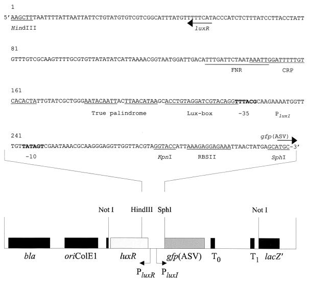
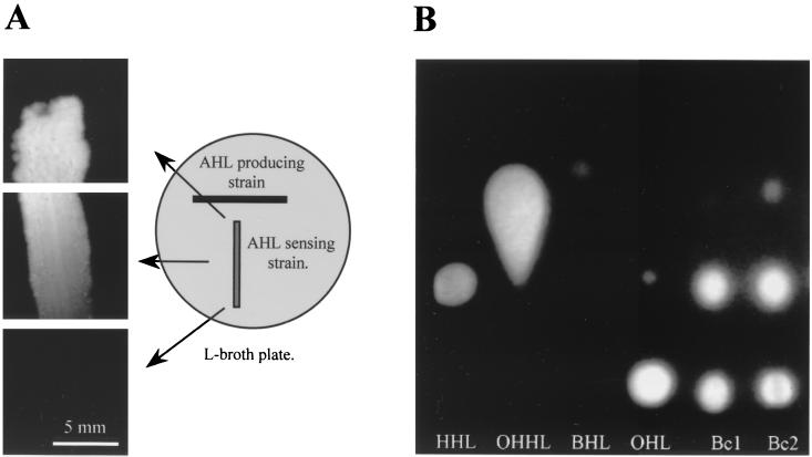
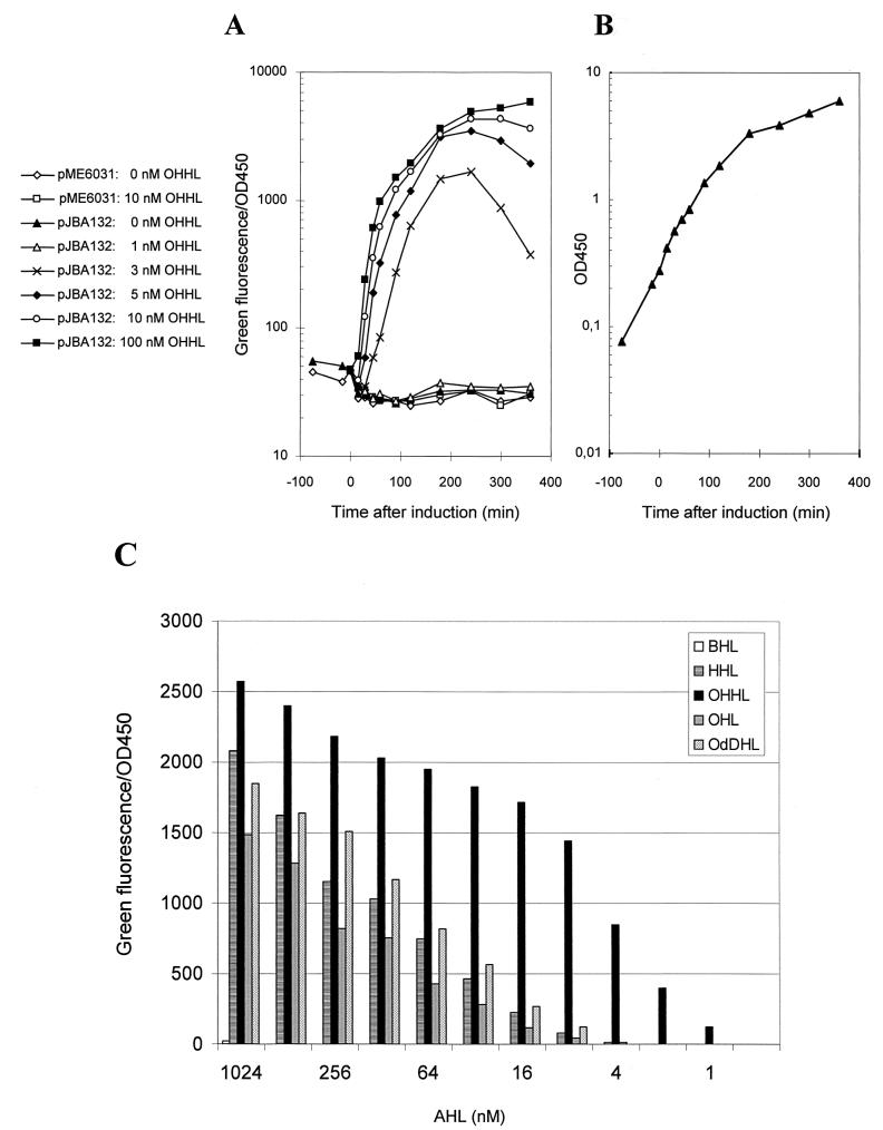
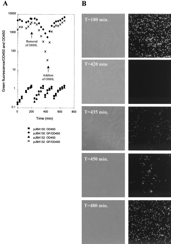
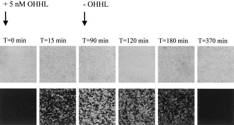
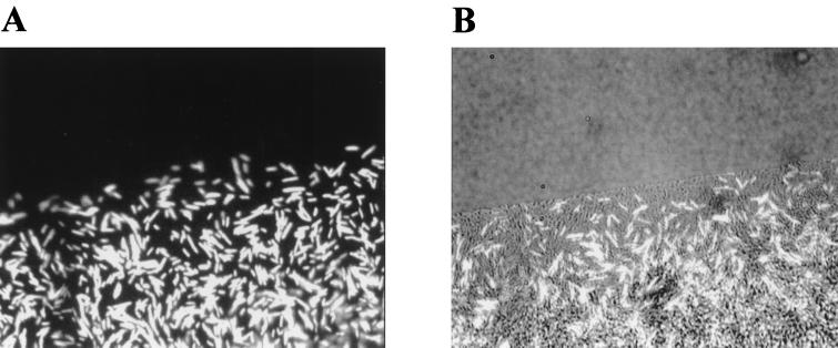
In order to perform single-cell analysis and online studies of N-acyl homoserine lactone (AHL)-mediated communication among bacteria, components of the Vibrio fischeri quorum sensor encoded by luxR-P(luxI) have been fused to modified versions of gfpmut3* genes encoding unstable green fluorescent proteins. Bacterial strains harboring this green fluorescent sensor detected a broad spectrum of AHL molecules and were capable of sensing the presence of 5 nM N-3-oxohexanoyl-L-homoserine lactone in the surroundings. In combination with epifluorescent microscopy, the sensitivity of the sensor enabled AHL detection at the single-cell level and allowed for real-time measurements of fluctuations in AHL concentrations. This green fluorescent AHL sensor provides a state-of-the-art tool for studies of communication between the individuals present in mixed bacterial communities.


Figures

FIG. 1
FIG. 1
Schematic drawing of pJBA89 and the DNA nucleotide sequence of the PCR-amplified luxR-PluxI-RBSII fragment used for the construction of pJBA88, pJBA89, pJBA130, and pJBA132. Arrows indicate directions of transcription of luxR and gfp(ASV). The positions of FNR, cAMP-CRP binding sites and the lux-box sequences are indicated, and the −35 and −10 regions of the luxI promoter (PluxI) are shown in bold letters (38). Important restriction sites are underlined.
FIG. 2
FIG. 2
(A) Epifluorescent image of the AHL sensor JM105 harboring pJBA89 cross-streaked with the AHL producer S. liquefaciens MG1. (B) Fluorescent image of a TLC plate used to separate extracts of B. cepacia (strains DM50180 [Bc1] and ATCC 25416 [Bc2]) and the reference compounds HHL, OHHL, BHL, and OHL overlaid with the AHL sensor (see Materials and Methods for details).
FIG. 3
FIG. 3
(A) Dose-response of the AHL sensor MT102 harboring pJBA132 to OHHL. The strain was grown at 30°C in LB medium. In the early-mid-log phase (t = 0 min), the culture was divided into six subcultures and OHHL was added as indicated. The plot shows specific green fluorescence as a function of time. (B) Bacterial growth of a single representative culture. (C) Sensitivity to different AHL molecules (see Materials and Methods for details).
FIG. 4
FIG. 4
OHHL down- and upshift experiment with two AHL sensors, MT102 harboring pJBA130 (encoding stable GFPmut3∗) and MT102 harboring pJBA132 [encoding the unstable GFP(ASV)]. The cultures were grown with 10 nM OHHL. At 180 min, OHHL was washed away, and the cultures were grown in fresh OHHL-free medium. At 420 min, 10 nM OHHL was added to the cultures. (A) Growth and specific green fluorescence. Arrows indicate the time of AHL removal (downshift) and the time of AHL readdition. (B) Phase-contrast (left) and epifluorescence (right), microscopy images of selected culture samples of MT102 harboring pJBA132 withdrawn during the AHL down- and upshift.
FIG. 5
FIG. 5
Glass surface-attached biofilm of the AHL sensor S. liquefaciens MG44 harboring pJBA132 in a flow cell. At 0 min, 5 nM OHHL was added to the medium flow. At 90 min, the chamber was shifted to medium without OHHL (−OHHL). (Upper panels) Phase-contrast images; (lower panels) epifluorescent images.
FIG. 6
FIG. 6
Binary swarming colony consisting of S. liquefaciens MG44 harboring pJBA132 and S. ficaria. Shown are an epifluorescence image (A) and a combined epifluorescence and phase-contrast image (B) of the moving colony.

Similar articles

Cited by

References

    1. Andersen J B, Sternberg C, Poulsen L K, Bjørn S P, Givskov M, Molin S. New unstable variants of green fluorescent protein for studies of transient gene expression in bacteria. Appl Environ Microbiol. 1998;64:2240–2246. - PMC - PubMed
    1. Bertani G. Studies on lysogenesis. I. The mode of phage liberation by lysogenic Escherichia coli. J Bacteriol. 1951;62:293–300. - PMC - PubMed
    1. Chalfie M, Tu Y, Euskirchen G, Ward W W, Prasher D C. Green fluorescent protein as marker for gene expression. Science. 1994;263:802–805. - PubMed
    1. Christensen B B, Sternberg C, Andersen J B, Eberl L, Møller S, Givskov M, Molin S. Establishment of new genetic traits in a microbial biofilm community. Appl Environ Microbiol. 1998;64:2247–2255. - PMC - PubMed
    1. Clark D J, Maaløe O. DNA replication and the division cycle in Escherichia coli. J Mol Biol. 1967;23:99–112.

Publication types

MeSH terms

LinkOut - more resources