

Expression of cmg1, an exo-beta-1,3-glucanase gene from Coniothyrium minitans, increases during sclerotial parasitism
- PMID: 11157256
- PMCID: PMC92660
- DOI: 10.1128/AEM.67.2.865-871.2001
Expression of cmg1, an exo-beta-1,3-glucanase gene from Coniothyrium minitans, increases during sclerotial parasitism
Abstract
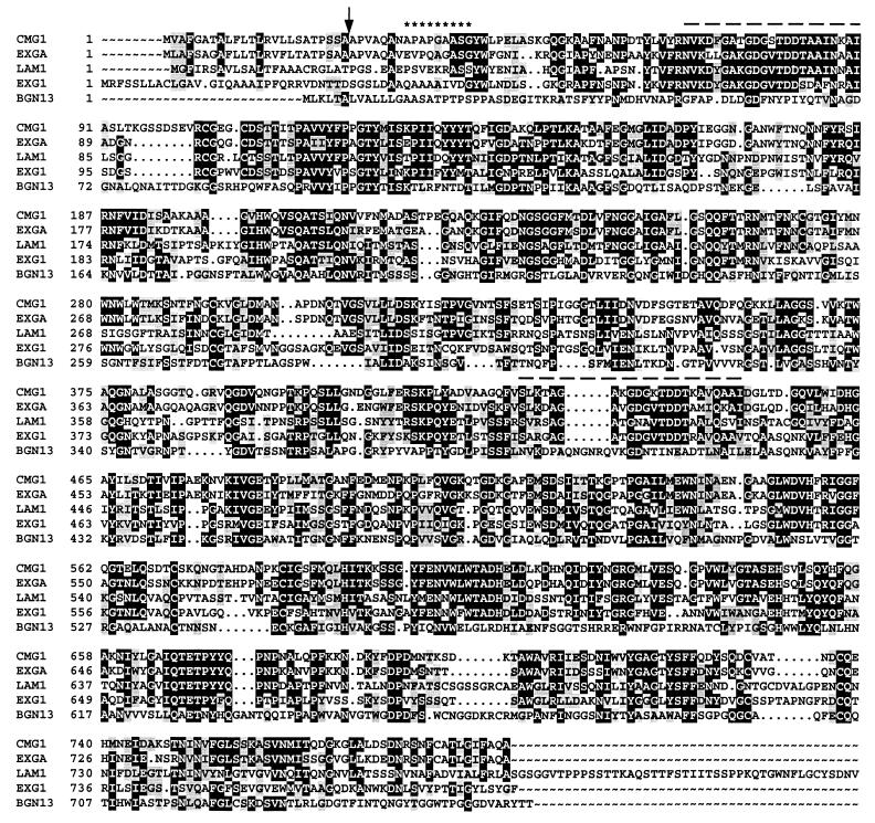
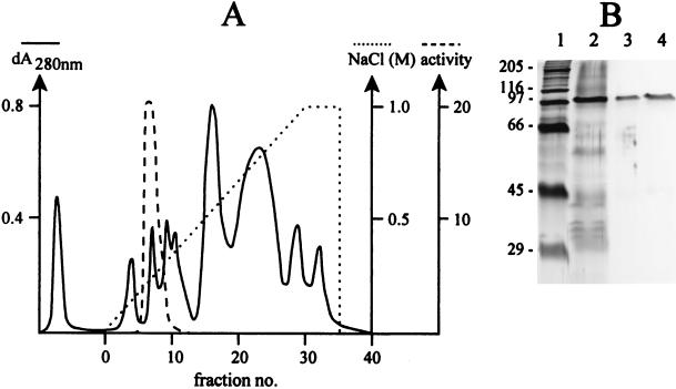
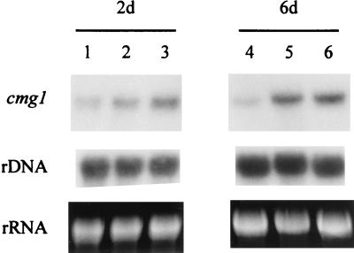
During sclerotial infection of Sclerotinia sclerotiorum the mycoparasite Coniothyrium minitans penetrates through the host cell wall, which contains beta-1,3-glucan as its major component. A PCR-based strategy was used to clone a beta-1,3-glucanase-encoding gene, designated cmg1, from a cDNA library of the fungus. The nucleotide and deduced amino acid sequences of this gene showed high levels of similarity to the sequences of other fungal exo-beta-1,3-glucanase genes. The calculated molecular mass of the deduced protein (without the predicted 24-amino-acid N-terminal secretion signal peptide) was 83,346 Da, and the estimated pI was 4.73. Saccharomyces cerevisiae INVSc1 expressing the cmg1 gene secreted a approximately 100-kDa beta-1,3-glucanase enzyme (as determined by sodium dodecyl sulfate-polyacrylamide gel electrophoresis) into the culture medium. N-terminal sequence analysis of the purified recombinant enzyme revealed that the secreted enzyme starts at Ala-32, seven amino acids downstream from the predicted signal peptidase cleavage site. The purified recombinant glucanase inhibited in vitro mycelial growth of S. sclerotiorum by 35 and 85% at concentrations of 300 and 600 microg x ml(-1), respectively. A single copy of the cmg1 gene is present in the genome of C. minitans. Northern analyses indicated increases in the transcript levels of cmg1 due to both carbon starvation and the presence of ground sclerotia of S. sclerotiorum; only slight repression was observed in the presence of 2% glucose. Expression of cmg1 increased during parasitic interaction with S. sclerotiorum.
Figures


Similar articles
-
An exo-beta-(1,3)-glucanase of Candida albicans: purification of the enzyme and molecular cloning of the gene.J Gen Microbiol. 1993 Feb;139(2):325-34. doi: 10.1099/00221287-139-2-325. J Gen Microbiol. 1993. PMID: 8436950
-
Sequencing of a 1,3-1,4-beta-D-glucanase (lichenase) from the anaerobic fungus Orpinomyces strain PC-2: properties of the enzyme expressed in Escherichia coli and evidence that the gene has a bacterial origin.J Bacteriol. 1997 Oct;179(19):6028-34. doi: 10.1128/jb.179.19.6028-6034.1997. J Bacteriol. 1997. PMID: 9324248 Free PMC article.
-
Analysis of cDNA transcripts from Coniothyrium minitans reveals a diverse array of genes involved in key processes during sclerotial mycoparasitism.Fungal Genet Biol. 2007 Dec;44(12):1262-84. doi: 10.1016/j.fgb.2007.07.011. Epub 2007 Aug 10. Fungal Genet Biol. 2007. PMID: 17888694
-
Cytochemical localization of beta-(1----4)-D-glucans in plant and fungal cells using an exoglucanase-gold complex.Electron Microsc Rev. 1989;2(1):123-38. doi: 10.1016/0892-0354(89)90013-0. Electron Microsc Rev. 1989. PMID: 2518813 Review.
-
The fungal biocontrol agent Coniothyrium minitans: production by solid-state fermentation, application and marketing.Appl Microbiol Biotechnol. 2001 Jul;56(1-2):58-68. doi: 10.1007/s002530100678. Appl Microbiol Biotechnol. 2001. PMID: 11499948 Review.
Cited by
-
Crystal structure of glycoside hydrolase family 55 {beta}-1,3-glucanase from the basidiomycete Phanerochaete chrysosporium.J Biol Chem. 2009 Apr 10;284(15):10100-9. doi: 10.1074/jbc.M808122200. Epub 2009 Feb 4. J Biol Chem. 2009. PMID: 19193645 Free PMC article.
-
Identification of mycoparasitism-related genes in Clonostachys rosea 67-1 active against Sclerotinia sclerotiorum.Sci Rep. 2015 Dec 14;5:18169. doi: 10.1038/srep18169. Sci Rep. 2015. PMID: 26657839 Free PMC article.
-
Active site and laminarin binding in glycoside hydrolase family 55.J Biol Chem. 2015 May 8;290(19):11819-32. doi: 10.1074/jbc.M114.623579. Epub 2015 Mar 9. J Biol Chem. 2015. PMID: 25752603 Free PMC article.
-
Sclerotinia sclerotiorum Agglutinin Modulates Sclerotial Development, Pathogenicity and Response to Abiotic and Biotic Stresses in Different Manners.J Fungi (Basel). 2023 Jul 10;9(7):737. doi: 10.3390/jof9070737. J Fungi (Basel). 2023. PMID: 37504726 Free PMC article.
-
A perilipin gene from Clonostachys rosea f. Catenulata HL-1-1 is related to sclerotial parasitism.Int J Mol Sci. 2015 Mar 9;16(3):5347-62. doi: 10.3390/ijms16035347. Int J Mol Sci. 2015. PMID: 25761240 Free PMC article.
References
-
- Altschul S F, Gish W, Miller W, Myers E W, Lipman D P. Basic local alignment search tool. J Mol Biol. 1990;215:403–410. - PubMed
-
- Archambault C, Coloccia G, Kermashe S, Jabai-Hare S. Characterization of an endo-1,3-β-d-glucanase produced during the interaction between Stachybotrys elegans and its host Rhizoctonia solani. Can J Microbiol. 1998;44:989–997. - PubMed
-
- Archer D B, Peberdy J F. The molecular biology of secreted enzyme production by fungi. Crit Rev Biotechnol. 1997;17:273–306. - PubMed
-
- Bolar J P, Norelli J L, Wong K-W, Hayes C K, Harman G E, Aldwinckle H S. Expression of endochitinase from Trichoderma harzianum in transgenic apple increases resistance to apple scab and reduces vigor. Phytopathology. 2000;90:72–77. - PubMed
Publication types
MeSH terms
Substances
LinkOut - more resources
Full Text Sources
Medical
Research Materials
Miscellaneous
