. 2001 Feb;67(2):922-8.
doi: 10.1128/AEM.67.2.922-928.2001.

Portable system for microbial sample preparation and oligonucleotide microarray analysis

Affiliations

Portable system for microbial sample preparation and oligonucleotide microarray analysis

S G Bavykin et al. Appl Environ Microbiol. 2001 Feb.

Abstract

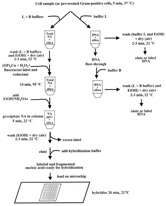
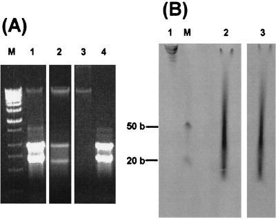
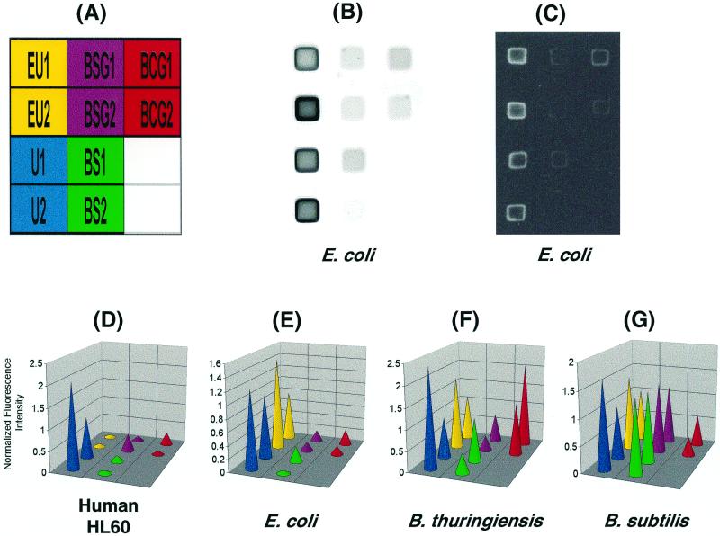
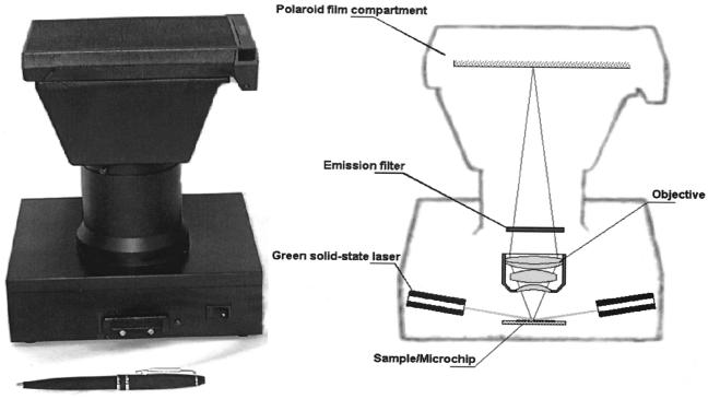
We have developed a three-component system for microbial identification that consists of (i) a universal syringe-operated silica minicolumn for successive DNA and RNA isolation, fractionation, fragmentation, fluorescent labeling, and removal of excess free label and short oligonucleotides; (ii) microarrays of immobilized oligonucleotide probes for 16S rRNA identification; and (iii) a portable battery-powered device for imaging the hybridization of fluorescently labeled RNA fragments with the arrays. The minicolumn combines a guanidine thiocyanate method of nucleic acid isolation with a newly developed hydroxyl radical-based technique for DNA and RNA labeling and fragmentation. DNA and RNA can also be fractionated through differential binding of double- and single-stranded forms of nucleic acids to the silica. The procedure involves sequential washing of the column with different solutions. No vacuum filtration steps, phenol extraction, or centrifugation is required. After hybridization, the overall fluorescence pattern is captured as a digital image or as a Polaroid photo. This three-component system was used to discriminate Escherichia coli, Bacillus subtilis, Bacillus thuringiensis, and human HL60 cells. The procedure is rapid: beginning with whole cells, it takes approximately 25 min to obtain labeled DNA and RNA samples and an additional 25 min to hybridize and acquire the microarray image using a stationary image analysis system or the portable imager.


Figures

FIG. 1
FIG. 1
Flowchart of the isolation, fractionation, fragmentation, and labeling of nucleic acids with subsequent removal of excess free label, using a silica minicolumn.
FIG. 2
FIG. 2
Nucleic acids isolated, fractionated, labeled with lissamine-rhodamine B, and fragmented on a silica syringe-operated column. (A) Isolated total nucleic acids (lane 1), partially fractionated DNA (lane 2), purified DNA (lane 3), and purified RNA (lane 4) from B. subtilis were analyzed by electrophoresis in 1% agarose. M, λ-HindIII DNA marker. (B) Total nucleic acids from B. thuringiensis fractionated in a denaturing 7.5% polyacrylamide gel (46) before (lane 1) and after (lanes 2 and 3) labeling and fragmentation; fluorescence of labeled and fragmented product before (lane 3) and after (lane 2) ethidium bromide staining. M, single-stranded 20- and 50-base size markers.
FIG. 3
FIG. 3
Hybridization of total nucleic acids with an oligonucleotide microarray. Total nucleic acids were isolated and labeled using the silica minicolumn. (A) The arrangement of probes (see Materials and Methods for a list of sequences) immobilized on the microarray for identification of U1 and U2 (“all life”), EU1 and EU2 (all eubacteria), BSG1 and BSG2 (B. subtilis group bacteria), BS1 and BS2 (B. subtilis spp), and BCG1 and BCG2 (B. cereus group bacteria). (B and C) Analysis of E. coli with a stationary microscope (B) and the portable imager (C). (D to G) Normalized fluorescent signal intensities for labeled total nucleic acids from human HL60 cells (D), E. coli (E), B. thuringiensis (F), and B. subtilis (G). Hybridization results were obtained with the stationary fluorescent microscope (B and D to G) or with the portable imager (C). Fluorescence intensities were quantified using Image, a custom LabVIEW program (National Instruments, Austin, Tex.).
FIG. 4
FIG. 4
Photograph and schematic of the portable imager.

Similar articles

Cited by

References

    1. Amann R I, Ludwig W, Schleifer K-H. Phylogenetic identification and in situ detection of individual microbial cells without cultivation. Microbiol Rev. 1995;59:143–169. - PMC - PubMed
    1. Barsky Y, Grammatin A, Ivanov A, Kreindlin E, Kotova E, Barskii V, Mirzabekov A. Wide-field luminescence microscopes for analyzing biological microchips. J Opt Technol. 1998;65:938–941.
    1. Beld M, Sol C, Goudsmit J, Boom R. Fractionation of nucleic acids into single-stranded and double-stranded forms. Nucleic Acids Res. 1996;24:2618–2619. - PMC - PubMed
    1. Boom R, Sol C J A, Salimans M M M, Jansen C L, Wertheim-van Dillen P M E, van der Noordaa J. Rapid and simple method for purification of nucleic acids. J Clin Microbiol. 1990;28:495–503. - PMC - PubMed
    1. Bowtell D D L. Options available—from start to finish—for obtaining expression data by microarray. Nat Genet. 1999;21:25–32. - PubMed

Publication types

MeSH terms

LinkOut - more resources