. 2001 Feb 1;97(3):652-9.
doi: 10.1182/blood.v97.3.652.

CD13/APN is activated by angiogenic signals and is essential for capillary tube formation

Affiliations

CD13/APN is activated by angiogenic signals and is essential for capillary tube formation

S V Bhagwat et al. Blood. .

Abstract

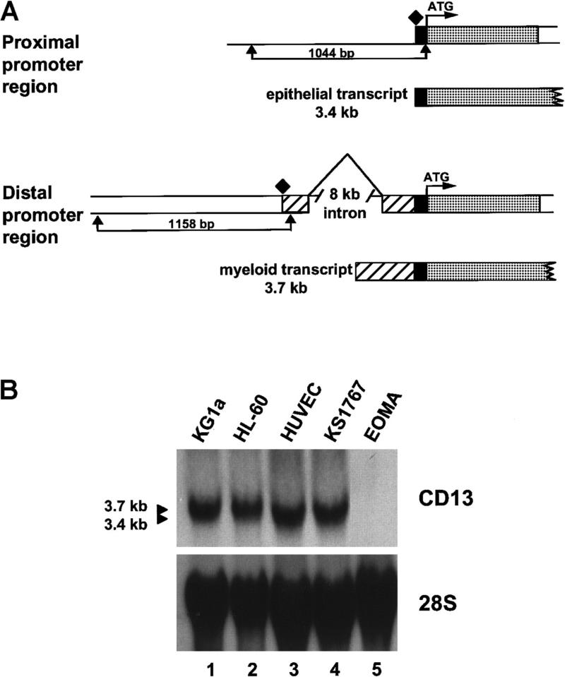
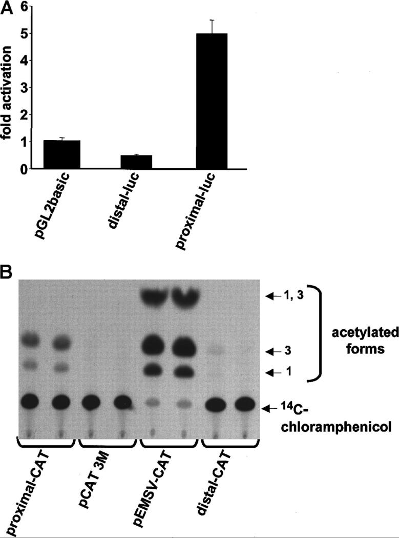
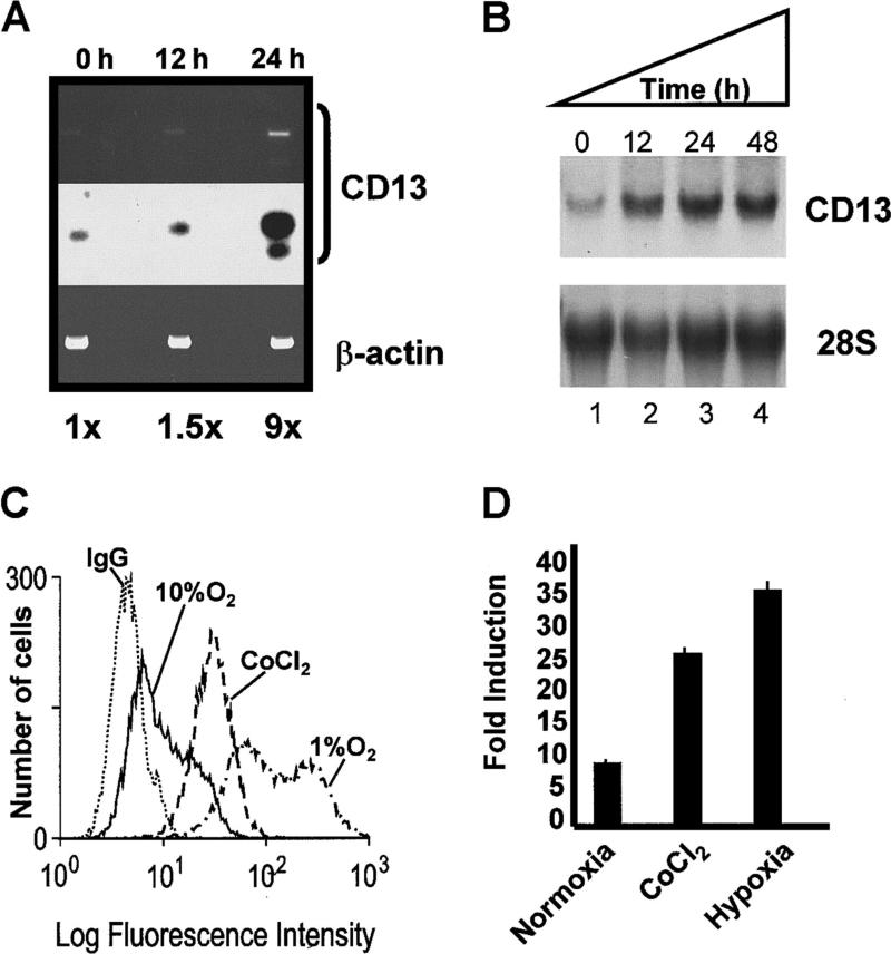
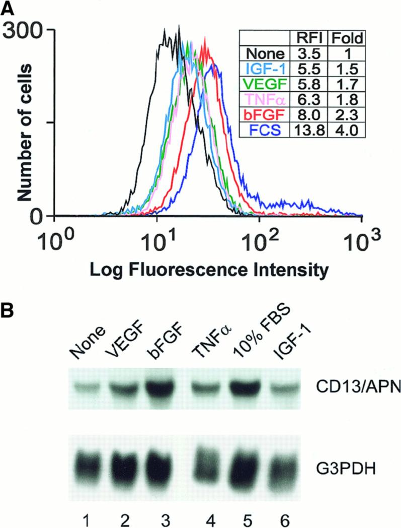
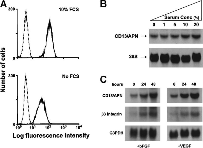
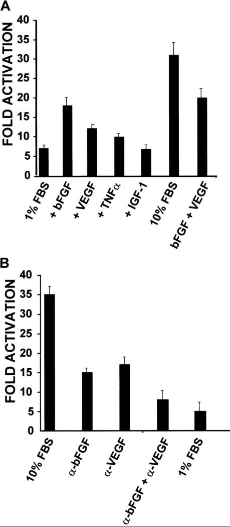
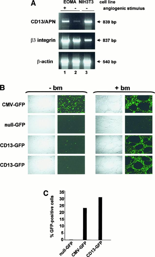
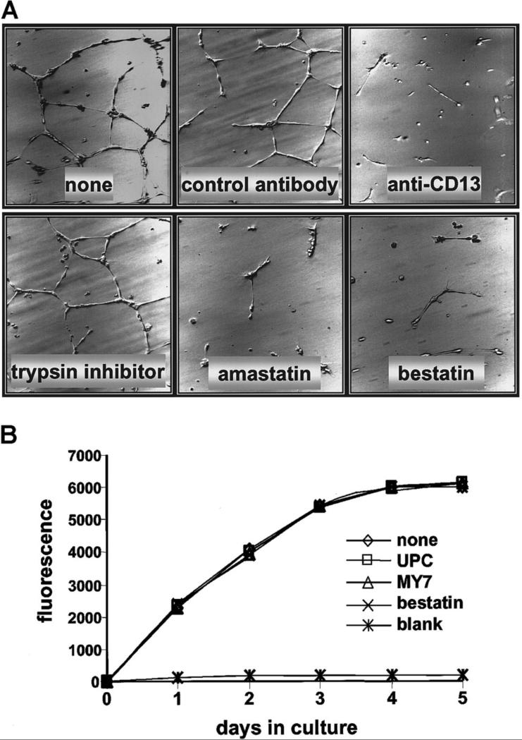
In the hematopoietic compartment, the CD13/APN metalloprotease is one of the earliest markers of cells committed to the myeloid lineage where it is expressed exclusively on the surface of myeloid progenitors and their differentiated progeny. CD13/APN is also found in nonhematopoietic tissues, and its novel expression on the endothelial cells of angiogenic, but not normal, vasculature was recently described. Treatment of animals with CD13/APN inhibitors significantly impaired retinal neovascularization, chorioallantoic membrane angiogenesis, and xenograft tumor growth, indicating that CD13/APN plays an important functional role in vasculogenesis and identifying it as a critical regulator of angiogenesis. To investigate the mechanisms of CD13/APN induction in tumor vasculature, the regulation of CD13/APN by factors contributing to angiogenic progression was studied. In this report, it is shown that endogenous CD13/APN levels in primary cells and cell lines are up-regulated in response to hypoxia, angiogenic growth factors, and signals regulating capillary tube formation during angiogenesis. Transcription of reporter plasmids containing CD13/APN proximal promoter sequences is significantly increased in response to the same angiogenic signals that regulate the expression of the endogenous gene and in human tumor xenografts, indicating that this fragment contains elements essential for the angiogenic induction of CD13/APN expression. Finally, functional antagonists of CD13/APN interfere with tube formation but not proliferation of primary vascular endothelial cells, suggesting that CD13/APN functions in the control of endothelial cell morphogenesis. These studies clearly establish the CD13/APN metalloprotease as an important regulator of endothelial morphogenesis during angiogenesis.


Figures

Figure 1
Figure 1. Characterization of CD13/APN endothelial cell transcripts
(A) Schematic diagram showing the CD13/APN promoter regions used by epithelial cells (proximal promoter, generating a 3.4-kb transcript, top) and myeloid cells and fibroblasts (distal promoter, generating a 3.7-kb transcript, bottom). The translation start site (ATG) is identical in transcripts from each cell type and is followed in genomic DNA by the protein coding sequences (■). The proximal (1044-bp) and distal (1158-bp) promoter fragments are delineated by arrows. Additional untranslated sequences found only in transcripts originating from the distal promoter, ■. Transcriptional start sites for each promoter are shown as diamonds. (B) CD13/APN expression in various tissues as determined by Northern blot analysis of total cellular RNA. The identical blot is shown after it was stripped and reprobed with a 28S probe as a control for RNA integrity and loading. Lanes are identical in each panel, showing that the CD13/APN transcript in the KS1767 endothelial cell line comigrates with that of the HUVECs (3.4-kb) and is smaller than that found in myeloid cells (HL-60, KG1a, 3.7 kb). CD13/APN transcripts are undetectable in EOMA cells.
Figure 2
Figure 2. The proximal promoter controls CD13/APN transcription in HUVECs and endothelial cell lines
(A) Plasmids containing the proximal or distal promoter fragments immediately upstream of the luciferase reporter gene were transiently transfected into primary HUVECs. Lysates of triplicate dishes were assayed at 24 hours for luciferase activity. pGL2basic refers to the promoterless control luciferase plasmid. (B) The identical experiment in KS1767 cells using CAT reporter plasmids. pEMSV-CAT refers to the positive control plasmid and pCAT3M to the promoterless control plasmid. Equal amounts of lysate (75 μg) were assayed in each lane.
Figure 3
Figure 3. Hypoxic conditions induce endogenous CD13/APN expression and proximalpromoter activity
(A) Endogenous CD13/APN mRNA in hypoxia-induced retinal neovessels is up-regulated. RNA isolated from retinas at 0, 12, and 24 hours in relative hypoxia was assayed for CD13/APN and control β-actin levels by RT-PCR. The Southern blot of RT-PCR products (middle panel) was quantitated by phosphorimager analysis. (B) Cobalt chloride treatment induces endogenous CD13/APN in endothelial cell lines. Serum-starved KS1767 cells were treated with cobalt chloride (100 μM) for 12, 24, and 48 hours, and total cellular CD13/APN RNA was assessed. (C) Hypoxia and cobalt chloride treatment induces CD13/APN cell-surface protein levels. Serum-starved KS1767 cells were treated with cobalt chloride (100 μM) or incubated in 1% or 10% oxygen for 24 hours and analyzed for CD13/APN expression with the MY7 monoclonal antibody or negative control immunoglobulin G (IgG) by flow cytometry. (D) Hypoxia induces the CD13/APN proximal promoter. Twenty-four hours after transient transfection of KS1767 cells in 1% serum with 3 μg of proximal promoter plasmid or pGL2basic vector alone and 1 μg of MAP1-SEAP control, cells were subjected to normoxia, hypoxia (1% oxygen), or cobalt chloride (100 μM) treatment for 24 hours before assay for luciferase activity. Results are shown as fold induction over identically treated cells transfected with the promoterless plasmid pGL2basic.
Figure 4
Figure 4. Angiogenic factors induce CD13/APN cell surface expression in primary endothelial cells
(A) Primary HUVECs were serum starved for 24 hours, then cultured without serum (none), with the indicated angiogenic growth factors, or with 10% fetal calf serum (FCS), stained for CD13/APN expression with an anti-CD13/APN monoclonal antibody (MY7) and analyzed by flow cytometry. The relative fluorescence index (RFI) and fold induction over untreated cells of each sample are indicated. (B) RNA isolated and probed for CD13/APN or 28S RNA expression shows that treatment of primary HUVECs with certain angiogenic factors induces CD13/APN mRNA (VEGF [25 ng/mL], bFGF [50 ng/mL], TNFα [10 ng/mL], and IGF-1 [50 ng/mL]).
Figure 5
Figure 5. Endogenous CD13/APN is induced in response to serum concentra- tion and angiogenic factors
Expression of CD13/APN protein (A) or mRNA (B) in KS1767 cells is dependent on serum concentration. Serum-starved cells were stimulated with the indicated concentrations of FBS or left untreated (no fetal calf serum [FCS]) for 24 hours. CD13/APN protein expression was measured by flow cytometry; relative transcript levels in total RNA were assayed by Northern blot analysis. (C) bFGF and VEGF induce time-dependent increases in CD13/APN mRNA expression in KS1767 cells similar to that seen with the angiogenic marker, β3 integrin. Total RNA was isolated at the indicated time intervals after addition of bFGF (50 ng/mL) or VEGF (25 ng/mL) to serum-starved cells and hybridized with the indicated probes.
Figure 6
Figure 6. CD13/APN promoter constructs are induced in response to serum concentration and angiogenic factors
(A) KS1767 cells in 1% serum were transiently transfected with 3 μg reporter plasmids containing the proximal promoter or promoterless vector and 1 μg MAP1-SEAP control, followed by stimulation with the indicated angiogenic factors alone or in combination (bFGF, VEGF, TNFα, or IGF-1). Cells were harvested and assayed for luciferase activity at 24 hours. Results are shown as fold activation over the activity of pGL2 basic control plasmid. (B) Anti-bFGF and anti-VEGF neutralizing antibodies inhibit CD13/APN promoter activity in KS1767 cells. Twenty-four hours after transfection, KS1767 cells in 10% serum were incubated with 20 μg/mL of goat IgG, antihuman bFGF, antihuman VEGF, or both anti-bFGF and anti-VEGF for 24 hours and assayed for luciferase activity. Results are shown as fold activation over identical treatment of cells transfected with the promoterless plasmid pGL2basic.
Figure 7
Figure 7. Expression of CD13/APN is induced during capillary tube formation in vitro and tumor progression in vivo
(A) RT-PCR analysis of 10 μg total RNA from EOMA cells cultured for 24 hours with (+, lane 1) or without (−, lane 2) reconstituted basement membrane matrix (Matrigel) or NIH3T3 fibroblast positive control cells (lane 3) using primers specific for CD13/APN, β3-integrin and β-actin. (B) A representative experiment showing EOMA cells stably transfected with the indicated plasmid constructs cultured with (+ bm) or without (-bm) basement membrane matrix (Matrigel) to stimulate endothelial morphogenesis. Confocal images were acquired 18 to 24 hours after incubation. CMV-GFP (positive control line with GFP driven by the constitutive CMV promoter); null-GFP (promoterless negative control cell line); CD13-GFP (images of 2 independent cell lines containing the CD13/APN proximal promoter driving GFP expression). (C) Flow cytometric analysis of GFP-expressing cells in single-cell suspensions of EOMA xenografts. Data are expressed as percentage GFP-positive cells and numbers are representative of 2 independent experiments.
Figure 8
Figure 8. CD13/APN antagonists inhibit HUVEC capillary tube formation but not proliferation
(A) HUVECs were plated on Matrigel basement membrane preparations and incubated with either the CD13/APN inhibitors amastatin or bestatin, or the anti-CD13/APN monoclonal antibody MY7; or the negative-control trypsin inhibitor or isotype-matched monoclonal antibodies. Plates were incubated for 24 hours before analysis. (B) HUVECs were plated on tissue culture dishes with anti-CD13/APN monoclonal antibody MY7 or isotype-matched control antibodies UPC10, and proliferation was assessed by accumulation of the fluorescent REDOX indicator at the indicated time intervals. The blank condition contained no cells.

Similar articles

Cited by

References

    1. Hanahan D, Folkman J. Patterns and emerging mechanisms of the angiogenic switch during tumorigenesis. Cell. 1996;86:353–364. - PubMed
    1. Pasqualini R, Koivunen E, Ruoslahti E. A peptide isolated from phage display libraries is a structural and functional mimic of an RGD-binding site on integrins. J Cell Biol. 1995;130:1189–1196. - PMC - PubMed
    1. Pasqualini R, Koivunen E, Kain R, et al. Aminopeptidase N is a receptor for tumor-homing peptides and a target for inhibiting angiogenesis. Cancer Res. 2000;60:722–727. - PMC - PubMed
    1. Hogg N, Horton MJ. Myeloid antigens: new and previously defined clusters. In: McMichael AJ, editor. Leukocyte Typing III: Proceedings of the Third International Workshop on Human Leukocyte Differentiation Antigens. Oxford University Press; New York, NY: 1987. pp. 576–621.
    1. Look AT, Ashmun RA, Shapiro LH, Peiper SC. Human myeloid plasma membrane glycoprotein CD13 (gp150) is identical to aminopeptidase N. J. Clin Invest. 1989;83:1299–1306. - PMC - PubMed

Publication types

MeSH terms