Comparative Study
. 2001 Feb 1;20(3):362-71.
doi: 10.1093/emboj/20.3.362.

Influenza B virus NS1 protein inhibits conjugation of the interferon (IFN)-induced ubiquitin-like ISG15 protein

Affiliations
Comparative Study

Influenza B virus NS1 protein inhibits conjugation of the interferon (IFN)-induced ubiquitin-like ISG15 protein

W Yuan et al. EMBO J. .

Abstract

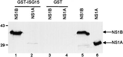
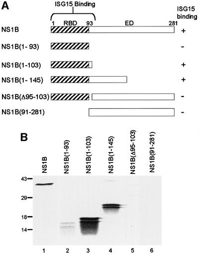
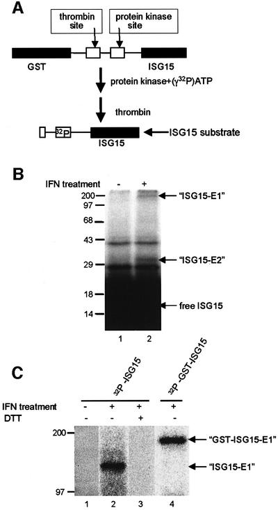
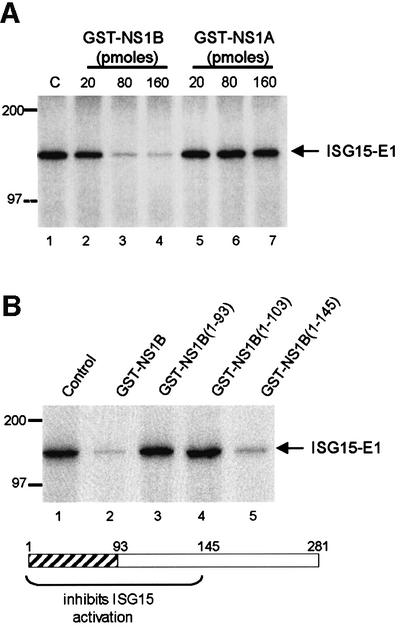
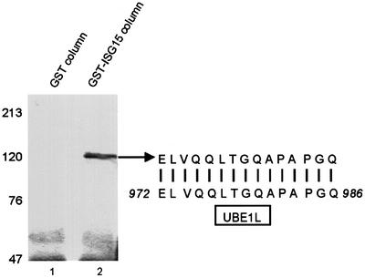
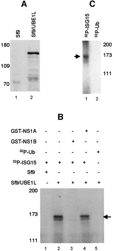
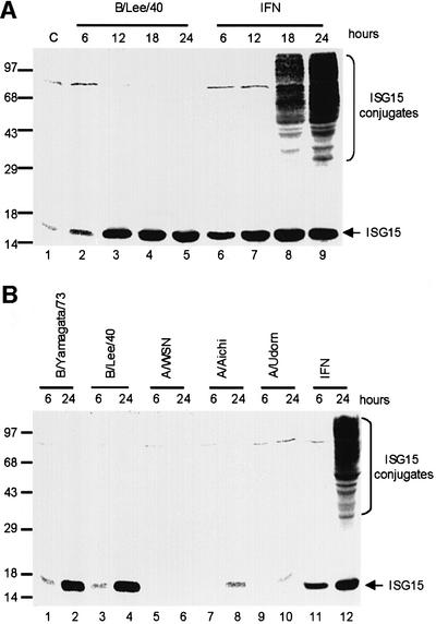
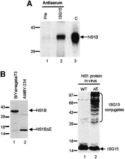
Of the several hundred proteins induced by interferon (IFN) alpha/beta, the ubiquitin-like ISG15 protein is one of the most predominant. We demonstrate the novel way in which the function of the ISG15 protein is inhibited by influenza B virus, which strongly induces the ISG15 protein: a specific region of the influenza B virus NS1 protein, which includes part of its effector domain, blocks the covalent linkage of ISG15 to its target proteins both in vitro and in infected cells. We identify UBE1L as the E1 enzyme that catalyzes the first activation step in the conjugation of ISG15, and show that the NS1B protein inhibits this activation step in vitro. Influenza A virus employs a different strategy: its NS1 protein does not bind the ISG15 protein, but little or no ISG15 protein is produced during infection. We discuss the likely basis for these different strategies.


Figures

None
Fig. 1. The NS1B, but not the NS1A protein binds to the ISG15 protein in vitro. GST–ISG15 protein (1 µg) (lanes 1 and 2) or GST (1 µg) (lanes 3 and 4) was incubated with 20 µl of 35S-labeled NS1B protein (lanes 1 and 3) and NS1A protein (lanes 2 and 4) in the presence of 20 µl of glutathione–Sepharose 4B beads. The labeled proteins eluted from the beads were analyzed by electrophoresis on a 12% SDS–polyacrylamide gel. Lanes 5 and 6: 2 µl of the indicated 35S-labeled NS1 protein. The mobilities of molecular weight markers are shown on the left.
None
Fig. 2. Identification of the ISG15 binding site on the NS1B protein. (A) Schematic diagram of the fragments of the NS1B protein that were used in the binding assay. RBD, RNA-binding domain; ED, effector domain. (B) Binding assay. GST–ISG15 protein (1 µg) was incubated with 104 c.p.m. of 35S-labeled full-length NS1B protein (lane 1), or the indicated fragment of the NS1B protein (lanes 2–6) in the presence of 20 µl of glutathione–Sepharose 4B beads. The same amount of each radiolabeled NS1B fragment was added to the binding assay. The labeled proteins eluted from the beads were analyzed by electrophoresis on a 12% SDS–polyacrylamide gel. The doublets seen with the NS1B fragments (lanes 2–4) were present both before and after the binding assay. The mobilities of molecular weight markers are shown on the left.
None
Fig. 3. In vitro assay for the E1-like enzyme that activates ISG15. (A) Schematic diagram of the strategy for preparing the 32P-labeled ISG15 substrate. (B) An extract (40 µg) from IFN-treated A549 cells (lane 2) or from untreated A549 cells (lane 1) was incubated with 106 c.p.m. (70 pmol) of 32P-labeled ISG15 protein in a 50 µl reaction, as described in Materials and methods. After a 2 h incubation at 37°C, the reactions were terminated by adding SDS sample buffer lacking DTT, and subjected to electrophoresis on a 12.5% SDS–PAGE gel in the absence of a reducing agent. The putative ISG15–E1 and ISG15–E2 adducts are denoted by arrows, and the mobilities of molecular weight markers are shown on the left. (C) An extract (40 µl) from IFN-treated A549 cells (lanes 2–4) or from untreated A549 cells (lane 1) was incubated for 2 h with 106 c.p.m. of 32P-labeled ISG15 protein (lanes 1–3) or 32P-labeled GST–ISG15 protein (lane 4) in a 50 µl reaction. The reactions were terminated by adding SDS sample buffer lacking DTT (lanes 1, 2 and 4) or containing DTT (lane 3), and analyzed on a 7.5% SDS–PAGE gel. The putative GST–ISG15–E1 and ISG15–E1 adducts are denoted by arrows.
None
Fig. 4. Identification of the region of the NS1B protein that is required for inhibition of the in vitro ISG15-coupling reaction. (A) The NS1B protein, and not the NS1A protein, inhibits the formation of the ISG15–E1-like intermediate in vitro. 32P-labeled ISG15 protein (106 c.p.m., 70 pmol) was pre-incubated with the indicated amounts of GST–NS1B (lanes 2–4) or GST–NS1A (lanes 5–7) prior to the incubation with the extract from IFN-treated A549 cells. C (lane 1), pre-incubation of ISG15 in the absence of an NS1 protein. (B) The N-terminal 145 amino acid region of the NS1B protein is required for inhibition of the coupling of ISG15 to the E1-like enzyme. 32P-labeled ISG15 protein (106 c.p.m., 70 pmol) was pre-incubated with 160 pmol of either full-length NS1B protein (lane 2), or the indicated fragment of the NS1B protein (lanes 3–5) prior to incubation with the extract from IFN-treated A549 cells. Control (lane 1): pre-incubation of ISG15 in the absence of an NS1 protein. The hatched region in the NS1B protein is the RNA-binding domain.
None
Fig. 5. Identification of the E1 enzyme that activates ISG15. (A) Cell extracts from IFN-treated A549 cells were subjected to affinity selection on a glutathione–Sepharose 4B column containing either GST (lane 1) or the GST–ISG15 fusion protein (lane 2). The eluate was resolved by 7.5% SDS–PAGE, followed by staining with Coomassie Blue. The 110 kDa protein species from lane 2 was analyzed by MALDI-MS, and one of the tryptic peptides was microsequenced by automated Edman degradation. The sequence of this peptide is compared to the amino acid sequence of UBE1L (amino acids 972–986).
None
Fig. 6. The recombinant UBE1L protein catalyzes the activation of ISG15. (A) The UBE1L protein is synthesized in insect cells infected with the recombinant virus (lane 2). Lane 1: uninfected Sf9 cells. Extracts from uninfected and infected cells were analyzed by SDS–PAGE followed by western analysis using anti-GST antibody. (B) Extracts from uninfected Sf9 insect cells (lane 1) or Sf9 cells infected with the recombinant baculovirus expressing the GST–UBE1L protein (lane 2) were incubated with 106 c.p.m. (70 pmol) of 32P-labeled ISG15, as described in the legend of Figure 3. Prior to incubation with the extract from cells infected with the recombinant baculovirus, the 32P-labeled ISG15 protein was pre-incubated with 160 pmol of either GST–NS1B (lane 3) or GST–NS1A (lane 4). In lane 5, 32P-labeled ubiquitin (Ub) was incubated with the extract from cells infected with the recombinant baculovirus. Labeled Ub was prepared using a plasmid containing the Ub sequence cloned into pGEX-2TK (kindly provided by Jon Huibregtse). The reaction products were analyzed by gel electrophoresis as described in the legend of Figure 3. The labeled ISG15–GST–UBE1L adduct is denoted by the arrow. (C) The GST–UBE1L protein was purified from the extract of insect cells infected with the recombinant baculovirus using glutathione–Sepharose, and the purified protein was incubated with 106 c.p.m. of either 32P-labeled ISG15 (lane 1) or 32P-labeled ubiquitin (Ub) (lane 2).
None
Fig. 7. (A) The ISG15 protein is synthesized in cells infected by influenza B virus, but the conjugation of ISG15 to its target proteins does not occur. A549 cells were infected with 20 p.f.u./cell influenza B/Lee/40 virus (lanes 2–5), or were treated with 1000 U/ml IFN-β (lanes 6–9). At the indicated time points, the cells were collected and proteins were resolved by 12% SDS–PAGE. The proteins were transferred to a BA S-85 filter membrane, which was probed with rabbit anti-ISG15 antiserum. C (lane 1), untreated A549 cells. (B) The ISG15 protein is not synthesized in cells infected by influenza A virus. A549 cells were infected with the indicated influenza B virus (lanes 1–4) or the indicated A virus (lanes 5–10) at 20 p.f.u./cell, or were treated with 1000 U/ml IFN-β (lanes 11 and 12). At the indicated time points, the cells were collected, and proteins were resolved by SDS–PAGE followed by western analysis using rabbit anti-ISG15 antiserum.
None
Fig. 8. (A) The NS1B protein is physically associated with the ISG15 protein in influenza B virus-infected cells. A549 cells infected with influenza B/Lee/40 virus were collected at 18 h post-infection, and were immunoprecipitated with either pre-immune mouse serum (lane 1) or mouse anti-ISG15 antiserum (lane 2). The immunoprecipitated proteins were resolved by 12% SDS–PAGE, followed by western blotting using rabbit anti-NS1B antiserum. C (lane 3), purified NS1B protein run on the same gel. (B) The NS1B protein inhibits the coupling of ISG15 to its target proteins in influenza B virus-infected cells. Left panel: the AWBY-234 mutant of B/Yamagata/1/73 synthesizes a truncated NS1B protein. A549 cells were infected with either influenza virus B/Yamagata/1/73 (lane 1) or its AWBY-234 mutant (lane 2) at 20 p.f.u./cell. At 18 h after infection, proteins were resolved by gel electrophoresis, followed by western blotting using rabbit anti-NS1B antiserum. Right panel: conjugation of ISG15 to its target proteins occurs in cells infected with the AWBY-234 mutant (ΔE NS1 protein; lane 2), but not in cells infected with wild-type virus (WT NS1 protein; lane 1). At 18 h after infection, proteins were resolved by gel electrophoresis, followed by western blotting using rabbit anti-ISG15 antiserum.

References

    1. Beattie E.J., Tartaglia,J. and Paoletti,E. (1991) Vaccinia virus-encoded eIF-2α homolog abrogates the antiviral effects of interferon. Virology, 183, 419–422. - PubMed
    1. Briedis D. and Lamb,R.A. (1982) Influenza B virus genome: sequences and structural organization of RNA segment 8 and the mRNAs coding for the NS1 and NS2 proteins. J. Virol., 42, 186–193. - PMC - PubMed
    1. Carritt B. et al. (1992) A gene from human chromosome region 3p21 with reduced expression in small cell lung cancer. Cancer Res., 52, 1536–1541. - PubMed
    1. Chen Z. and Krug,R.M. (2000) Selective nuclear export of viral mRNAs in influenza-virus-infected cells. Trends Microbiol., 8, 376–383. - PubMed
    1. Chen Z., Li,Y. and Krug,R.M. (1999) Influenza A virus NS1 protein targets poly(A)-binding protein II of the cellular 3′ end processing machinery. EMBO J., 18, 2273–2283. - PMC - PubMed

Publication types