. 2001 Feb 1;20(3):387-96.
doi: 10.1093/emboj/20.3.387.

The human cytomegalovirus gene product US6 inhibits ATP binding by TAP

Affiliations

The human cytomegalovirus gene product US6 inhibits ATP binding by TAP

E W Hewitt et al. EMBO J. .

Abstract

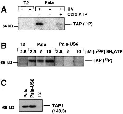
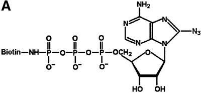
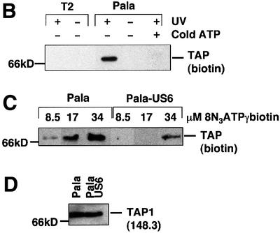
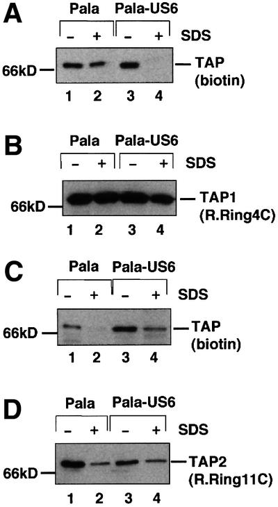
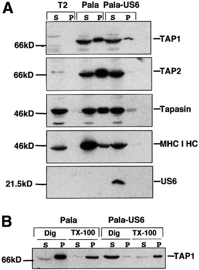
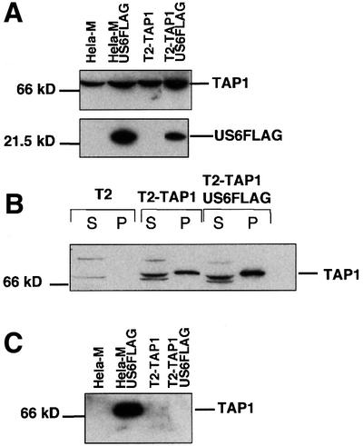
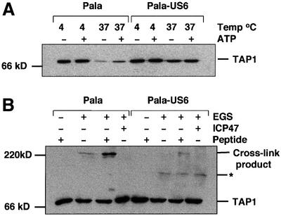
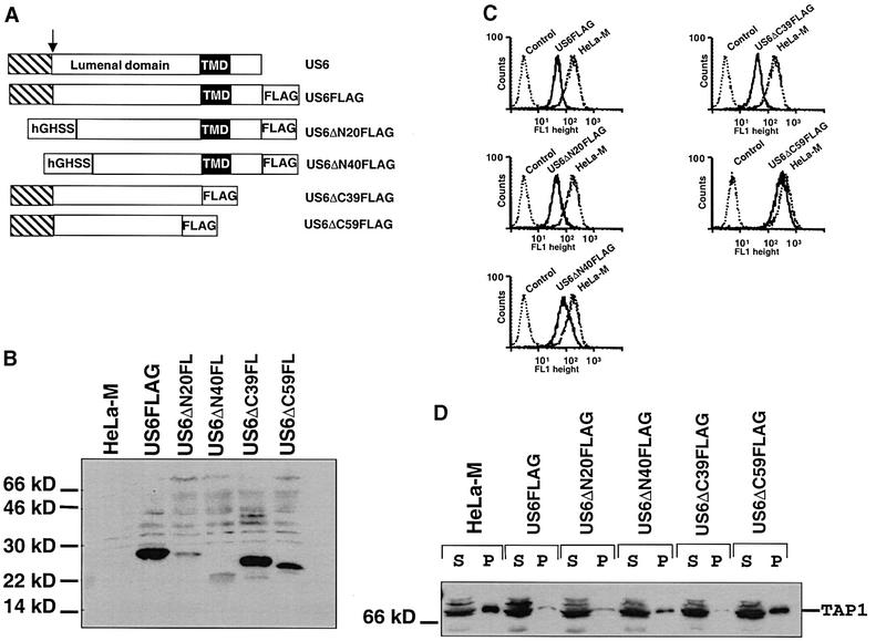
Human cytomegalovirus (HCMV) encodes several genes that disrupt the major histocompatibility complex (MHC) class I antigen presentation pathway. We recently described the HCMV-encoded US6 gene product, a 23 kDa endoplasmic reticulum (ER)-resident type I integral membrane protein that binds to the transporter associated with antigen processing (TAP), inhibits peptide translocation and prevents MHC class I assembly. The functional consequence of this inhibition is to prevent the cell surface expression of class I bound viral peptides and their recognition by HCMV-specific cytotoxic T cells. Here we describe a novel mechanism of action for US6. We demonstrate that US6 inhibits the binding of ATP by TAP1. This is a conformational effect, as the ER lumenal domain of US6 is sufficient to inhibit ATP binding by the cytosolic nucleotide binding domain of TAP1. US6 also stabilizes TAP at 37 degrees C and prevents conformational rearrangements induced by peptide binding. Our findings suggest that the association of US6 with TAP stabilizes a conformation in TAP1 that prevents ATP binding and subsequent peptide translocation.


Figures

None
Fig. 1. Photoaffinity labelling of TAP with [α-32P]8N3ATP is inhibited by US6. (A) [α-32P]8N3ATP labelling of TAP. Digitonin cell lysates from T2 and Pala cells (106 per reaction) were labelled with [α-32P]8N3ATP (as described in Materials and methods) in the presence (+) or absence (–) of UV light and cold ATP (1 mM). Following a TAP1 (148.3) immunoprecipitation, eluted proteins were separated on a 10% SDS–polyacrylamide gel prior to exposure to a phosphoimager screen. (B and C) US6 inhibits TAP labelling by [α-32P]8N3ATP. Lysates from T2-, Pala- and US6-transfected Pala cells were photolabelled with 2.5, 5 and 10 µM [α-32P]8N3ATP, immunoprecipitated as described above and separated on a 10% SDS–polyacrylamide gel prior to exposure to a phosphoimager screen (B). A parallel immunoprecipitation with a TAP1 (148.3) mAb from unlabelled cell lysates was performed, and probed with TAP1 (R.RING4C) antisera (C).
None
Fig. 2. Photoaffinity labelling of TAP with 8N3ATPγbiotin is inhibited by US6. (A) Chemical structure of 8N3ATPγbiotin. (B) 8N3ATPγbiotin specifically labels TAP. Digitonin lysates of T2 and Pala cells (106 per reaction) were labelled with 34 µM 8N3ATPγbiotin (as described in Materials and methods) in the presence (+) or absence (–) of UV light and cold ATP (136 µM). Following a TAP1 (148.3) mAb immunoprecipitation, eluted proteins were separated on a 10% SDS–polyacrylamide gel, transferred to PVDF membranes and probed with Extravidin-HRP conjugate (Sigma). Reactive bands were detected by chemiluminescence. (C and D) US6 inhibits TAP photolabelling by 8N3ATPγbiotin. Digitonin lysates of Pala- and US6-transfected Pala cells (106 per reaction) were photolabelled with 8.5, 17 and 34 µM 8N3ATPγbiotin, immunoprecipitated with a TAP1 (148.3) mAb (as described above), and eluted proteins were separated on a 10% SDS–polyacrylamide gel, transferred to PVDF membranes and visualized with Extravidin-HRP (C). A parallel immunoprecipitation with a TAP1 (148.3) mAb from unlabelled cell lysates was performed, and probed with TAP1 (R.RING4C) antisera (D).
None
Fig. 3. US6 inhibits 8N3ATPγbiotin photolabelling of TAP1, and promotes ATP labelling of TAP2. (AD) Digitonin extracts from Pala- and US6-transfected Pala cells (2 × 10⁶ per reaction) were photolabelled with 68 µM 8N3ATPγbiotin and each lysate was divided into four. In two samples (lanes 2 and 4), the TAP heterodimer was dissociated by incubation at 37°C for 15 min in 0.5% SDS (+); the other two samples (lanes 1 and 3) were not heated with SDS prior to immunoprecipitation (–). (A and C) TAP was immunoprecipitated with either the TAP1 mAb (148.3) (A) or the TAP2 mAb (435.3) (C). Immunoprecipitates were separated on 10% SDS–polyacrylamide gels and labelled products detected with Extravidin-HRP. Parallel immunoprecipitations with the TAP1 (148.3) mAb (B) or the TAP2 (435.3) mAb (D) were performed, and separated proteins transferred onto PVDF membranes and probed with TAP1 (R.RING4C) antisera (B) or TAP2 (R.RING11C) antisera (D).
None
Fig. 4. US6 inhibits the binding of TAP to ATP-agarose. (A) US6 inhibits ATP binding by TAP and TAP-associated proteins. Digitonin extractions of T2, Pala and Pala-US6 cells were incubated with ATP-agarose for 1 h. The final ATP concentration was 13 µM. Agarose bound (P) and unbound (S) proteins separated on either 10 or 12% SDS–polyacrylamide gels. Immunoblots of the ATP-agarose fractions were probed with TAP1- (148.3), TAP2- (435.3), MHC class I heavy chain (HC10)-, tapasin (R.gp48N)- and US6 (R.US6N)-specific antibodies. (B) US6 inhibition of ATP binding by TAP is lost following TAP–US6 dissociation in Triton X-100. Pala- and US6-transfected Pala cells were extracted in 1% digitonin (Dig) and, after removal of the nuclear fraction, each lysate was divided into two aliquots. Triton X-100 (TX-100) (1% final) was added to one aliquot and samples were incubated with ATP-agarose for 0.5 h. Equal cell equivalents of the ATP-agarose bound pellet (P) and unbound supernatant (S) fractions were separated on a 10% SDS–polyacrylamide gel, transferred onto PVDF membranes and probed with TAP1 (148.3) antibodies.
None
Fig. 5. US6 does not inhibit ATP binding by single chain TAP1. (A) Expression of US6FLAG in T2-TAP1 cells. Cell lysates were run on 10 and 12% SDS–polyacrylamide gels and immunoblotted with FLAG (M2) and TAP1 (148.3) antibodies. (B) US6 does not inhibit ATP binding by single chain TAP1. Digitonin extracts of T2, T2-TAP1 and T2-TAP1-US6FLAG cells were incubated with ATP-agarose for 1 h, and ATP-agarose bound (P) and unbound supernatant (S) fractions separated on a 10% SDS–polyacrylamide gel, transferred onto PVDF membranes and probed with TAP1 (148.3) antibodies. (C) US6 does not associate with single chain TAP1. Digitonin cell lysates were immunoprecipitated with the FLAG mAb M2. Eluted proteins were separated on a 10% SDS–polyacrylamide gel and immunoblotted with TAP1 (R.RING4C)-specific antisera.
None
Fig. 6. US6 stabilizes TAP at 37°C but inhibits peptide-stimulated chemical cross-linking. (A) US6 stabilizes TAP at 37°C. Membranes from Pala- and US6-transfected Pala cells were incubated at either 4 or 37°C for 1 h in the presence (+) or absence (–) of 2 mM ATP (as described in Materials and methods). Lysates were digitonin solubilized, immunoprecipitated with TAP1 (148.3) mAb and after separation on a 10% SDS–polyacrylamide gel, immunoblotted with TAP1 (R.RING4C) antisera. (B) Peptide-stimulated chemical cross-linking of the TAP heterodimer is inhibited by US6. Membranes from Pala- and US6-transfected Pala cells were incubated in the presence (+) or absence (–) of 10 µM HLA-B27 (SRYWAIRTR) or ICP47 peptides for 1 h at 4°C before cross-linking with EGS. The cross-linked membranes were resolved on a 6% SDS–polyacrylamide gel and immunoblotted with TAP1 (148.3) mAb. Asterisk, an unidentified band.
None
Fig. 7. The C-terminus of the US6 lumenal domain is essential for function. (A) Schematic representation of the construction of the US6 N- and C-terminal deletions. The US6 N-terminal signal sequence is indicated by a hatched box, the arrow indicates the site of signal peptide cleavage and the transmembrane domain is indicated by a black box. The human growth hormone signal sequence is indicated by hGHSS. (B) Expression of the deletion constructs. Cell lysates were prepared and analysed by immunoblotting with the FLAG mAb M2. (C) Cytofluorometric analysis of cell surface MHC class I expression in HeLa-M cells expressing the US6 deletions. Cells were stained with the monoclonal mAb W6/32 and a FITC-conjugated secondary antibody. In the control, HeLa-M cells were only stained with the secondary antibody. (D) ATP-agarose binding of TAP1 in the HeLa-M cell transfectants. Digitonin extracts of HeLa-M cells were incubated with ATP-agarose for 1 h, and ATP-agarose bound pellet (P) and unbound supernatant (S) fractions separated on a 10% SDS–polyacrylamide gel, transferred onto PVDF membranes and probed with TAP1 (148.3) antibodies.

Similar articles

Cited by

References

    1. Abele R. and Tampe,R. (1999) Function of the transport complex TAP in cellular immune recognition. Biochim. Biophys. Acta, 1461, 405–419. - PubMed
    1. Ahn K., Meyer,T.H., Uebel,S., Sempe,P., Djaballah,H., Yang,Y., Peterson,P.A., Fruh,K. and Tampe,R. (1996) Molecular mechanism and species specificity of TAP inhibition by herpes simplex virus ICP47. EMBO J., 15, 3247–3255. - PMC - PubMed
    1. Ahn K., Gruhler,A., Galocha,B., Jones,T.R., Wiertz,E.J., Ploegh,H.L., Peterson,P.A., Yang,Y. and Fruh,K. (1997) The ER-luminal domain of the HCMV glycoprotein US6 inhibits peptide translocation by TAP. Immunity, 6, 613–621. - PubMed
    1. Ambudkar S.V., Lelong,I.H., Zhang,J., Cardarelli,C.O., Gottesman,M.M. and Pastan,I. (1992) Partial purification and reconstitution of the human multidrug-resistance pump: characterization of the drug-stimulatable ATP hydrolysis. Proc. Natl Acad. Sci. USA, 89, 8472–8476. - PMC - PubMed
    1. Androlewicz M.J. and Cresswell,P. (1994) Human transporters associated with antigen processing possess a promiscuous peptide-binding site. Immunity, 1, 7–14. - PubMed

Publication types

MeSH terms

Substances