Comparative Study
. 2001 Feb 1;20(3):433-45.
doi: 10.1093/emboj/20.3.433.

Structural analysis of adenylate cyclases from Trypanosoma brucei in their monomeric state

Affiliations
Comparative Study

Structural analysis of adenylate cyclases from Trypanosoma brucei in their monomeric state

B Bieger et al. EMBO J. .

Erratum in

  • EMBO J 2001 Sep 17;20(18):5302

Abstract

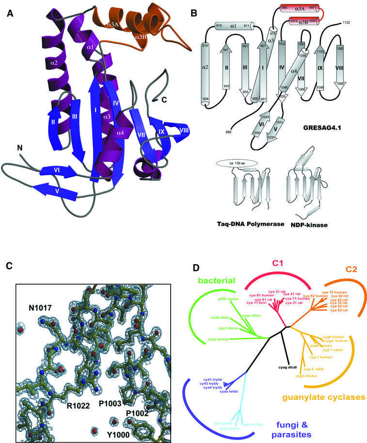
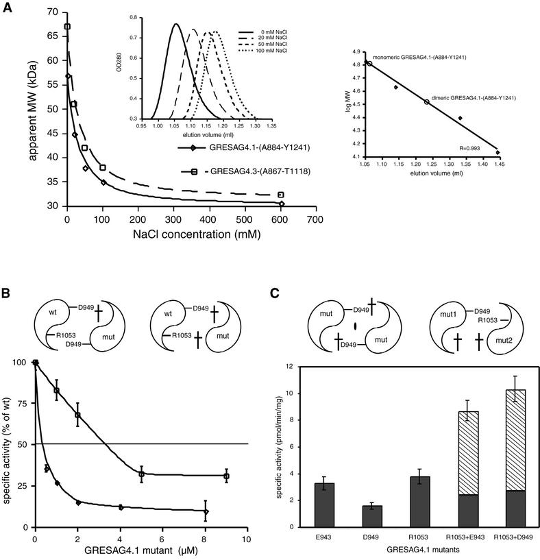
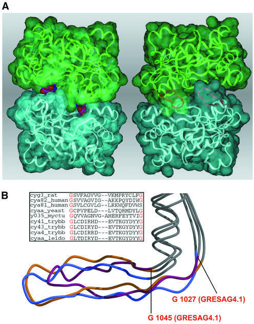
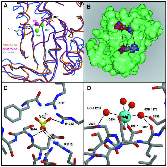
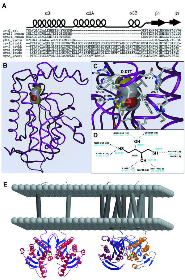
Cyclic AMP is a major trigger of the differentiation process of Trypanosoma brucei, a bloodstream parasite causing sleeping sickness. Its generation in trypanosomes is accomplished by a unique battery of membrane-bound adenylate cyclases (ACs). We have determined the high-resolution X-ray structures of the catalytic domains of two trypanosomal ACs (tACs), GRESAG4.1 and GRESAG4.3. The tAC domains are structurally highly related to the AC domains of higher eukaryotes, but also comprise a highly conserved structural element near the active site, the Delta-subdomain. A cavity below the Delta-subdomain might correspond to an allosteric regulator site as indicated by the stereospecific binding of a single (2S,3S)-1,4- dimercapto-2,3-butanediol molecule. In three different crystal forms, the tAC domains are exclusively observed in a monomeric, catalytically inactive state. Biochemical analysis and the mutagenesis profile of GRESAG4.1 confirmed a common catalytic mechanism of tACs that involves transient dimerization of the AC domain. A low dimerization tendency might play a regulatory role in T. brucei if the activation of tACs is similarly driven by ligand-induced dimerization as in membrane-bound guanylate cyclases.


Figures

None
Fig. 1. (A) Overall structure of GRESAG4.1-(A884–T1131). The Δ-subdomain between α3 and β4 that is absent in other ACs is highlighted in orange. (B) Topologies of tACs, DNA polymerase and NDP kinase. The first half of the GRESAG4.1 AC domain (β1–β4, α1, α2) superimposes at 73 Cα positions with Taq DNA polymerase (r.m.s.d. 1.9 Å) and at 42 positions with NDP kinase from D.melanogaster (r.m.s.d. 2.3 Å). The active center is only in ACs and DNA polymerases placed along β1–β4, while NDP kinases harbor the active site in a loop region. (C) σA-weighted 2FobsFcalc electron density map of GRESAG4.1 at 1.46 Å resolution (contouring level 1.7 σ). (D) Dendrogram showing the phylogenetic relationship of ACs and GCs. For its calculation, 40 protein sequences of nucleotidyl cyclases were aligned using the structural alignment between the mammalian C1A and C2A domains and GRESAG4.1 and the multiple and pairwise sequence alignments as computed by PSI-Blast (Altschul et al., 1997) and Clustal_X (Thompson et al., 1997). A non-redundant subset of 22 nucleotidyl cyclase sequences was selected from the resulting multiple sequence alignment to calculate the phylogenetic relationships using PAUPSCRIPT. The sequences of the AC domains of GRESAG4.1 and GRESAG4.3 correspond to the SWISSPROT entries cy41_trybb and cy43_trybb. Figure 1A and Figures 3–5 were made with MOLSCRIPT (Kraulis, 1991) and RASTER3D (Merrit and Murphy, 1994).
None
Fig. 2. The monomer–dimer equilibrium of tAC domains. (A) Analytical gel filtrations were carried out in the presence of increasing NaCl concentrations on a SMART chromatography station (Pharmacia). Each run was made with 50 µg of purified protein in 20 mM Tris–HCl pH 8.0, flow rate 100 µl/min. The continuous shift of the retention factor (Rf) (left inlay) indicates a dynamic equilibrium between a monomeric and a dimeric species. At low salt, the Rf value corresponds to the molecular weight of a tAC dimer while in the presence of 600 mM NaCl only the monomeric species is present. The gel filtration column S75 PC 3/20 was calibrated (right inlay) using bovine serum albumin (67.0 kDa), ovalbumin (43.0 kDa), chymotrypsinogen A (25 kDa) and ribonuclease A (13.7 kDa). (B) Inhibition of wild-type GRESAG4.1 activity by addition of the mutant D949A (squares) and the double mutant D949A/R1053A (diamonds). AC assays were carried out at a protein concentration of 2 nM (4 nM) for the D949A (D949A/R1053A) mutant as previously described (Bieger and Essen, 2000). (C) Specific activities of the mutants E943A, D949A and R1053A and the equimolar mixtures of E943A with R1053A and D949A with R1053A.
None
Fig. 3. The dimerization of tACs. (A) Model of the tAC dimer as shown from the dorsal (left) and the ventral site (right). Whereas the ventral site is blocked by the pairing of the arm regions (purple), the dorsal site allows substrate access as indicated by ATP molecules occupying both active sites (CPK models). (B) Structural comparison between the arm region of GRESAG4.1 (purple), the C1 (yellow) and C2 domain (blue). The inlay shows the sequence divergence between tACs and other nucleotidyl cyclases in this region.
None
Fig. 4. The catalytic surface of tACs. (A) Superposition of GRESAG4.1 (orange, sulfate), GRESAG4.3 (purple, magnesium) and the mammalian C1A domain (blue, metal centers green, ATPαS) along the active site surface. (B) Model for the binding of ATP (CPK model) to the catalytic surface of a tAC monomer (green). (C) Binding of a sulfate anion (sticks) to the catalytic surface of GRESAG4.1. (D) Magnesium binding site in the monomeric form of GRESAG4.3. The coordination sphere of the magnesium ion (blue) consists of two conserved aspartates and four water ligands. Putative H-bonds are shown as dotted lines.
None
Fig. 5. The Δ-subdomain. (A) Multiple sequence alignment of nucleotidyl cyclases along the β4–α3 insertion. (B) Stereospecific binding of d-DTT in GRESAG4.1. (C and D) Interactions between d-DTT and its binding site below the Δ-subdomain. All residues that make contacts with d-DTT are strictly conserved among other tACs. (E) Models of tAC dimers (left) and mammalian C1C2 heterodimers (right) in their native membrane context. The Δ-subdomain of tACs is distally located from the membrane.

References

    1. Abrahams J.P. and Leslie,A.G.W. (1996) Methods used in the structure determination of bovine mitochondrial F1-ATPase. Acta Crystallogr. D, 52, 30–42. - PubMed
    1. Alexandre S., Paindavoine,P., Tebabi,P., Pays,A., Halleux,S., Steinert,M. and Pays,E. (1990) Differential expression of a family of putative adenylate/guanylate cyclase genes in Trypanosoma brucei. Mol. Biochem. Parasitol., 43, 279–288. - PubMed
    1. Altschul S.F., Madden,T.L., Schäffer,A.A., Zhang,J., Zhang,Z., Miller,W. and Lipman,D.J. (1997) Gapped BLAST and PSI-BLAST: a new generation of protein database search programs. Nucleic Acids Res., 25, 3389–3402. - PMC - PubMed
    1. Artymiuk P.J., Poirrette,A.R., Rice,D.W. and Willett,P. (1997) A polymerase I palm in adenylyl cyclase? Nature, 388, 33–34. - PubMed
    1. Barzu O. and Danchin,A. (1994) Adenylyl cyclases: a heterogeneous class of ATP-utilizing enzymes. Prog. Nucleic Acid Res. Mol. Biol., 49, 241–283. - PubMed

Publication types