. 2001 Jan 15;15(2):226-40.
doi: 10.1101/gad.855001.

The retinoic acid-metabolizing enzyme, CYP26A1, is essential for normal hindbrain patterning, vertebral identity, and development of posterior structures

Affiliations

The retinoic acid-metabolizing enzyme, CYP26A1, is essential for normal hindbrain patterning, vertebral identity, and development of posterior structures

S Abu-Abed et al. Genes Dev. .

Abstract

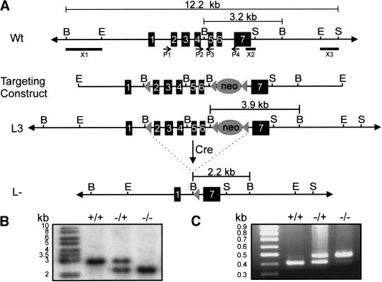
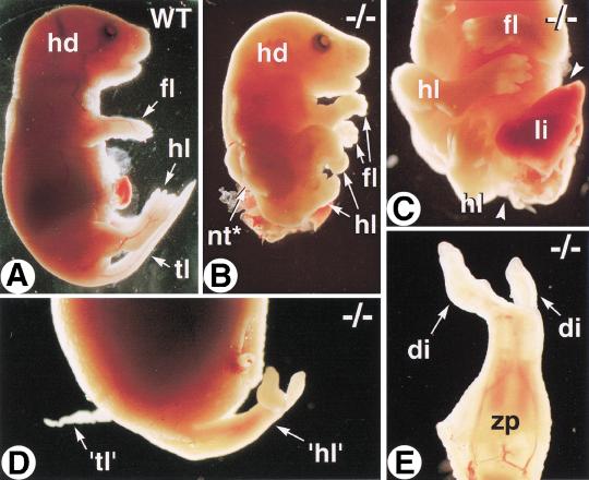
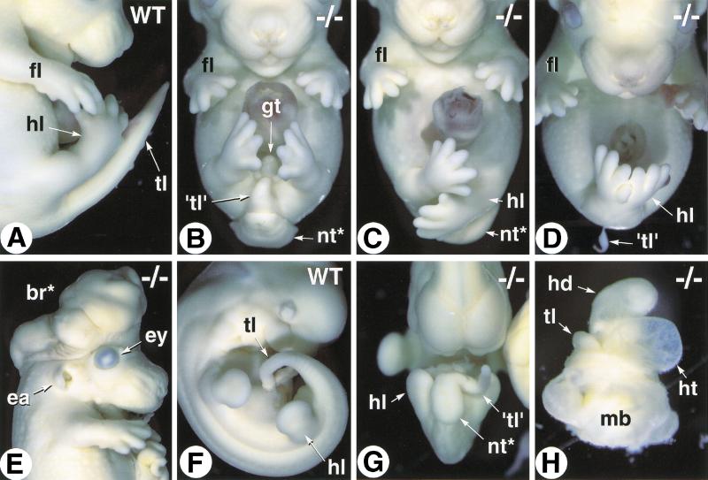
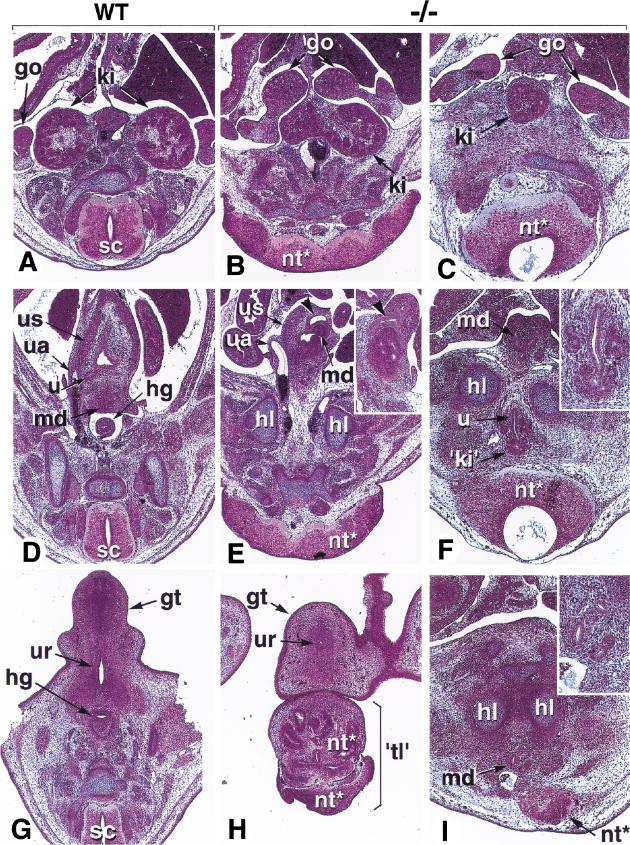
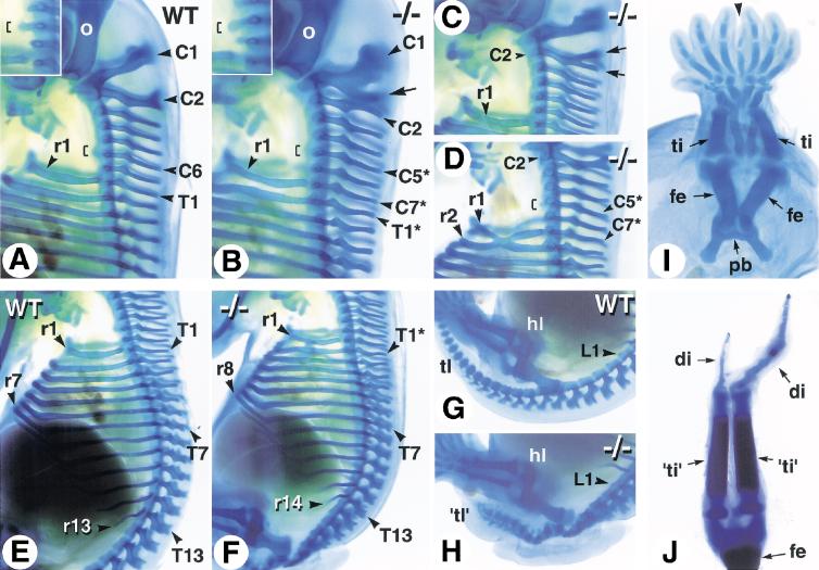
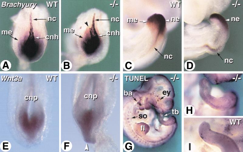
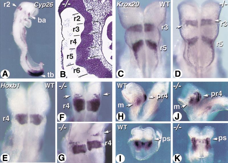
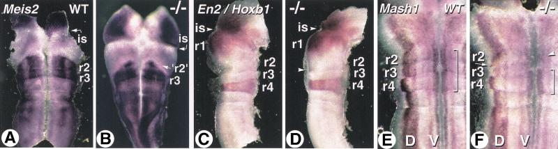
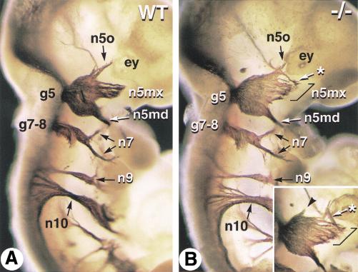
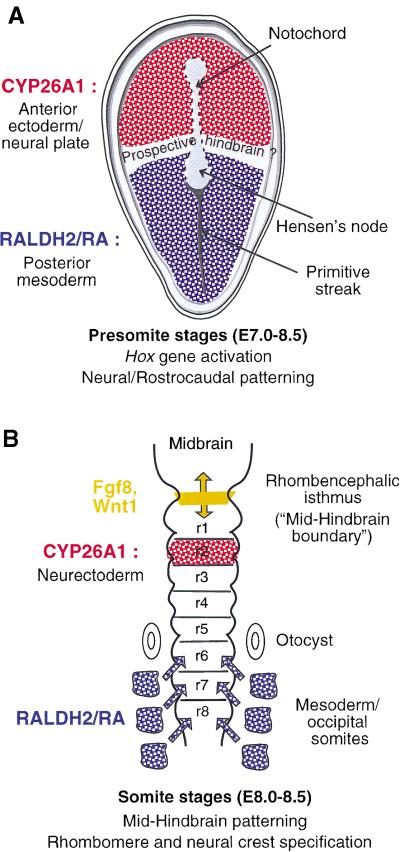
The active derivative of vitamin A, retinoic acid (RA), is essential for normal embryonic development. The spatio-temporal distribution of embryonic RA results from regulated expression of RA-synthesizing retinaldehyde dehydrogenases and RA-metabolizing cytochrome P450s (CYP26). Excess RA administration or RA deficiency results in a complex spectrum of embryonic abnormalities. As a first step in understanding the developmental function of RA-metabolizing enzymes, we have disrupted the murine Cyp26A1 gene. We report that Cyp26A1-null mutants die during mid-late gestation and show a number of major morphogenetic defects. Spina bifida and truncation of the tail and lumbosacral region (including abnormalities of the kidneys, urogenital tract, and hindgut) are the most conspicuous defects, leading in extreme cases to a sirenomelia ("mermaid tail") phenotype. Cyp26A1 mutants also show posterior transformations of cervical vertebrae and abnormal patterning of the rostral hindbrain, which appears to be partially posteriorly transformed. These defects correlate with two major sites of Cyp26A1 expression in the rostral neural plate and embryonic tail bud. Because all of the Cyp26A1(-/-) abnormalities closely resemble RA teratogenic effects, we postulate that the key function of CYP26A1 is to maintain specific embryonic areas in a RA-depleted state, to protect them against the deleterious effect of ectopic RA signaling.


Figures

Figure 1
Figure 1
Targetted excision of exons 2–6 of the Cyp26A1 locus by using the Cre-loxP system. (A) Schematic representation of the Cyp26A1 wild-type locus (Wt, top), targeting construct (middle), targeted allele (L3, middle), and null allele (L–, bottom). Exons are represented by solid rectangles. The targeting construct is flanked by 2.8 kb of 5′ homology and 4.3 kb of 3′ homology and contains novel 5′ BglII restriction sites preceding the subcloned antisense loxP sites (solid triangle) and PGK-neo cassette (also flanked by loxP sites) in introns 1 and 6, respectively. (B) Southern blot analysis of yolk sac genomic DNA of progeny from Cyp26A1L–/ + matings by using a BglII digest. Cre-mediated excision of exons 2–6 was verified by using a 565-bp probe that hybridizes to a 3.2- and 2.2-kb fragment in the case of the wild-type (WT or +) and deleted alleles (L– or –), respectively. (C) PCR analysis of yolk sac genomic DNA. Four primers (P1–P4) used for PCR identification of the Cyp26A1 alleles are indicated by arrows below the Wt locus. P1 and P4 amplify a 484-bp fragment from the null allele (−), whereas P2 and P3 amplify a 414-bp fragment from the WT allele (+). (B) BglII; (E) EcoRI; (encircled B) newly created BglII site; (S) SspI. Note that not all BglII and SspI restriction sites are shown.
Figure 2
Figure 2
Morphological abnormalities of E18.5 Cyp26A1−/− fetuses. Profile views of wild-type (WT) (A) and Cyp26A1−/− (B) littermates. (C) Posterior body region of the same mutant. Arrowheads indicate the open abdominal wall, with protruding viscera. (D) Profile view of the abnormal caudal region of a second Cyp26A1−/− mutant. (E) Detail of the abnormal sirenomelic hindlimb ('hl'). (di) digit; (hd) head; (fl) forelimb; (hl) hindlimb; (li) liver; (nt*) open neural tube (spina bifida); (tl) tail; ('tl') abnormal tail rudiment; (zp) zeugopod.
Figure 3
Figure 3
Variability of the Cyp26A1−/− phenotype. (A) Close-up of the caudal region of an E14.5 wild-type (WT) embryo (profile view). (B–D) E14.5 Cyp26A1−/− embryos with increasing degrees of caudal truncation and hindlimb malposition and malformations (frontal views). (E) E13.5 Cyp26A1−/− embryo with exencephaly. (F,G) Abnormal twisting of the tail bud and opening of the neural tube in an E10.5 Cyp26A1−/− embryo (G, compare with WT littermate, F). (H) E10.5 Cyp26A1−/− mutant showing arrested development at a stage equivalent to WT E8.5 (before embryonic turning) and a markedly dilated heart tube. (br*) exencephalic brain tissue; (ea) ear; (ey) eye; (gt) genital tubercle; (hd) head; (ht) heart; (fl) forelimb; (hl) hindlimb; (mb) extraembryonic membranes; (nt*) open neural tube; (tl) tail; ('tl') tail rudiment.
Figure 4
Figure 4
Histological defects of Cyp26A1−/− embryos. Three successive section planes are shown, at the level of the kidneys (A–C), urogenital sinus (developing urinary bladder; D–F), and genital tubercle (G–I). (A,D,G) E14.5 control embryo. (B,E,H) E14.5 Cyp26A1−/− embryo with a 'mild' (nonsirenomelic) phenotype. (C,F,I) E13.5 Cyp26A1−/− embryo with sirenomelic hindlimbs. Panel E and its inset shows the intra-abdominal ending of the hindgut as a dilated ampulla within the urogenital sinus wall (arrowheads). Panel F and I insets are higher magnifications of the abnormal kidney rudiment and mesonephric ducts, respectively. (go) gonad; (gt) genital tubercle; (hg) hindgut; (hl) hindlimb; (ki) kidney; (md) mesonephric ducts; (nt*) abnormal neural tissue; (sc) spinal cord; ('tl') abnormal tail; (ua) umbilical artery; (u) ureter; (ur) urethral groove; (us) urogenital sinus.
Figure 5
Figure 5
Skeletal defects of Cyp26A1−/− mutants. Whole-mount alcian blue cartilage stainings were performed at E14.5. (A,E,G) Profile views of the cervical (A), thoracic (E), and lumbosacral (G) region of control embryos. (B–D) Abnormal patterning of the cervical and upper thoracic vertebrae in Cyp26A1−/− mutants. Insets in (A) and (B) are close-ups focused on the tuberculum anterior (brackets), which is a characteristic structure of C6 in wild-type embryos. The arrow in (B) points to a supernumerary spinous process on C1. In C, such a supernumerary process is branched on C2 (arrows). (F) Except for the presence of a supernumerary rib on C7, the mutant rib cage appears normally patterned. (H) Disorganized and poorly differentiated vertebral cartilages in a Cyp26A1−/− embryo, caudally to the first lumbar (L1) vertebra. Note, however, the normal position and pattern of the hindlimb. (I) Partial fusion of skeletal elements in the sirenomelic hindlimbs of an E13.5 Cyp26A1−/− embryo. (J) Skeletal pattern of the rudimentary “hindlimbs” of the E18.5 mutant shown in Figure 2, D and E. (C1–C7) cervical vertebrae; (di) digit; (fe) femur; (hl) hindlimb; (pb) pelvic bone; (r1–r14) ribs; (T1–T13) thoracic vertebrae; (L1) first lumbar vertebra; (ti) tibia. The mutant vertebrae have been numbered according to the wild-type formula, and transformed vertebrae are indicated by asterisks.
Figure 6
Figure 6
Molecular analysis of tail development in Cyp26A1−/− embryos. (A–D) Whole-mount in situ hybridization of Brachyury transcripts in wild-type (WT) (A,C) and Cyp26A1−/− (B,D) embryos. A,B are dorsal views of the caudal extremity of E8.5 embryos. Arrowhead in B shows abnormal bifurcation of the caudal portion of the notochord. C,D are views of the tail bud of E9.5 embryos. (E,F) Comparison of Wnt3a transcript distribution in the caudal neural plate of E8.5 WT (E) and Cyp26A1−/− (F) embryos. The arrowhead points to the abnormally thin caudal extremity of the neuroepithelium in the mutant embryo. (G) TUNEL labeling of an E10.5 Cyp26A1−/− embryo. (H,I) comparative views of the tail buds of the same mutant (H) and a littermate control (I). (ba) branchial arch; (cnh) chordoneural hinge; (cnp) caudal neural plate; (ey) eye; (fl) forelimb bud; (me) caudal/tail-bud mesoderm; (nc) notochord; (ne) tail-bud neuroepithelium; (so) somites; (tb) tail bud.
Figure 7
Figure 7
Abnormal patterning of the rostral hindbrain in Cyp26A1−/− embryos. (A) Expression pattern of Cyp26A1 in a wild-type E8.5 embryo, detected by whole-mount in situ hybridization. (B) Sagittal section through the hindbrain region of an E9.5 Cyp26A1−/− embryo. Hematoxylin-eosin staining. (C,D) Whole-mount in situ hybridization of Krox20 transcripts in E8.5 wild-type (WT) control (C) and Cyp26A1−/− (D) embryos. Dorsal views of the hindbrain region. Arrows point to patches of unlabeled cells in mutant r3. (E–G) Hoxb1 transcript distribution in the hindbrain of E8.5 WT (E) and Cyp26A1−/− (F,G) embryos. Arrows show ectopic Hoxb1 expression rostrally to r4 in mutants. (H–K) EphA2 transcript distribution in E8.25 WT (H,I) and Cyp26A1−/− (J,K) embryos. Profile views (H,J) show the distinct EphA2 expression domains in neuroepithelium and head mesenchyme, whereas dorsal views after removal of the caudal region show the hindbrain region (I,K). Note that EphA2 transcripts extend beyond the preotic sulcus (ps) in the mutant embryo. (ba) branchial arch; (m) mesenchyme; (pr4) prospective rhombomere 4; (r2–r6) rhombomeres; (tb) tail bud.
Figure 8
Figure 8
Molecular alterations in the Cyp26A1−/− rostral hindbrain. (A,B) Flatmounts of the mid-hindbrain region of E9.5 control (A) and Cyp26A1−/− (B) embryos hybridized with a Meis2 probe. Note that this mutant embryo was exencephalic. (C,D) Double labeling with Engrailed-2 (En2) and Hoxb1 probes on E10.5 control (C) and mutant (D) embryos. The mid-hindbrain region was bisected and one of its halves is shown in flatmount (floor plate is oriented to the right). (E,F) Details of flatmounted hindbrains from E10.5 control (E) and Cyp26A1−/− (F) embryos hybridized with a Mash1 probe. The specific distribution of Mash1 transcripts in dorsal (D) and ventral (V) neuronal columns is thus seen. The bracket indicates more prominent ventral expression in wild-type (WT) rhombomeres 2–4. In the mutant, this ventral column is disrupted at the level of r3–r2, where only patches of cells show higher Mash1 expression (arrowhead). The arrow points to ectopic dorsally located Mash1-expressing cells in the caudal portion of mutant r2. (is) rhombencephalic isthmus; (r1–r4) rhombomeres.
Figure 9
Figure 9
Abnormal trigeminal nerve patterning in Cyp26A1−/− embryos. Whole-mount immunostaining with an antineurofilament monoclonal antibody was performed on E10.5 wild-type (WT) (A) and Cyp26A1−/− (B) embryos. Neurofilament-labeled nerve tracts are visualized after partial removal of the surface ectoderm. An inset in (B) shows the contralateral trigeminal ganglion of the mutant embryo (in reverse orientation). Brackets indicate an enlarged maxillary nerve component in the mutant embryo. Asterisks point to abnormal routes of some putative ophthalmic nerve fibers, and an arrowhead points to a small ectopic rostral nerve tract. (ey) eye; (g5) trigeminal ganglion; (g7–8) facial-acoustic ganglia; (n5md/mx/o) mandibular, maxillary and ophthalmic branches of the trigeminal nerve; (n7) facial nerve branches; (n9) glossopharyngeal nerve; (n10) vagus nerve.
Figure 10
Figure 10
Schematic representation of the complementary RALDH2 retinoic acid (RA)-synthesizing and CYP26A1 RA-metabolizing activities during early embryo patterning (A) and hindbrain development (B). See the main text for references concerning the various expression patterns.

References

    1. Abu-Abed SS, Beckett BR, Chiba H, Chithalen JV, Jones G, Metzger D, Chambon P, Petkovich M. Mouse P450RAI (CYP26) expression and retinoic acid-inducible retinoic acid metabolism in F9 cells are regulated by retinoic acid receptor γ and retinoid X receptor α. J Biol Chem. 1998;273:2409–2415. - PubMed
    1. Burke AC, Nelson CE, Morgan BA, Tabin C. Hox genes and the evolution of vertebrate axial morphology. Development. 1995;121:333–346. - PubMed
    1. Chambon P. A decade of molecular biology of retinoic acid receptors. FASEB J. 1996;10:940–954. - PubMed
    1. Chazaud C, Chambon P, Dollé P. Retinoic acid is required in the mouse embryo for left-right asymmetry determination and heart morphogenesis. Development. 1999;126:2589–2596. - PubMed
    1. Conlon RA. Retinoic acid and pattern formation in vertebrates. Trends Genet. 1995;11:314–319. - PubMed

Publication types

MeSH terms

LinkOut - more resources