. 2001 Jan 15;15(2):241-53.
doi: 10.1101/gad.840301.

Genetic evidence for a role for Src family kinases in TNF family receptor signaling and cell survival

Affiliations

Genetic evidence for a role for Src family kinases in TNF family receptor signaling and cell survival

L Xing et al. Genes Dev. .

Abstract

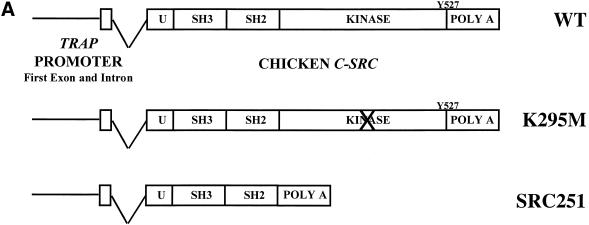
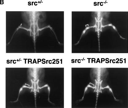
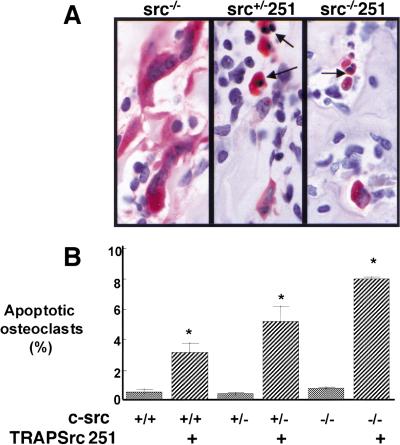
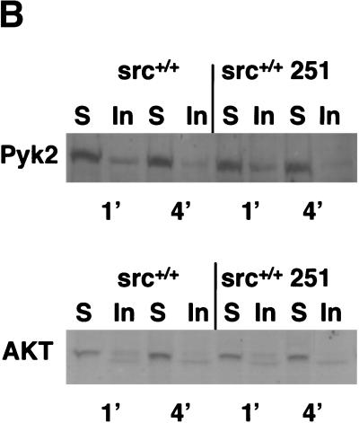
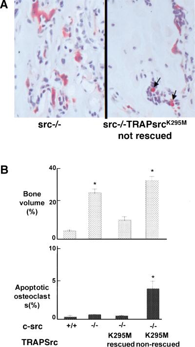
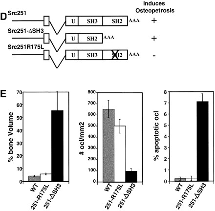
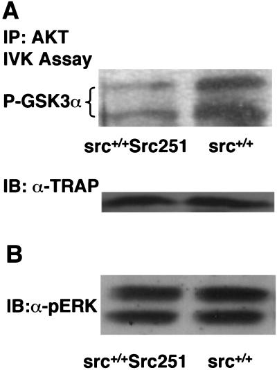
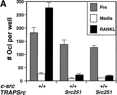
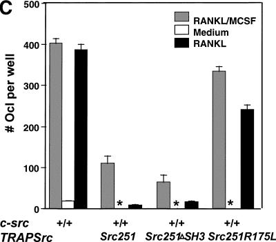
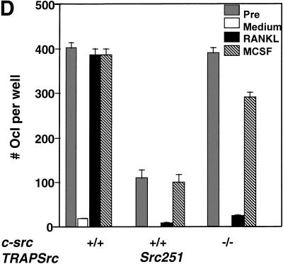
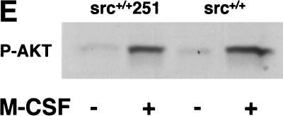
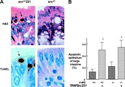
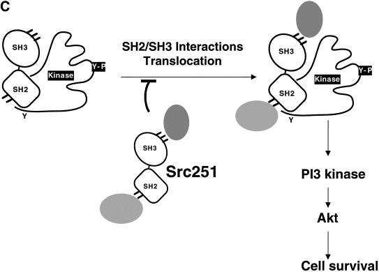
Mutant src(-/-) mice have osteopetrosis resulting from defective osteoclasts, the cells that resorb bone. However, signaling pathways involving Src family members in osteoclasts remain unclear. We demonstrate that expression of a truncated Src molecule, Src251, lacking the kinase domain, induces osteopetrosis in wild-type and src(+/-) mice and worsens osteopetrosis in src(-/-) mice by a novel mechanism, increased osteoclast apoptosis. Induction of apoptosis by Src251 requires a functional SH2, but not an SH3, domain and is associated with reduced AKT kinase activity. Expression of Src251 dramatically reduces osteoclast survival in response to RANKL/TRANCE/OPGL, providing evidence that Src family kinases are required in vivo for survival signaling pathways downstream from TNF family receptors.


Figures

Figure 1
Figure 1
Src251 induces osteopetrosis. (A) Structure of TRAPSrc transgenic constructs. The TRAP promoter, first exon, and intron were fused to the ATG of wild-type (WT) and mutant versions of the chicken c-src gene. (U) Unique region; (SH3) Src homology 3 region; (SH2) Src homology 2 region; (Kinase) kinase domain. Crosses overlay domains containing inactivating mutations. (B) Radiographic analyses of Src251 mice. X-rays, frontal view. Brackets indicate regions that demonstrate increased bone density. (C) Histological analyses of tibia. Sections were stained both with Hematoxylin and Eosin (H&E) and for TRAP activity. (Upper panel) low power (4× magnification). (Lower panel) intermediate power (20×), showing TRAP-positive osteoclasts. (D) Histomorphometric analyses of mice. Genotype is indicated on the X axis. Percentage bone volume (amount of bone matrix as a percentage of the cancellous bone) is indicated on the Y axis. Osteoclasts were counted as TRAP-positive cells.
Figure 1
Figure 1
Src251 induces osteopetrosis. (A) Structure of TRAPSrc transgenic constructs. The TRAP promoter, first exon, and intron were fused to the ATG of wild-type (WT) and mutant versions of the chicken c-src gene. (U) Unique region; (SH3) Src homology 3 region; (SH2) Src homology 2 region; (Kinase) kinase domain. Crosses overlay domains containing inactivating mutations. (B) Radiographic analyses of Src251 mice. X-rays, frontal view. Brackets indicate regions that demonstrate increased bone density. (C) Histological analyses of tibia. Sections were stained both with Hematoxylin and Eosin (H&E) and for TRAP activity. (Upper panel) low power (4× magnification). (Lower panel) intermediate power (20×), showing TRAP-positive osteoclasts. (D) Histomorphometric analyses of mice. Genotype is indicated on the X axis. Percentage bone volume (amount of bone matrix as a percentage of the cancellous bone) is indicated on the Y axis. Osteoclasts were counted as TRAP-positive cells.
Figure 2
Figure 2
TRAPSrc251 transgenic mice show increased osteoclast apoptosis. (A) Histological analyses, high power (100×), showing TRAP-positive osteoclasts. Osteoclasts in Src251 mice are intensely staining with pyknotic nuclei characteristic of apoptotic cells (arrows). (B) Percentage of osteoclasts that were apoptotic.
Figure 3
Figure 3
Src251 is detergent-insoluble in osteoclasts. (A) Osteoclasts were differentiated from bone marrow in culture, then lysed gently in 0.5% Triton X-100 for varying times (2-min lysis is shown here; similar results were obtained at other time points) and the remaining detergent-insoluble fraction then lysed with RIPA. Src was immunoprecipitated from detergent-soluble and -insoluble fractions and analyzed by immunoblotting (Kaplan et al. 1994). (B) Detergent solubility of Pyk2 and AKT are not altered by expression of Src251. Osteoclasts were lysed as above, and lysates were immunoblotted for Pyk2 and AKT. Similar results were seen at 1-, 2-, and 4-min detergent solubility lysis conditions (1- and 4-min conditions are shown here). (S) Detergent-soluble fraction; (In) detergent-insoluble fraction.
Figure 3
Figure 3
Src251 is detergent-insoluble in osteoclasts. (A) Osteoclasts were differentiated from bone marrow in culture, then lysed gently in 0.5% Triton X-100 for varying times (2-min lysis is shown here; similar results were obtained at other time points) and the remaining detergent-insoluble fraction then lysed with RIPA. Src was immunoprecipitated from detergent-soluble and -insoluble fractions and analyzed by immunoblotting (Kaplan et al. 1994). (B) Detergent solubility of Pyk2 and AKT are not altered by expression of Src251. Osteoclasts were lysed as above, and lysates were immunoblotted for Pyk2 and AKT. Similar results were seen at 1-, 2-, and 4-min detergent solubility lysis conditions (1- and 4-min conditions are shown here). (S) Detergent-soluble fraction; (In) detergent-insoluble fraction.
Figure 4
Figure 4
Other Src mutants induce apoptosis of osteoclasts. (A) Histological analyses of src−/− mice expressing the K295M kinase-inactive mutant that were not rescued. Arrows indicate apoptotic osteoclasts. (B) Bone volume and percentage apoptotic osteoclasts in wild-type (WT), src−/−, and src−/− mice that showed rescue or did not show rescue by TRAPSrcK295M. (C) Relative levels of expression of Src251 and SrcK295M transgenes. Lysates from osteoclast cultures of Line V Src251 and Line NN SrcK295M transgenics were normalized for TRAP levels, Src-immunoprecipitated with monclonal 327, and immunoblotted with anti-avian specific Src monoclonal EC10, which only reacts with the transgenic Src (left panel), or monoclonal 327, (right panel) which reacts only with both endogenous and transgenic Src. Similar results were seen with other transgenic lines. (D) Phenotype of mutants affecting the SH3 and SH2 domains of Src251. (E) Bone histomorphometry, osteoclast numbers, and percent apoptotic osteoclasts from mutants affecting the SH3 and SH2 domains of Src251.
Figure 4
Figure 4
Other Src mutants induce apoptosis of osteoclasts. (A) Histological analyses of src−/− mice expressing the K295M kinase-inactive mutant that were not rescued. Arrows indicate apoptotic osteoclasts. (B) Bone volume and percentage apoptotic osteoclasts in wild-type (WT), src−/−, and src−/− mice that showed rescue or did not show rescue by TRAPSrcK295M. (C) Relative levels of expression of Src251 and SrcK295M transgenes. Lysates from osteoclast cultures of Line V Src251 and Line NN SrcK295M transgenics were normalized for TRAP levels, Src-immunoprecipitated with monclonal 327, and immunoblotted with anti-avian specific Src monoclonal EC10, which only reacts with the transgenic Src (left panel), or monoclonal 327, (right panel) which reacts only with both endogenous and transgenic Src. Similar results were seen with other transgenic lines. (D) Phenotype of mutants affecting the SH3 and SH2 domains of Src251. (E) Bone histomorphometry, osteoclast numbers, and percent apoptotic osteoclasts from mutants affecting the SH3 and SH2 domains of Src251.
Figure 5
Figure 5
Src251 osteoclasts have reduced AKT kinase activity. (A) Osteoclasts were differentiated from bone marrow in culture, then lysed, immunoprecipitated with anti-AKT serum, and in vitro kinase assays were performed using GST–GSK3α as a substrate. Products were detected using an anti-phospho GSK-3 antibody by immunoblotting. After normalization for AKT protein levels, AKT activity was observed to be decreased twofold. In this experiment, twofold lower levels of AKT protein were also observed. (Lower panel) Lysates were normalized by immunoblotting for TRAP protein. (B) Osteoclasts were lysed as above and TRAP-normalized lysates were immunoblotted with anti-active ERK. Equivalent levels of total ERK were observed in these samples.
Figure 6
Figure 6
Src251 blocks osteoclast responses to RANKL but not to M-CSF. (A) Osteoclasts were differentiated in culture, then washed twice with PBS and left in either complete media or media plus RANKL overnight. Wells (in quadruplicate) were stained for TRAP activity either before (pre) or after treatment (media or RANKL), and TRAP-positive multinucleated cells were counted. (B) AKT activity in response to RANKL. Osteoclast cultures were serum starved, treated with RANKL for 10 min, lysed, and AKT-immunoprecipitated; and AKT kinase activity was evaluated using GST–GSK3α as a substrate. (C) WT, Src251, Src251-ΔSH3, and Src251-R175L osteoclasts were treated with RANKL overnight, as in A. Src251-ΔSH3 blocks cell survival in response to RANKL, paralleling its induction of osteopetrosis. (D) Osteoclasts from src−/− and Src251 mice both respond poorly to RANKL, but respond normally to M-CSF. Osteoclasts differentiated in culture were treated as above or cultured in media with M-CSF overnight. (E) Src251 does not block AKT activation in response to M-CSF. Osteoclast cultures were serum starved, then treated with M-CSF for 10 min, lysed, and immunoblotted with anti-pS473-AKT to monitor AKT activation.
Figure 6
Figure 6
Src251 blocks osteoclast responses to RANKL but not to M-CSF. (A) Osteoclasts were differentiated in culture, then washed twice with PBS and left in either complete media or media plus RANKL overnight. Wells (in quadruplicate) were stained for TRAP activity either before (pre) or after treatment (media or RANKL), and TRAP-positive multinucleated cells were counted. (B) AKT activity in response to RANKL. Osteoclast cultures were serum starved, treated with RANKL for 10 min, lysed, and AKT-immunoprecipitated; and AKT kinase activity was evaluated using GST–GSK3α as a substrate. (C) WT, Src251, Src251-ΔSH3, and Src251-R175L osteoclasts were treated with RANKL overnight, as in A. Src251-ΔSH3 blocks cell survival in response to RANKL, paralleling its induction of osteopetrosis. (D) Osteoclasts from src−/− and Src251 mice both respond poorly to RANKL, but respond normally to M-CSF. Osteoclasts differentiated in culture were treated as above or cultured in media with M-CSF overnight. (E) Src251 does not block AKT activation in response to M-CSF. Osteoclast cultures were serum starved, then treated with M-CSF for 10 min, lysed, and immunoblotted with anti-pS473-AKT to monitor AKT activation.
Figure 7
Figure 7
Src251 induces apoptosis of intestinal epithelial cells. (A) Histological analyses of large intestine (100×). Arrows indicate apoptotic epithelial cells. (Upper panels) H&E-stained sections; (lower panels) TUNEL staining showing apoptotic cells. (B) Percentage apoptotic large-intestine epithelial cells. Numbers were obtained from 3000 epithelial cells counted in each of two sections. (C) Model for action of Src251. Src251 is a particularly effective dominant-negative molecule because it efficiently blocks interactions of the Src homology protein interaction domains.
Figure 7
Figure 7
Src251 induces apoptosis of intestinal epithelial cells. (A) Histological analyses of large intestine (100×). Arrows indicate apoptotic epithelial cells. (Upper panels) H&E-stained sections; (lower panels) TUNEL staining showing apoptotic cells. (B) Percentage apoptotic large-intestine epithelial cells. Numbers were obtained from 3000 epithelial cells counted in each of two sections. (C) Model for action of Src251. Src251 is a particularly effective dominant-negative molecule because it efficiently blocks interactions of the Src homology protein interaction domains.

References

    1. Bolen JB, Veillette A, Schwartz AM, DeSeau V, Rosen N. Activation of pp60c-src protein kinase activity in human colon carcinoma. Proc Natl Acad Sci. 1987;84:2251–2255. - PMC - PubMed
    1. Boyce B, Yoneda T, Lowe C, Soriano P, Mundy G. Requirement of pp60c-src expression for osteoclasts to form ruffled borders and to resorb bone in mice. J Clin Invest. 1992;90:1622–1627. - PMC - PubMed
    1. Brown MT, Cooper JA. Regulation, substrates, and functions of Src. Biochim Biophys Acta. 1996;1287:121–149. - PubMed
    1. Cartwright CA, Kamps MP, Meisler AI, Pipas JM, Eckhart W. pp60c-src activation in human colon carcinoma. J Clin Invest. 1989;83:2025–2033. - PMC - PubMed
    1. Clark EA, Brugge JS. Redistribution of activated pp60c-src to integrin-dependent cytoskeletal complexes in thrombin-stimulated platelets. Mol Cell Biol. 1993;13:1863–1871. - PMC - PubMed

Publication types

MeSH terms

LinkOut - more resources