. 2001 Feb 5;152(3):531-43.
doi: 10.1083/jcb.152.3.531.

The cytokinesis gene KEULE encodes a Sec1 protein that binds the syntaxin KNOLLE

Affiliations

The cytokinesis gene KEULE encodes a Sec1 protein that binds the syntaxin KNOLLE

F F Assaad et al. J Cell Biol. .

Abstract

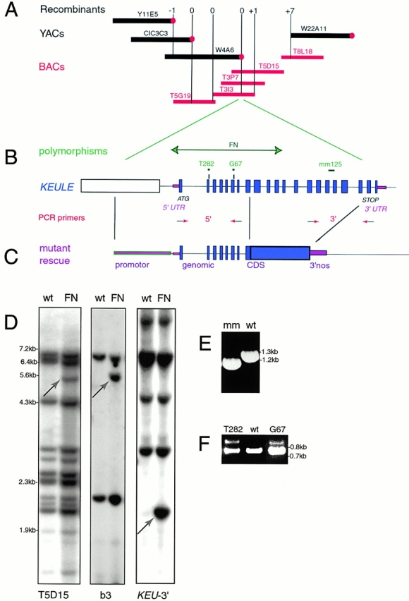
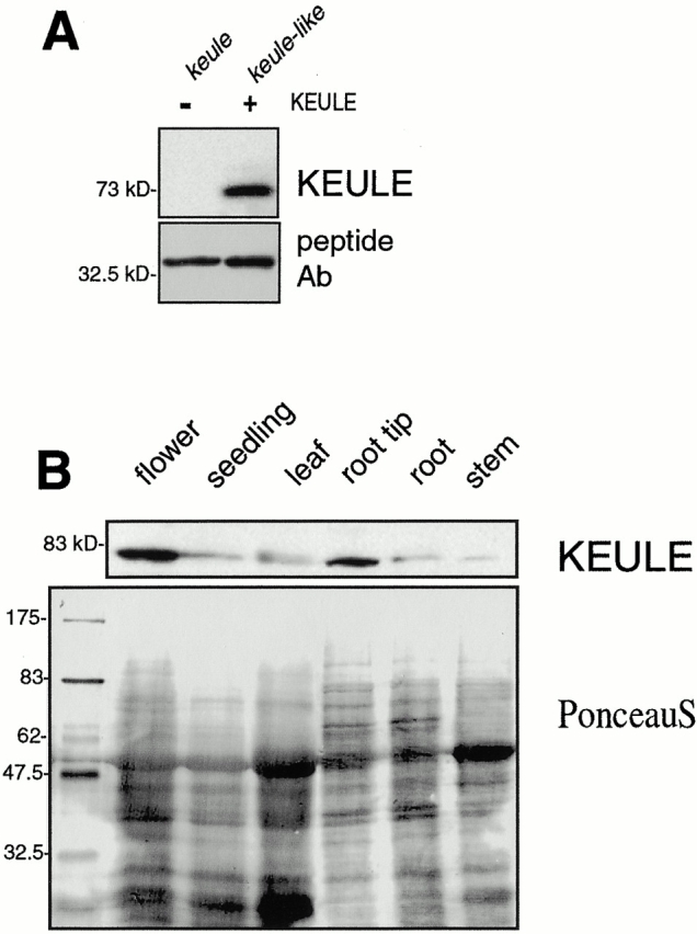
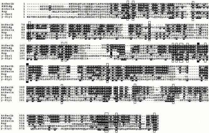
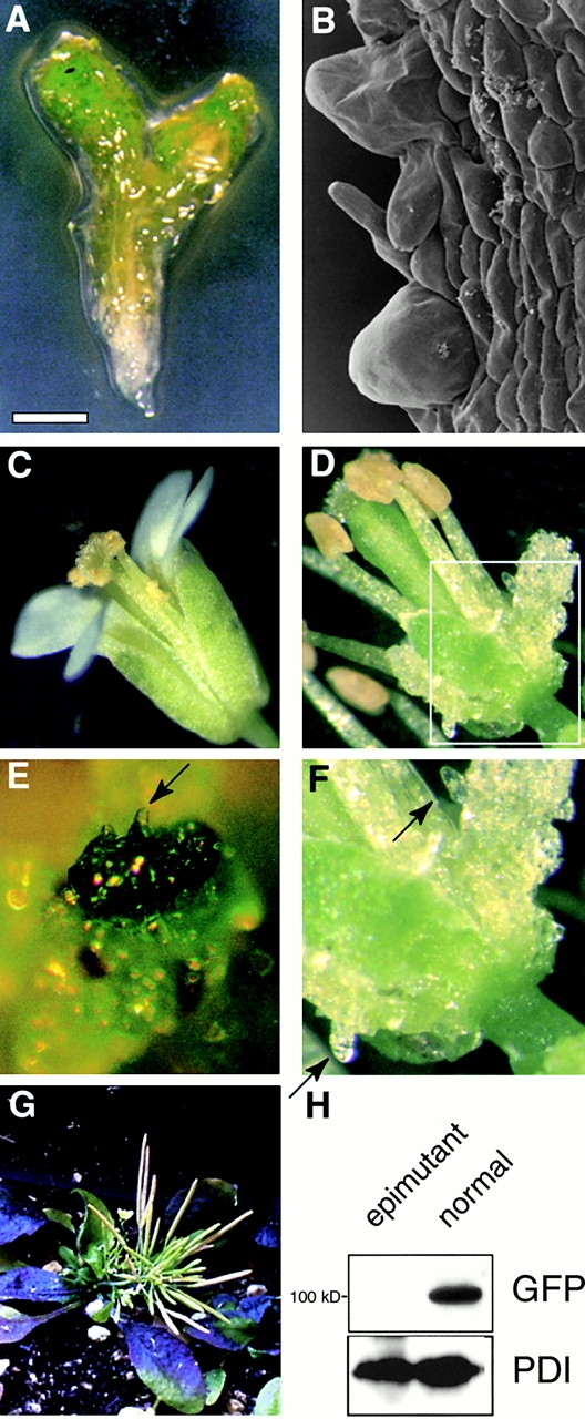
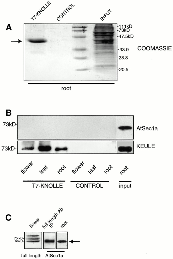
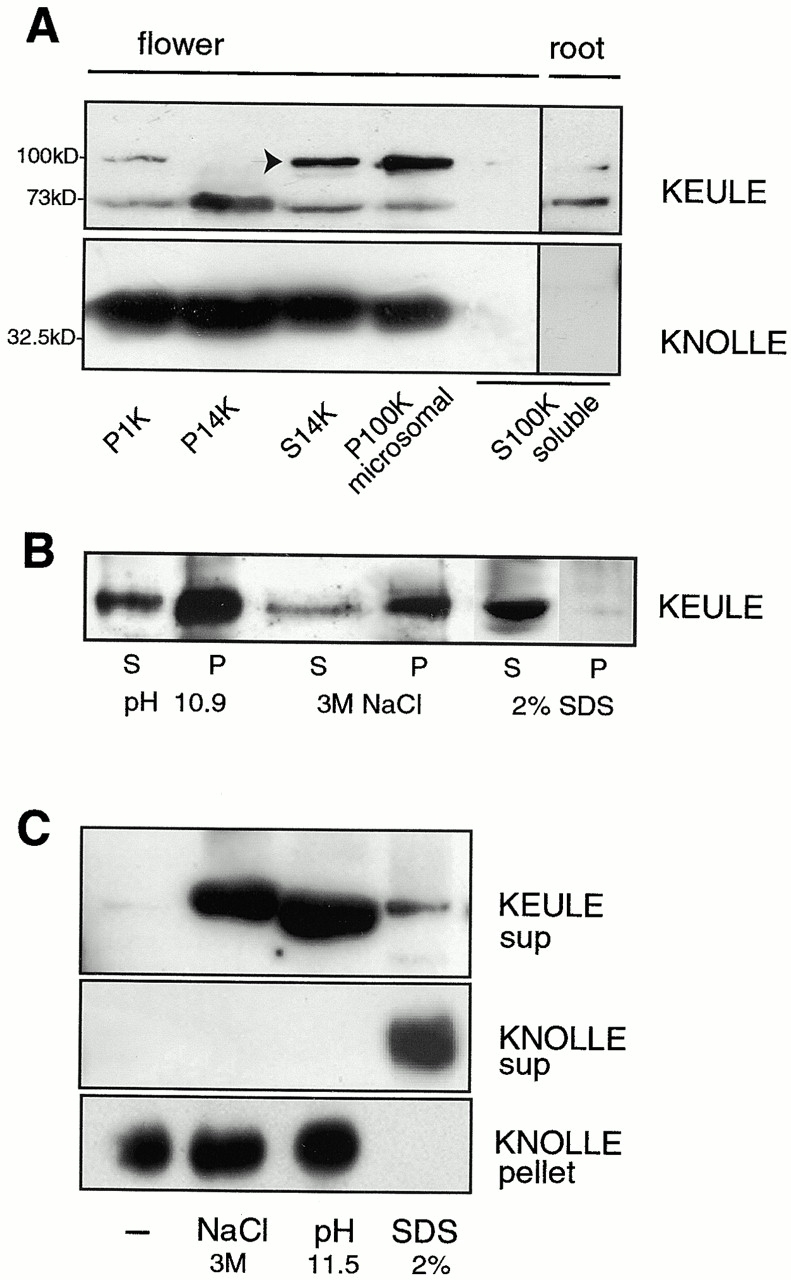
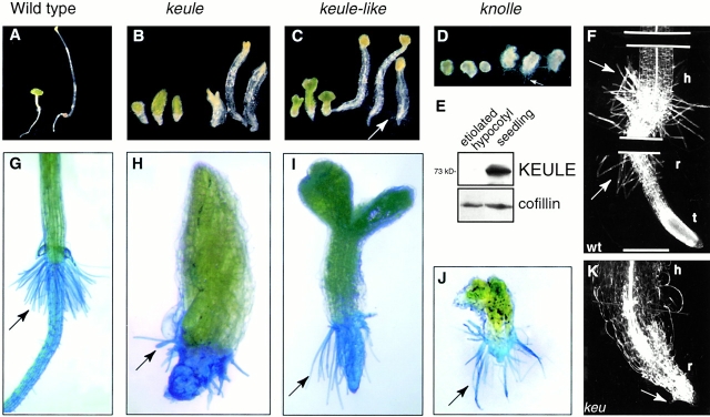
KEULE is required for cytokinesis in Arabidopsis thaliana. We have positionally cloned the KEULE gene and shown that it encodes a Sec1 protein. KEULE is expressed throughout the plant, yet appears enriched in dividing tissues. Cytokinesis-defective mutant sectors were observed in all somatic tissues upon transformation of wild-type plants with a KEULE-green fluorescent protein gene fusion, suggesting that KEULE is required not only during embryogenesis, but at all stages of the plant's life cycle. KEULE is characteristic of a Sec1 protein in that it appears to exist in two forms: soluble or peripherally associated with membranes. More importantly, KEULE binds the cytokinesis-specific syntaxin KNOLLE. Sec1 proteins are key regulators of vesicle trafficking, capable of integrating a large number of intra- and/or intercellular signals. As a cytokinesis-related Sec1 protein, KEULE appears to represent a novel link between cell cycle progression and the membrane fusion apparatus.


Figures

Figure 2
Figure 2
Cloning and molecular characterization of KEULE. (A) Selected portion of the chromosome walk spanning the KEULE region. The BACs (red bars) span the gap in the YAC contig (black bars), which was otherwise only covered by the large (600–800 kb) CIC YACS CIC12A9, CIC12H10, and CIC9G11. The vertical, purple lines represent RFLP markers used for mapping and correspond to polymorphic YAC or BAC ends (filled circles represent left ends of YACs). The numbers above the bars represent the number of recombinants between the marker and keule. The −1 to +1 recombination interval corresponds to 130 kb. Y, YUP; EW, Eric Ward YACs. All BACs are from the Texas A&M University library. Not to scale. (B) Structure of the KEULE locus. The KEULE gene spans 5 kb and includes 21 exons (filled, blue boxes) and 20 introns. The upstream gene (b3 cDNA, below) is represented as an open rectangle. The intergenic region is very small (only 352 bp). Mutations in four alleles are indicated. The two breakpoints in the fast-neutron allele lie in the intergenic region, and in the middle of the KEULE gene. The EMS-induced T282 and G67 mutations lie at intron/exon junctions. MM125 is a small x-ray–induced 156-bp deletion which spans 72 bp of coding and 72 bp of intron sequences. Arrows with red arrowheads represent primers used for amplifying the 5′ and 3′ ends of the coding region. (C) A construct sufficient for mutant rescue consists of genomic sequences fused to cDNA sequences as shown. (D) Fast-neutron–induced sequence polymorphisms at the KEULE locus. Southern blots of wild-type Lansdberg DNA (wt) or of DNA heterozygous for fast-neutron (FN)-induced keule mutations were probed with the BAC T5D15 (left) with the adjacent cDNA (b3, middle) or with a PCR product corresponding to the 3′ end of the KEULE gene (right). Dra1 polymorphisms (green-headed arrows) are shown. The 3′ end of the KEULE gene detects a Dra1 polymorphism distinct from the one detected by b3. (E) PCR analysis of genomic DNA of seedlings homozygous for the MM125 x-ray–induced allele of KEULE detects a deletion at the 3′ end of the coding sequences (3′ PCR primers used). (F) RT-PCR analysis of heterozygous mutant seedlings from the EMS-induced alleles T282 and G67 reveals a small increase in exon length (98 and 85 bp expected, respectively) at the 5′ end of the coding sequences.
Figure 1
Figure 1
Cytokinesis defects in keule embryos A and B depict histological embryo sections (A) wild-type (triangular) (B) keule (delayed in their development). Note the large, irregularly shaped multinucleate cells in keule mutants. Bar: (A) 50 μm; (B) 20 μm.
Figure 5
Figure 5
KEULE antibody and expression of the KEULE protein visualized by Western analysis. (A) The peptide antibody specifically recognizes a 73-kD band in keule-like mutants (described in Materials and Methods) but no band of the size of KEULE in keule mutants. Bottom panel shows the dominant contaminating band revealed by this peptide antibody. Both lanes are loaded with 50 mutant seedlings (left, fast-neutron–induced keule allele; right, cytokinesis-defective line G235) homogenized in 10 μl of sample buffer. (B) KEULE is expressed throughout the plant (as seen after longer exposures of the blot; expression in root shown in Fig. 7 B and in seedling in Fig. 8 C) and appears enriched in dividing tissues of Arabidopsis, namely the root tips and inflorescence meristems, designated “flower” in the figure. The membrane was stained with Ponceau S to monitor loading.
Figure 3
Figure 3
Sequence alignment of the KEULE protein. KEULE has two closely related homologues in Arabidopsis, AtSec1a and AtSec1b. These three proteins are clearly much closer to each other than to their homologues in other organisms. The Sec1 signature was defined before the publication of plant Sec1s as consisting of 23 highly conserved residues, boxed in this figure. Note that KEULE contains 21 of these 23 residues. The two residues that do not fit in this signature are identical in all three Arabidopsis Sec1s and in one instance, they are replaced by a conserved amino acid (I in lieu of L, KEULE amino acid 269). Rop, Drosophila nSec1 homologue; Sly1, yeast Sec1 homologue involved in ER-Golgi transport; accession numbers are to be found in Halachmi and Lev 1996. Plant Sec1 homologue sequence data are available from GenBank/EMBL/DDBJ under accession nos.: KEULE, AF331066; AtSec1a, AF335539; AtSec1b, CAB40953. Note that we do not show the entire proteins but only the domains with higher degrees of conservation. Black boxes highlight identical residues and gray boxes shade conserved residues.
Figure 4
Figure 4
KEULE is required for cytokinesis in somatic cells throughout the plant life cycle. (A) keule seedling, with its characteristic bloated surface. (B) Scanning electron micrograph of a keule seedling (hypocotyl), showing the bloated cells at the surface layer. (C) Wild-type flower. D–G are epimutations at the keule locus due most likely to cosuppression in transformants harboring a P35S::GFP-KEULE gene fusion. (D) Sectors are seen on petals and sepals, but not on the carpels or stamens. (E) Sector taking over the entire apical meristem. (F) Bloated, keule-like surface cells (arrows) in somatic sector boxed in D. (G) Lines with large numbers of mutant sectors (such as E) matured as a cluster or large rosette of fertile silliques. (H) Using an anti-GFP antibody, we fail to detect the GFP-KEULE fusion protein in cytokinesis-defective sectored plants (epimutant), but do see it in nonsectored siblings (normal). Anti-protein disulfide isomerase (PDI) was used as a loading control. Bar: (A) 300 μm; (B) 45 μm; (E) 200 μm; (F) 170 μm.
Figure 7
Figure 7
KEULE and KNOLLE interact. T7-KNOLLE was bacterially overexpressed and bound to α-T7 agarose beads. In the control lanes, bacterially expressed GST-KNOLLE was used in lieu of T7-KNOLLE. The loaded beads were incubated with protein extracts from flowers, leaves, and root lengths. 10% of the bead-bound proteins (T7-KNOLLE, CONTROL) or 1% of the plant extracts (INPUT) are loaded. (A) Coomassie-stained gel of beads incubated with root extract, or of root extract. Arrow points to T7-KNOLLE. GST-KNOLLE fails to bind the α-T7 agarose beads. (B) Westerns were probed with a peptide antibody against the highly conserved KEULE homologue AtSec1a (top) and with the KEULE peptide antibody (bottom). Longer exposures of the upper panel reveal the AtSec1a band in all six lanes, with no differential behavior between the experiment and negative control. The input lanes are loaded with the root extracts used for this experiment. (C) Specificity of the AtSec1a antibody. The AtSec1a peptide antibody recognizes a band at the expected size, 66 kD (arrow), in plant extracts (root). To confirm that this indeed corresponds to AtSec1a, we show that the antibody cross-reacts with a 66-kD protein immunoprecipitated (IP) by an antibody raised against the full length KEULE protein (middle). As expected based on sequence analysis (see Fig. 3), the full length antibody reveals three bands in plant extracts; the lower one presumambly corresponds to AtSec1a (left).
Figure 6
Figure 6
KEULE is peripherally associated with membranes. (A) KEULE appears to be membrane associated: KEULE is present in both the heavy membrane (P14K) and microsomal (P100) fractions. Longer exposure of the blot shows that KEULE is found in the soluble fraction as well. KEULE is shown to be in the soluble fraction of roots (right). As a control, the syntaxin KNOLLE is shown to be present in the membrane but not the soluble (S100K) fractions (bottom). P, pellet; S, supernatant. Note: not equiloaded, the membrane fractions are fivefold more concentrated than the soluble fractions. The arrow points to the KEULE-specific band which migrates anomalously at 100 kD in this sample (run with <100 mM DTT). It is not clear why this band is absent in the P14K fraction. (B and C) Membrane association is peripheral. KEULE can be released from the microsomes (P100K) if these are incubated with high salt (3 M NaCl), if the pH is increased (0.1 M NaCO3, pH 10.9–11.5), or with 2% SDS. (B) Solubilzation is complete with 2% SDS but only partial with high salt and high pH. (C) KEULE and KNOLLE differ with respect to the nature of their membrane association. In contrast to KEULE, KNOLLE was released from the microsomal fraction by SDS but not by high salt or high pH. (Although it appears that less KEULE protein is released from membranes by SDS than by high salt or high pH, this is most likely an artefact due to incomplete deoxycholate/TCA precipitation in the presence of SDS; see Materials and Methods for sample preparation. As in B, the majority of the KEULE protein remains in the pellets; not shown as grossly overexposed.) sup, supernatant.
Figure 8
Figure 8
Cell elongation and root hair growth in keule mutants. (A–D) Mutant seedlings were germinated in the light (left) or dark (right). keule (B), keule-like (C), and knolle (D) mutants are capable of elongation, but not to the same extent as wild-type (A). (E) KEULE is only weakly expressed in etiolated hypocotyls. Anticofillin antibody was used as a load control. (F–K) Root hairs are absent (K) or stunted and radially swollen (H) in keule mutants, but of normal length in other cytokinesis-defective mutants. Root hairs are stained with methylene blue. Each keule allele exhibits a range of phenotypes, though the range may differ in a given allele. In contrast to keule mutants, keule-like (I) and knolle (J) mutants grow long root hairs. (F and K) Clearing preparation of wild-type (F) and keule (K) seedling. Note that the basal portion of keule seedlings have root-like characteristics. Arrows point to root hairs. h, hypocotyl; r, root; t, root tip. KEULE alleles: MM125 in B and H; G67 in K. Bars: (F) 270 μm; (K) 200 μm.

References

    1. Assaad F.F., Tucker K.L., Signer E.R. Epigenetic repeat-induced gene silencing (RIGS) in Arabidopsis . Plant Mol. Biol. 1993;22:1067–1085. - PubMed
    1. Assaad F., Mayer U., Wanner G., Jürgens G. The keule gene is involved in cytokinesis in Arabidopsis . Mol. Gen. Genet. 1996;253:267–277. - PubMed
    1. Assaad F.F., Lukowitz W., Mayer U., Jürgens G. Cytokinesis in somatic plant cells. Plant Physiol. Biochem. 1997;35:175–182.
    1. Bassham D.C., Raikhel N.V. An Arabidopsis VPS45 homolog implicated in protein transport to the vacuole. Plant Physiol. 1998;117:407–415. - PMC - PubMed
    1. Bock J.B., Klumperman J., Davanger S., Scheller R. Syntaxin 6 functions in trans-Golgi network vesicle trafficking. Mol. Biol. Cell. 1997;8:1261–1271. - PMC - PubMed

Publication types

MeSH terms