. 2001 Feb 5;152(3):563-78.
doi: 10.1083/jcb.152.3.563.

Identification and characterization of an escorter for two secretory adhesins in Toxoplasma gondii

Affiliations

Identification and characterization of an escorter for two secretory adhesins in Toxoplasma gondii

M Reiss et al. J Cell Biol. .

Abstract

The intracellular protozoan parasite Toxoplasma gondii shares with other members of the Apicomplexa a common set of apical structures involved in host cell invasion. Micronemes are apical secretory organelles releasing their contents upon contact with host cells. We have identified a transmembrane micronemal protein MIC6, which functions as an escorter for the accurate targeting of two soluble proteins MIC1 and MIC4 to the micronemes. Disruption of MIC1, MIC4, and MIC6 genes allowed us to precisely dissect their contribution in sorting processes. We have mapped domains on these proteins that determine complex formation and targeting to the organelle. MIC6 carries a sorting signal(s) in its cytoplasmic tail whereas its association with MIC1 involves a lumenal EGF-like domain. MIC4 binds directly to MIC1 and behaves as a passive cargo molecule. In contrast, MIC1 is linked to a quality control system and is absolutely required for the complex to leave the early compartments of the secretory pathway. MIC1 and MIC4 bind to host cells, and the existence of such a complex provides a plausible mechanism explaining how soluble adhesins act. We hypothesize that during invasion, MIC6 along with adhesins establishes a bridge between the host cell and the parasite.


Figures

Figure 1
Figure 1
Illustration of the structural domains of the proteins and the recombinant mutants used in this study. (A) Schematic representation of the structural domains on the micronemal proteins MIC1, MIC3, MIC4, MIC6, and the major tachyzoite surface antigen SAG1. The proteolytic cleavages of the micronemal proteins are indicated with two types of symbols distinguishing posttranslational and postexocytosis cleavages. Sequence data for MIC1, MIC4, and MIC6 are available from GenBank/EMBL/DDBJ under accession numbers Z71786, AF143487, and AF110270, respectively. (B) Schematic representation of the constructs used in this study. The epitope tags Ty-1 and myc are represented by a black box. The color code of the diverse domains is as described in A. Schematic drawing of pTMIC6Ty-1, pTMIC6ΔCD, pTMIC6GPI, pTMIC6ΔEGF-1, -2, or -3, pTMIC6ΔAD, pTMIC4Ty-1, pTMIC4ΔA5-6, pTMIC4ΔA3-6, pM2SAG1TM-CD, and pM2MIC1myc.
Figure 2
Figure 2
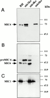
Disruption of MIC1, MIC4, and MIC6 genes in T. gondii tachyzoites by double homologous recombination. Western blot analysis of an equal loading of whole cell lysates corresponding to 5 × 10⁶ tachyzoites from RH, mic1ko, mic4ko, and mic6ko. In A–C, membranes were probed with rabbit antibodies anti-MIC4, anti-MIC6, and mAb anti-MIC-1, respectively.
Figure 4
Figure 4
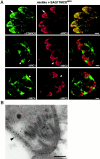
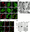
The CD of MIC6 contains sorting signals for the micronemes targeting. (A) IFA analysis by confocal microscopy on monolayers of HFF cells infected with mic6ko mutant expressing stably SAG1TM-CDMIC6. The subcellular distribution of the SAG1TM-CDMIC6 fusion protein was detected by using antibodies raised against the CD of MIC6. The colocalization of SAG1TM-CDMIC6 with MIC2 is illustrated by the yellow color in the merged image and indicated by arrows. In this genetic background, both MIC1 and MIC4 were missorted and accumulated in the vacuolar space, as indicated by arrowheads. (B) SAG1TM-CDMIC6 accumulates precisely in the micronemes as demonstrated by immunoelectron microscopy using anti-SAG1 antibodies. SAG1 is detected by gold particles and, when GPI anchored, was found in its normal location at the parasite surface (arrowheads). In contrast, SAG1TM-CDMIC6 was found in the micronemes (arrow). As micronemes are thinner than the section, some of them were not exposed to the antibody and were not labeled. Bars: (A) 1 μm; (B) 0.2 μm.
Figure 5
Figure 5
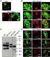
Rescue of mic6ko and evidence for a direct interaction between MIC1 MIC4 and MIC6. (A) Subcellular distribution of endogenous MIC1 and MIC6Ty-1 were analyzed by confocal microscopy in mic6ko and mic6ko expressing MIC6Ty1. The missorting of MIC1 to the vacuolar space (arrowhead) in mic6ko was reverted in mic6ko expressing MIC6Ty-1. MIC6Ty-1 localized to the micronemes as detected by mAb anti-Ty1 (arrows). (B) Expression of pTMIC6ΔCD in mic6ko was analyzed by IFA using anti-MIC6 raised against the EGF domains. MIC6ΔCD lacking the microneme sorting signals was retained in a perinuclear region (arrowheads). Similarly, endogenous MIC1 and MIC4 accumulated in the same compartments. The absence of accumulation of MIC4 in the vacuolar space or MIC1 targeting to the micronemes were observed by double IFA with the two markers GRA3 and MIC2, respectively. (C) MIC6GPI is covalently linked to a GPI and, as a consequence, anchored at the plasma membrane (arrowhead) of the parasites in mic6ko. The localization of MIC6GPI at the plasma membrane caused the quantitative redistribution of endogenous MIC1 and MIC4 to the surface of the parasites (arrowheads). MIC6GPI and MIC4 were excluded from the micronemes as seen by the lack of colocalization with MIC2. Moreover, MIC1 and MIC4 colocalized perfectly. (D) RH and transformed parasites expressing MIC6Ty-1, MIC6GPI, and MIC6ΔCD were analyzed by Western blot with anti-MIC6. The two processed forms of MIC6 detectable in RH were also present in the rescued parasites expressing MIC6Ty-1. Two forms of MIC6GPI are detectable suggesting that the NH2-terminal processing occurred. Bars, 1 μm.
Figure 3
Figure 3
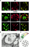
MIC1 and MIC4 are mistargeted in mic6ko and accumulate in the dense granules and the parasitophorous vacuole. (A) Mistargeting of MIC1 and MIC4 was analyzed by confocal microscopy of HFF cells infected with mic6ko and compared with RH. In wild-type parasites, T101F7 mAb anti-MIC1 and polyclonal anti-MIC4 showed perfect colocalization of the two proteins and a typical apical micronemes staining. In contrast, MIC4 did not colocalize with the other micronemal protein MIC2 (mAb T34A11) in mic6ko parasites but instead accumulated in dense granules and in the parasitophorous vacuolar space. Under mild fixation conditions, the vacuolar signal of MIC4 is lost, and the remaining MIC4 colocalizes with the dense granule marker GRA3 (mAb T26H11, in red). (B) The vacuolar accumulation of MIC4 in mic6ko was confirmed by immunolocalization of MIC4 in mic6ko. The label is over the dense material found at the posterior end of a tachyzoite fixed early after invasion, typical of dense granules exocytosis. T, tachyzoite; V, parasitophorous vacuole; H, host cell. (C) Schematic representation of the secretory pathway in a tachyzoite. The nucleus (gray), the ER (dark green), the Golgi apparatus (light green), the apicoplast (pink), the dense granules (blue), the rhoptries (yellow), and the micronemes (red). (D) Schematic representation eight parasites arranged in rosette within the parasitophorous vacuole after three cell divisions. Bars: (A) 1 μm; (B) 0.2 μm.
Figure 6
Figure 6
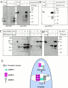
MIC1, MIC4, and MIC6 physically interact. (A) Immunoprecipitation of a complex containing MIC1, MIC4, and MIC6 from wild-type parasite lysate (RH lysate) by anti-MIC1 mAbs. The immunoprecipitates were shown to contain also MIC6 and MIC4 by Western blot analysis using rabbit polyclonals anti-MIC6 or anti-MIC4. The two processed forms of MIC6 and the two forms of MIC4 are indicated by arrows. Controls included immunoprecipitation in absence of lysate or in absence of the mAb anti-MIC1. (B) Coimmunoprecipitation of MIC4 with anti-MIC1 in cell lysates of mic6ko parasites showed that they interact in absence of MIC6. (C) Coimmunoprecipitation of MIC1 (detected with mAb anti-myc) and MIC4 using the polyclonals anti-CDMIC6 from lysates of mic1ko parasites expressing MIC1myc. The immunoprecipitation was carried out with increasing amount of antibodies (5, 20, and 40 μl). (D) Coimmunoprecipitation of MIC4 with anti-MIC6 from RH lysate. (E) Model. A complex between MIC1, MIC4, and MIC6 forms in the early compartments of the secretory pathway and ensures the proper targeting of MIC1 and MIC4 to the micronemes via the interaction of MIC6 cytoplasmic tail with the sorting machinery. During its transport, MIC6 is processed at the NH2 terminus and looses the first EGF-like domain. Upon contact with host cells, an increase in free intracellular calcium stimulates the discharge of the microneme contents. The complex of MICs is liberated at the surface of the parasites, potentially anchored within the plasma membrane via MIC6.
Figure 7
Figure 7
The EGF-3 domain of MIC6 is necessary for accurate targeting of MIC1 and MIC4 to the micronemes. (A) Western blot analysis of cell lysates from RH and recombinant parasites expressing MIC6ΔEGF1, MIC6ΔEGF2, and MIC6ΔEGF3 with anti-CDMIC6 antibodies (raised against the CD of MIC6). MIC6ΔEGF1 and MIC6ΔEGF3 were still subjected to NH2-terminal cleavage, whereas a single form of MIC6ΔEGF2 was detectable, suggesting that the cleavage site has been deleted by removing the EGF-2 domain. (B) IFA analysis by confocal microscopy. MIC6ΔEGF-3 expressed in mic6ko was accurately targeted to the micronemes, as detected with anti-CDMIC6, and colocalized with MIC2 (arrows). In this mutant, MIC4 was only partially sorted to the micronemes (arrowhead), with a significant amount of protein still accumulating in the dense granules. MIC1 and MIC4 colocalized (arrows). Bar, 1 μm.
Figure 8
Figure 8
Expression of MIC4 deletion mutants in mic4ko. The NH2-terminal region of MIC4 containing the first two apple domains was correctly sorted to the micronemes. (A) Western blot analysis of cell lysates from RH and recombinant parasites expressing MIC4ΔA5-6 or MIC4ΔA3-6. The migration of MIC4 and the truncated forms on SDS-PAGE were slower than their predicted molecular weight. (B) IFA analysis by confocal microscopy of parasites expressing MIC4ΔA5-6 and MIC4ΔA3-6. Proper targeting to the micronemes was confirmed by colocalization with MIC3. Bar, 1 μm.
Figure 9
Figure 9
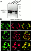
Analysis of mic1ko. MIC1 is necessary for MIC6 and MIC4 to leave the ER and Golgi. (A) Subcellular distribution of MIC4 and MIC6 in mic1ko strain analyzed by IFA. MIC3 was faithfully sorted to the micronemes, whereas MIC4 and MIC6 were retained in the early compartments of the secretory pathway. Two vacuoles stained with anti-MIC4 and three vacuoles stained with anti-MIC6 are presented here to illustrate the various compartments where the two proteins are retained. A double IFA of MIC4 and MIC6 in mic1ko indicated that in all vacuoles, both proteins colocalized perfectly as shown in the merged image. (B) Immunolocalization of MIC6 and MIC4 in mic1ko by electron microscopy revealed various patterns of distribution in the early secretory compartments. (a) MIC4 was detected in the nuclear envelope (arrowheads) and in the successive stacks of the Golgi (arrows). N, nucleus; G, Golgi apparatus. (b) MIC6 stagged in the cis-Golgi. (c) MIC4 stagged in the ER (arrows) and nuclear envelope (arrowheads). (C) The mic1ko mutant expressing MIC1myc showed rescue of the phenoptype by IFA. In presence of MIC1myc, both MIC4 and MIC6 were quantitatively sorted to the micronemes. A slight overexpression of MIC1 led to some leakage to the vacuolar space as detected by anti-MIC1 mAb. (D) Western blot analysis of lysates form RH, mic1ko, and mic1ko complemented with MIC1myc using either anti-MIC1sera or anti-myc mAb. A slight increase in the size of MIC1myc was apparent compared with endogenous MIC1, due to the presence of the myc tag. Bars: (A and C) 1 μm; (B) 0.2 μm.
Figure 10
Figure 10
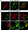
Trafficking of MIC6GPI and SAG1TM-CDMIC6 in mic1ko. Pools of parasites expressing MIC6TyGPI and SAG1Ty1TM-CDMIC6 were analyzed by IFA. (A) MIC6TyGPI localized predominantly to the perinuclear region as detected by the anti–Ty-1 antibodies (arrowhead). The endogenous MIC4 was retained in the early compartments of the secretory pathway (arrow). (B) SAG1TM-CDMIC6 was accurately sorted to the micronemes as shown by the extensive colocalization with the microneme marker MIC7 (rabbit polyclonal anti-MIC7 raised against the EGF-like domains; Meissne, M., and D. Soldati, unpublished results). The vacuole on the left was not transformed with SAG1TM-CDMIC6 and showed the absence of background with the mAb anti-Ty-1. The pool of transformants was analyzed with the anti-CDMIC6 revealing that the endogenous MIC6 (green, arrows) is retained in the ER and Golgi. Bar, 1 μm.

References

    1. Achbarou A., Mercereau-Puijalon O., Autheman J.M., Fortier B., Camus D., Dubremetz J.F. Characterization of microneme proteins of Toxoplasma gondii . Mol. Biochem. Parasitol. 1991;47:223–233. - PubMed
    1. Baldi D.L., Andrews K.T., Waller R.F., Roos D.S., Howard R.F., Crabb B.S., Cowman A.F. RAP1 controls rhoptry targeting of RAP2 in the malaria parasite Plasmodium falciparum . EMBO (Eur. Mol. Biol. Organ.) J. 2000;19:2435–2443. - PMC - PubMed
    1. Barnwell J.W., Galinski M.R. Plasmodium vivaxa glimpse into the unique and shared biology of the merozoite. Ann. Trop. Med. Parasitol. 1995;89:113–120. - PubMed
    1. Bastin P., Bagherzadeh Z., Matthews K.R., Gull K. A novel epitope tag system to study protein targeting and organelle biogenesis in Trypanosoma brucei . Mol. Biochem. Parasitol. 1996;77:235–239. - PubMed
    1. Bermudes D., Dubremetz J.F., Achbarou A., Joiner K.A. Cloning of a cDNA encoding the dense granule protein GRA3 from Toxoplasma gondii . Mol. Biochem. Parasitol. 1994;68:247–257. - PubMed

Publication types

MeSH terms