. 2001 Feb 5;152(3):633-43.
doi: 10.1083/jcb.152.3.633.

Matrix attachment regulates Fas-induced apoptosis in endothelial cells: a role for c-flip and implications for anoikis

Affiliations

Matrix attachment regulates Fas-induced apoptosis in endothelial cells: a role for c-flip and implications for anoikis

F Aoudjit et al. J Cell Biol. .

Abstract

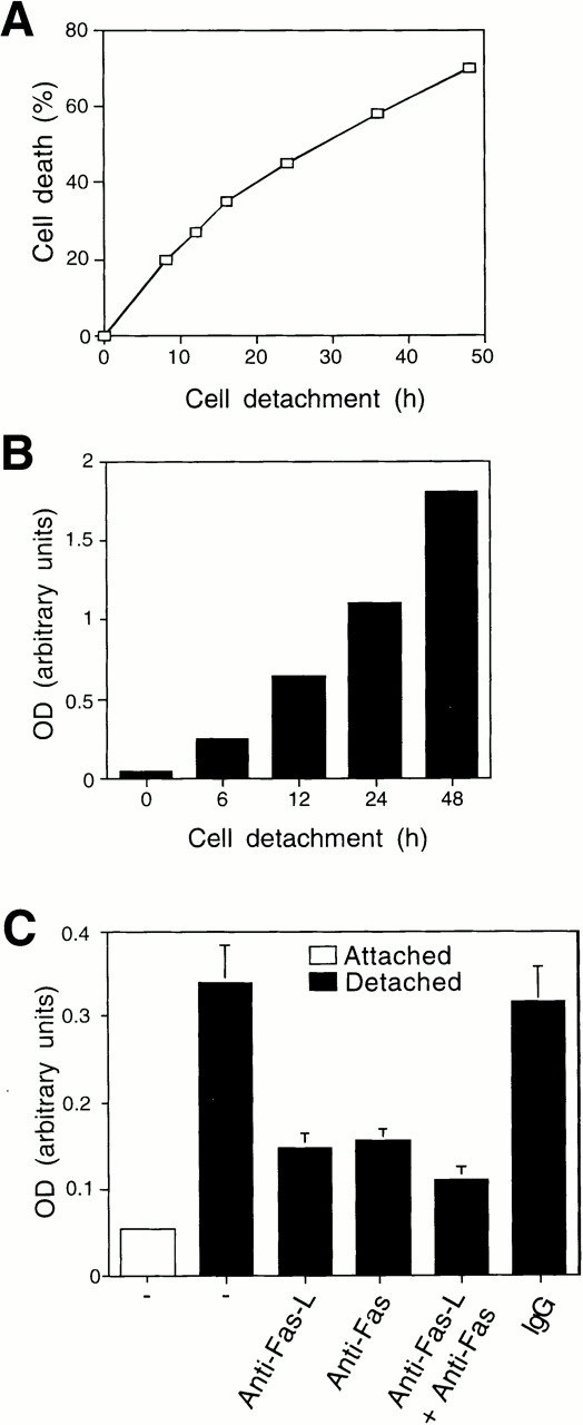
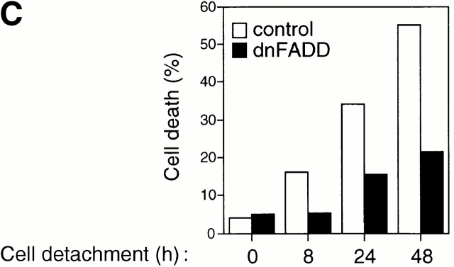
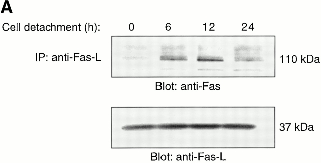
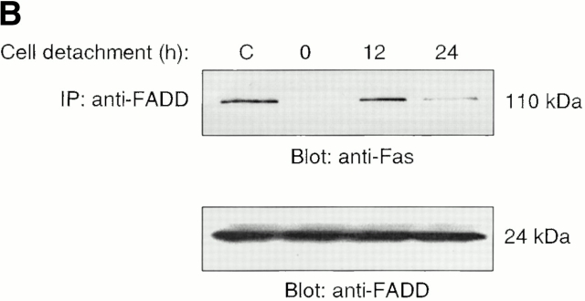
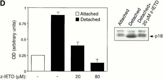
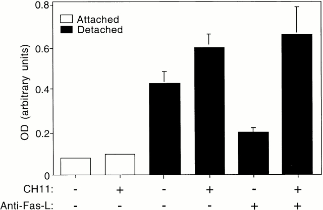
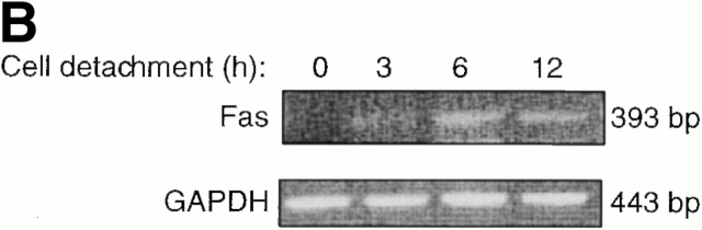
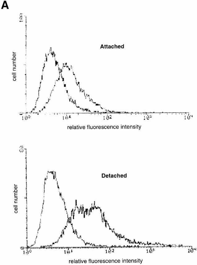
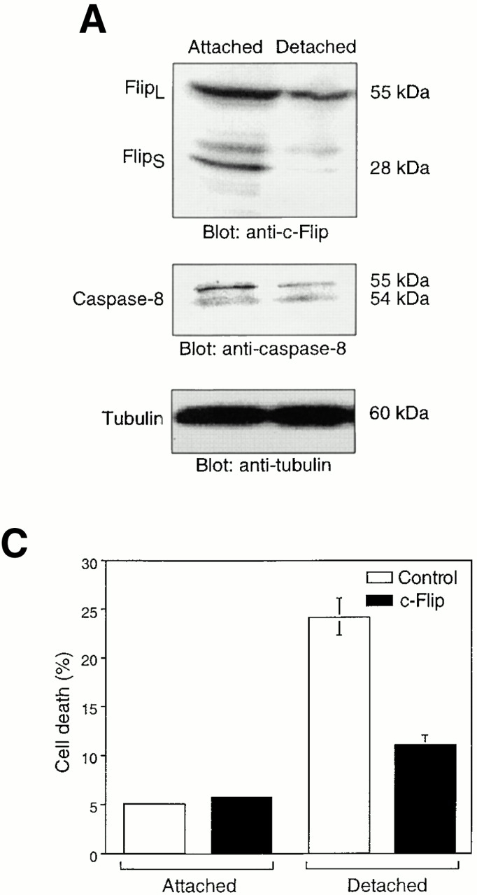
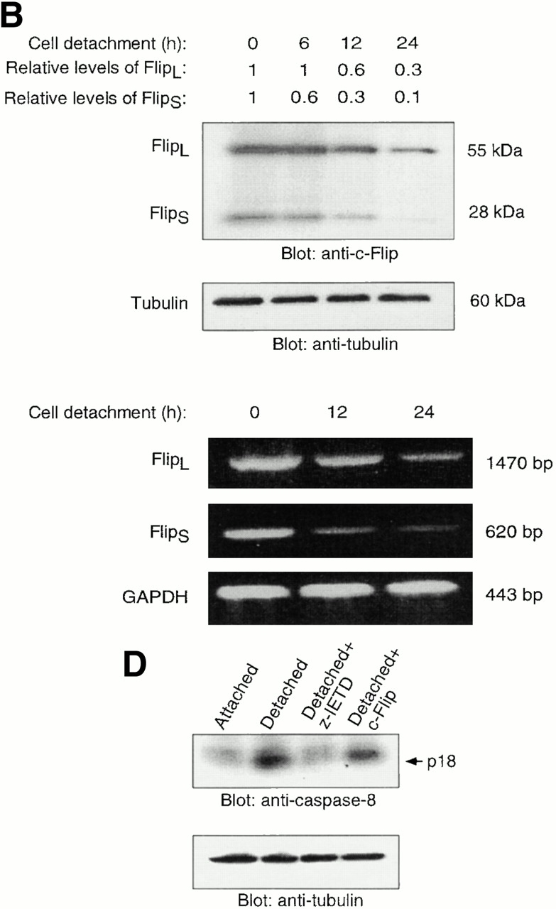
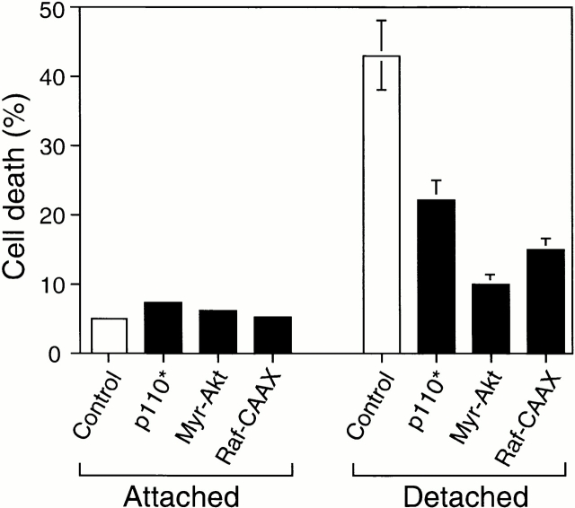
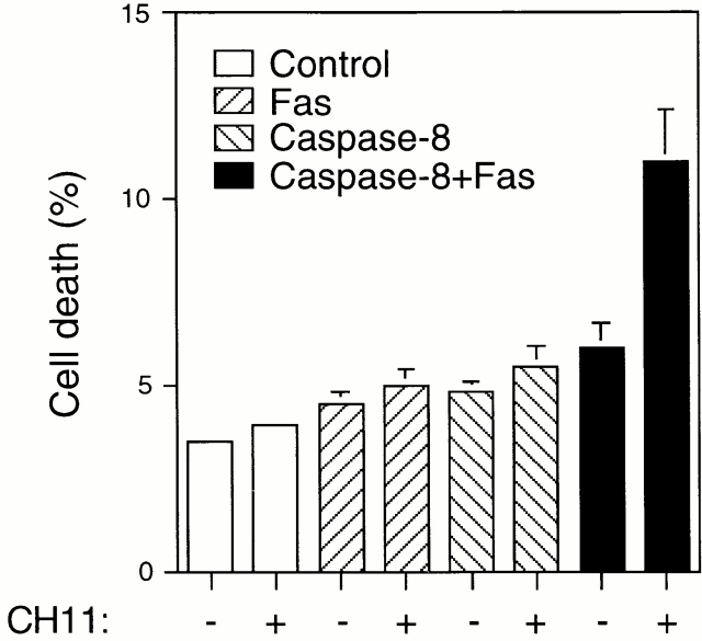
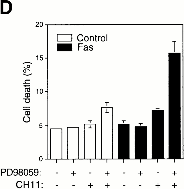
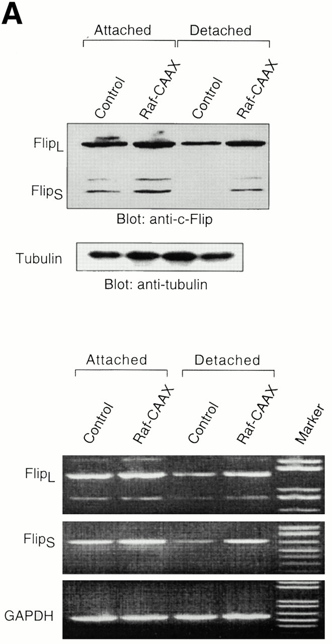
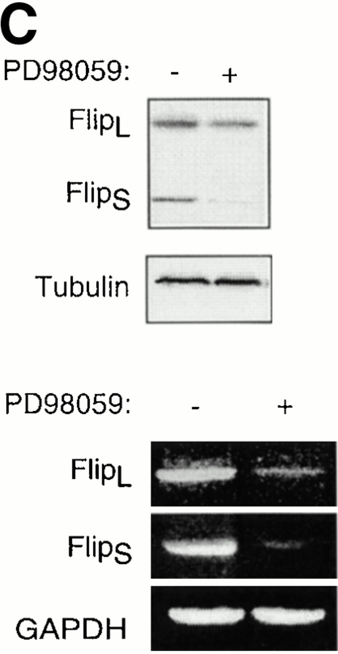
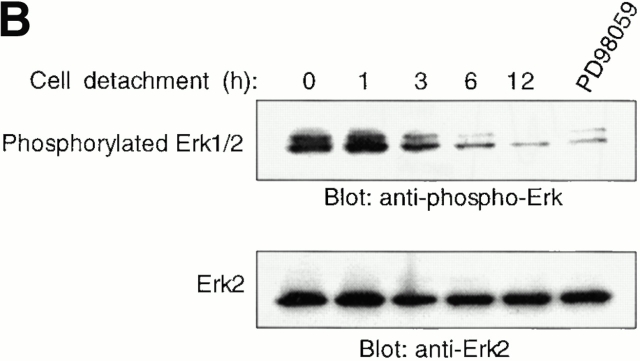
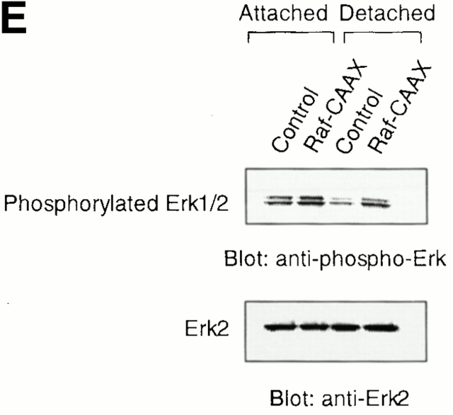
Survival of endothelial cells is critical for cellular processes such as angiogenesis. Cell attachment to extracellular matrix inhibits apoptosis in endothelial cells both in vitro and in vivo, but the molecular mechanisms underlying matrix-induced survival signals or detachment-induced apoptotic signals are unknown. We demonstrate here that matrix attachment is an efficient regulator of Fas-mediated apoptosis in endothelial cells. Thus, matrix attachment protects cells from Fas-induced apoptosis, whereas matrix detachment results in susceptibility to Fas-mediated cell death. Matrix attachment modulates Fas-mediated apoptosis at two different levels: by regulating the expression level of Fas, and by regulating the expression level of c-Flip, an endogenous antagonist of caspase-8. The extracellular signal-regulated kinase (Erk) cascade functions as a survival pathway in adherent cells by regulating c-Flip expression. We further show that detachment-induced cell death, or anoikis, itself results from activation of the Fas pathway by its ligand, Fas-L. Fas-L/Fas interaction, Fas-FADD complex formation, and caspase-8 activation precede the bulk of anoikis in endothelial cells, and inhibition of any of these events blocks anoikis. These studies identify matrix attachment as a survival factor against death receptor-mediated apoptosis and provide a molecular mechanism for anoikis and previously observed Fas resistance in endothelial cells.


Figures

Figure 1
Figure 1
Detachment-induced apoptosis in HUVECs is Fas/Fas-L dependent. HUVECs were detached and either kept in suspension or on polyhema-coated dishes in complete growth medium (EGM medium containing 2% FCS, 10 ng/ml hEGF, and 1 μg/ml hydrocortisone, supplemented with 12 μg/ml of bovine brain extract, 2 mM l-glutamine, 50 μg/ml streptomycin, and 50 U/ml penicillin) for the indicated time periods, and cell death was determined by (A) trypan blue exclusion or (B) DNA fragmentation analysis. Identical results were obtained under the two experimental conditions; results shown are for cells kept in suspension, and these experimental conditions were used in all subsequent experiments involving detachment-induced cell death. (C) HUVECs were kept adherent or in suspension for 12 h in the presence or absence of 5 μg/ml of inhibitory anti–Fas-L antibodies (NOK-2), inhibitory anti-Fas antibodies (ZB4), or with control antibodies (IgG). Apoptosis was determined by DNA fragmentation analysis. In A and B, results are shown for representative experiments independently carried out three times. In C, bars indicate SD in a representative experiment done in triplicate.
Figure 3
Figure 3
Cell detachment induces Fas/Fas-L interaction, DISC formation, and caspase-8 activation in HUVECs. Inhibition of anoikis by dnFADD and caspase-8 inhibitors. (A) Cell lysates were prepared from adherent HUVECs and from HUVECs that had been kept in suspension for the indicated times, and the lysates were subjected to immunoprecipitation analysis by anti–Fas-L antibodies (clone 33). Immunoprecipitates (IP) were analyzed by immunoblotting with anti-Fas antibodies (clone 13; top). The membranes were stripped and reprobed with anti–Fas-L antibodies (clone 33) to confirm equal amounts of Fas-L in the precipitates (bottom). Similar results were obtained when anti–Fas-L antibody G247-4 was used (data not shown). (B) The cell lysates were subjected to immunoprecipitations by anti-FADD antibodies. The immunoprecipitates were analyzed by immunoblotting with anti-Fas antibodies (clone 13; top), and the membranes were stripped and reprobed with anti-FADD antibodies to confirm equal amounts of FADD in the precipitates (bottom). C, Control cell lysate prepared from Jurkat T cells that had been activated by T cell receptor stimulation. (C) HUVECs were transiently transfected with an empty vector (control) or with an expression plasmid encoding dnFADD, together with plasmid coding for the green fluorescent protein (GFP). 24 h after transfection, the cells were either kept adherent or kept in suspension for the indicated time periods. The cells were then washed and propidium iodide was added for 20 min on ice. Apoptosis analysis by FACS® was carried out in the double positive cell population for propidium iodide and fluorescent GFP. In a separate experiment, we monitored apoptosis by staining the cells with Hoechst dye and found that results obtained with propidium iodide and Hoechst were indistinguishable from each other (not shown). Thus, the results obtained with the Hoechst dye confirm that cell death observed under our experimental conditions results from apoptosis, rather than necrosis. (D) HUVECs were left adherent or kept in suspension for 12 h in the presence or absence of the indicated concentrations of caspase-8 inhibitor z-IETD-fmk. In the left panel, apoptosis was determined by DNA fragmentation analysis; bars indicate SD in a representative experiment done in triplicate. In the right panel, cell lysates were prepared and analyzed by immunoblotting with an anti–caspase-8 antibody (Ab-1) that detects the p18 active form of the enzyme.
Figure 3
Figure 3
Cell detachment induces Fas/Fas-L interaction, DISC formation, and caspase-8 activation in HUVECs. Inhibition of anoikis by dnFADD and caspase-8 inhibitors. (A) Cell lysates were prepared from adherent HUVECs and from HUVECs that had been kept in suspension for the indicated times, and the lysates were subjected to immunoprecipitation analysis by anti–Fas-L antibodies (clone 33). Immunoprecipitates (IP) were analyzed by immunoblotting with anti-Fas antibodies (clone 13; top). The membranes were stripped and reprobed with anti–Fas-L antibodies (clone 33) to confirm equal amounts of Fas-L in the precipitates (bottom). Similar results were obtained when anti–Fas-L antibody G247-4 was used (data not shown). (B) The cell lysates were subjected to immunoprecipitations by anti-FADD antibodies. The immunoprecipitates were analyzed by immunoblotting with anti-Fas antibodies (clone 13; top), and the membranes were stripped and reprobed with anti-FADD antibodies to confirm equal amounts of FADD in the precipitates (bottom). C, Control cell lysate prepared from Jurkat T cells that had been activated by T cell receptor stimulation. (C) HUVECs were transiently transfected with an empty vector (control) or with an expression plasmid encoding dnFADD, together with plasmid coding for the green fluorescent protein (GFP). 24 h after transfection, the cells were either kept adherent or kept in suspension for the indicated time periods. The cells were then washed and propidium iodide was added for 20 min on ice. Apoptosis analysis by FACS® was carried out in the double positive cell population for propidium iodide and fluorescent GFP. In a separate experiment, we monitored apoptosis by staining the cells with Hoechst dye and found that results obtained with propidium iodide and Hoechst were indistinguishable from each other (not shown). Thus, the results obtained with the Hoechst dye confirm that cell death observed under our experimental conditions results from apoptosis, rather than necrosis. (D) HUVECs were left adherent or kept in suspension for 12 h in the presence or absence of the indicated concentrations of caspase-8 inhibitor z-IETD-fmk. In the left panel, apoptosis was determined by DNA fragmentation analysis; bars indicate SD in a representative experiment done in triplicate. In the right panel, cell lysates were prepared and analyzed by immunoblotting with an anti–caspase-8 antibody (Ab-1) that detects the p18 active form of the enzyme.
Figure 2
Figure 2
Cell detachment sensitizes HUVECs to Fas-mediated apoptosis. The ability of agonistic anti-Fas antibody (CH11) to induce apoptosis was analyzed in attached and detached HUVECs. The cells were incubated with or without 1 μg/ml of CH11 for 12 h in the presence or absence of blocking anti–Fas-L antibody (NOK-2), and apoptosis was determined by DNA fragmentation analysis. Bars indicate SD in a representative experiment done in triplicate.
Figure 4
Figure 4
Cell detachment upregulates Fas expression in HUVECs. (A) Detachment induces cell surface levels of Fas. HUVECs were kept adherent or in suspension for 12 h. FACS® analysis with anti-Fas antibody (UB2) was carried out as described in Materials and Methods. (B) Cell detachment increases the mRNA levels of Fas. HUVECs were kept adherent or in suspension for the indicated times, and Fas mRNA levels were determined by an RT-PCR analysis as described in Materials and Methods.
Figure 4
Figure 4
Cell detachment upregulates Fas expression in HUVECs. (A) Detachment induces cell surface levels of Fas. HUVECs were kept adherent or in suspension for 12 h. FACS® analysis with anti-Fas antibody (UB2) was carried out as described in Materials and Methods. (B) Cell detachment increases the mRNA levels of Fas. HUVECs were kept adherent or in suspension for the indicated times, and Fas mRNA levels were determined by an RT-PCR analysis as described in Materials and Methods.
Figure 5
Figure 5
Cell detachment results in downmodulation of c-Flip expression. Exogenous expression of c-Flip protects HUVECs from anoikis and prevents caspase-8 activation in detached cells. (A) Cell lysates were prepared from adherent HUVECs and from HUVECs that had been kept in suspension for 8 h, and the lysates were subjected to immunoblotting analysis by antibodies against c-Flip (top), caspase-8 (5F7; middle), and tubulin (bottom). (B) c-Flip expression was determined in attached cells or in cells that had been kept in suspension for the indicated times by immunoblot analysis (top two panels) and by RT-PCR (bottom three panels). As a control, protein expression levels in the lysates were analyzed by antitubulin immunoblotting (top), and mRNA expression levels by RT-PCR analysis with primers specific for GAPDH (bottom). The relative expression levels of Flip proteins as quantitated with a densitometric analysis are indicated. (C) Exogenous expression of c-Flip protects HUVECs from anoikis. HUVECs were cotransfected with an empty vector (control) or with plasmid encoding c-FlipL, together with plasmid coding for GFP. 24 h after transfection, the cells were either kept adherent or in suspension for 12 h. Apoptosis analysis by FACS® was carried out in the double positive cell population for propidium iodide and fluorescent GFP as described above. Bars indicate SD in a representative experiment done in triplicate. (D) Exogenous expression of c-Flip blocks caspase-8 activation in detached HUVECs. HUVECs were transiently transfected with an empty vector or with plasmid encoding c-FlipL. 24 h after transfection, the cells were kept adherent or in suspension for 12 h. In some of the experiments, the cells were simultaneously treated with 20 μM of the caspase-8 inhibitor z-IETD-fmk, as indicated. The cell lysates were analyzed by immunoblotting with an anti–caspase-8 antibody (Ab-1) that detects the p18 active form of the enzyme. The membrane was stripped and reprobed with an anti–α-tubulin control antibody to confirm equal loading (bottom).
Figure 5
Figure 5
Cell detachment results in downmodulation of c-Flip expression. Exogenous expression of c-Flip protects HUVECs from anoikis and prevents caspase-8 activation in detached cells. (A) Cell lysates were prepared from adherent HUVECs and from HUVECs that had been kept in suspension for 8 h, and the lysates were subjected to immunoblotting analysis by antibodies against c-Flip (top), caspase-8 (5F7; middle), and tubulin (bottom). (B) c-Flip expression was determined in attached cells or in cells that had been kept in suspension for the indicated times by immunoblot analysis (top two panels) and by RT-PCR (bottom three panels). As a control, protein expression levels in the lysates were analyzed by antitubulin immunoblotting (top), and mRNA expression levels by RT-PCR analysis with primers specific for GAPDH (bottom). The relative expression levels of Flip proteins as quantitated with a densitometric analysis are indicated. (C) Exogenous expression of c-Flip protects HUVECs from anoikis. HUVECs were cotransfected with an empty vector (control) or with plasmid encoding c-FlipL, together with plasmid coding for GFP. 24 h after transfection, the cells were either kept adherent or in suspension for 12 h. Apoptosis analysis by FACS® was carried out in the double positive cell population for propidium iodide and fluorescent GFP as described above. Bars indicate SD in a representative experiment done in triplicate. (D) Exogenous expression of c-Flip blocks caspase-8 activation in detached HUVECs. HUVECs were transiently transfected with an empty vector or with plasmid encoding c-FlipL. 24 h after transfection, the cells were kept adherent or in suspension for 12 h. In some of the experiments, the cells were simultaneously treated with 20 μM of the caspase-8 inhibitor z-IETD-fmk, as indicated. The cell lysates were analyzed by immunoblotting with an anti–caspase-8 antibody (Ab-1) that detects the p18 active form of the enzyme. The membrane was stripped and reprobed with an anti–α-tubulin control antibody to confirm equal loading (bottom).
Figure 7
Figure 7
Ectopic activation of the PI 3′-kinase/Akt and Erk pathways prevents anoikis in HUVECs. HUVECs were cotransfected with an empty vector (control) or with plasmids encoding activated forms of PI 3′-kinase (p110*), Akt (Myr-Akt), and c-Raf-1 (Raf-CAAX), together with a plasmid coding for GFP. After transfection, the cells were kept adherent or in suspension for 24 h. Apoptosis was determined by using FACS® in the double positive cell population for GFP and propidium iodide as described above. Bars indicate SD in an experiment done in triplicate.
Figure 6
Figure 6
Exogenous expression of both Fas and caspase-8 is required to render attached HUVECs sensitive to Fas-mediated apoptosis. HUVECs were transfected with an empty vector (control), or with plasmids encoding Fas and procaspase-8, together with plasmid coding for the GFP. 24 h after transfection, the cells were stimulated or not for an additional 24 h with 1 μg/ml of anti-Fas antibody CH11. Apoptosis analysis by FACS® was carried out in the double positive cell population for propidium iodide and fluorescent GFP as described above. Bars indicate SD in a representative experiment done in triplicate.
Figure 8
Figure 8
The Erk pathway functions as a survival pathway in attached HUVECs and modulates c-Flip expression. (A) Exogenous expression of activated Raf-1 enhances c-Flip expression in detached HUVECs. HUVECs were transfected with an empty vector (Control) or with a plasmid encoding activated form of c-Raf-1 (Raf-CAAX). After transfection, the cells were kept adherent or in suspension for 24 h. c-Flip expression was determined by immunoblot analysis (top) and by RT-PCR (bottom). (B) Cell detachment results in the inactivation of the MAPK/Erk pathway in a time-dependent manner. HUVECs were kept adherent or in suspension for the indicated times. Cell lysates were prepared and Erk activation was determined by an immunoblot analysis with an anti–phospho-Erk1/2 antibody (top). The membrane was stripped and reprobed with an anti-Erk2 antibody to confirm equal loading (bottom). As indicated, in one of the experiments, the effectiveness of the MEK inhibitor PD98059 was analyzed by treating adherent HUVECs for 16 h before cell lysis. (C) Inhibition of the Erk pathway in adherent HUVECs downregulates c-Flip expression. HUVECs were treated or not with the MEK inhibitor PD98059 for 16 h and c-Flip expression was determined by immunoblotting (top) and by RT-PCR (bottom). (D) Inhibition of the Erk pathway in adherent HUVECs sensitizes the cells to Fas-mediated apoptosis. HUVECs were transiently transfected with an empty control vector or with a plasmid encoding Fas, together with plasmid coding for GFP. After transfection, the cells were treated or not with 25 μM of the MEK inhibitor PD98059 in the presence or absence of 1 μg/ml of the anti-Fas antibody CH11 for 24 h. Apoptosis analysis by FACS® was carried out in the double positive cell population for propidium iodide and fluorescent GFP as described above. Bars indicate SD in a representative experiment done in triplicate. (E) Exogenous expression of activated Raf-1 enhances Erk phosphorylation in HUVECs. HUVECs were transfected with an empty vector (Control) or with a plasmid encoding activated form of c-Raf-1 (Raf-CAAX). After transfection, the cells were kept adherent or in suspension for 12 h. Cell lysates were prepared and Erk activation was determined by an immunoblot analysis with an anti–phospho-Erk1/2 antibody (top). The membrane was stripped and reprobed with an anti-Erk2 antibody to confirm equal loading (bottom).
Figure 8
Figure 8
The Erk pathway functions as a survival pathway in attached HUVECs and modulates c-Flip expression. (A) Exogenous expression of activated Raf-1 enhances c-Flip expression in detached HUVECs. HUVECs were transfected with an empty vector (Control) or with a plasmid encoding activated form of c-Raf-1 (Raf-CAAX). After transfection, the cells were kept adherent or in suspension for 24 h. c-Flip expression was determined by immunoblot analysis (top) and by RT-PCR (bottom). (B) Cell detachment results in the inactivation of the MAPK/Erk pathway in a time-dependent manner. HUVECs were kept adherent or in suspension for the indicated times. Cell lysates were prepared and Erk activation was determined by an immunoblot analysis with an anti–phospho-Erk1/2 antibody (top). The membrane was stripped and reprobed with an anti-Erk2 antibody to confirm equal loading (bottom). As indicated, in one of the experiments, the effectiveness of the MEK inhibitor PD98059 was analyzed by treating adherent HUVECs for 16 h before cell lysis. (C) Inhibition of the Erk pathway in adherent HUVECs downregulates c-Flip expression. HUVECs were treated or not with the MEK inhibitor PD98059 for 16 h and c-Flip expression was determined by immunoblotting (top) and by RT-PCR (bottom). (D) Inhibition of the Erk pathway in adherent HUVECs sensitizes the cells to Fas-mediated apoptosis. HUVECs were transiently transfected with an empty control vector or with a plasmid encoding Fas, together with plasmid coding for GFP. After transfection, the cells were treated or not with 25 μM of the MEK inhibitor PD98059 in the presence or absence of 1 μg/ml of the anti-Fas antibody CH11 for 24 h. Apoptosis analysis by FACS® was carried out in the double positive cell population for propidium iodide and fluorescent GFP as described above. Bars indicate SD in a representative experiment done in triplicate. (E) Exogenous expression of activated Raf-1 enhances Erk phosphorylation in HUVECs. HUVECs were transfected with an empty vector (Control) or with a plasmid encoding activated form of c-Raf-1 (Raf-CAAX). After transfection, the cells were kept adherent or in suspension for 12 h. Cell lysates were prepared and Erk activation was determined by an immunoblot analysis with an anti–phospho-Erk1/2 antibody (top). The membrane was stripped and reprobed with an anti-Erk2 antibody to confirm equal loading (bottom).

Similar articles

Cited by

References

    1. Almeida E.A., Ilic D., Han Q., Hauck C.R., Jin F., Kawakatsu H., Schlaepfer D.D., Damsky C.H. Matrix survival signalingfrom fibronectin via focal adhesion kinase to c-Jun NH2-terminal kinase. J. Cell Biol. 2000;149:741–754. - PMC - PubMed
    1. Aoudjit F., Vuori K. Engagement of the alpha2beta1 integrin inhibits Fas ligand expression and activation-induced cell death in T cells in a focal adhesion kinase-dependent manner. Blood. 2000;95:2044–2051. - PubMed
    1. Barberis L., Wary K.K., Fiucci G., Liu F., Hirsch E., Brancaccio M., Altruda F., Tarone G., Giancotti F.G. Distinct roles of the adaptor protein shc and focal adhesion kinase in integrin signaling to ERK. J. Biol. Chem. 2000;275:36532–36540. - PubMed
    1. Brooks P.C., Montgomery A.M., Rosenfeld M., Reisfeld R.A., Hu T., Klier G., Cheresh D.A. Integrin αvβ3 antagonists promote tumor regression by inducing apoptosis of angiogenic blood vessels. Cell. 1994;79:1157–1164. - PubMed
    1. Datta S.R., Brunet A., Greenberg M.E. Cellular survivala play in three Akts. Genes Dev. 1999;13:2905–2927. - PubMed

Publication types

MeSH terms