

Focal activation of a mutant allele defines the role of stem cells in mosaic skin disorders
- PMID: 11157989
- PMCID: PMC2195990
- DOI: 10.1083/jcb.152.3.645
Focal activation of a mutant allele defines the role of stem cells in mosaic skin disorders
Abstract
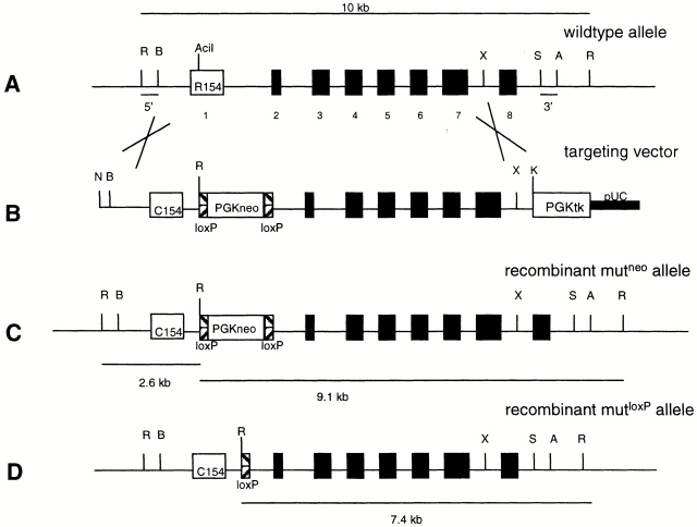
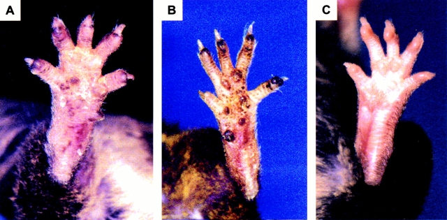
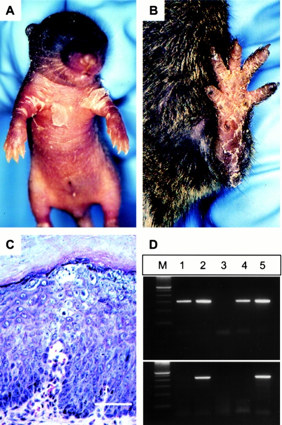
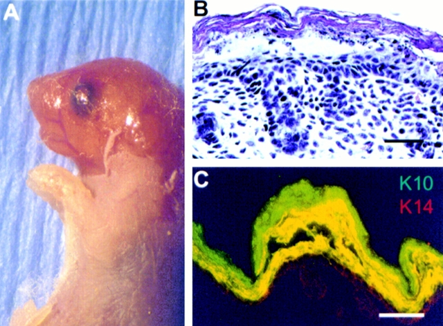
Stem cells are crucial for the formation and maintenance of tissues and organs. The role of stem cells in the pathogenesis of mosaic skin disorders remains unclear. To study the molecular and cellular basis of mosaicism, we established a mouse model for the autosomal-dominant skin blistering disorder, epidermolytic hyperkeratosis (MIM 113800), which is caused by mutations in either keratin K1 or K10. This genetic model allows activation of a somatic K10 mutation in epidermal stem cells in a spatially and temporally controlled manner using an inducible Cre recombinase. Our results indicate that lack of selective pressure against certain mutations in epidermal stem cells leads to mosaic phenotypes. This finding has important implications for the development of new strategies for somatic gene therapy of dominant genodermatoses.
Figures
References
-
- Bale S.J., Compton J.G., DiGiovanna J.J. Epidermolytic hyperkeratosis. Semin. Dermatol. 1993;12:202–209. - PubMed
-
- Berton T.R., Wang X.J., Zhou Z., Kellendonk C., Schutz G., Tsai S., Roop D.R. Characterization of an inducible, epidermal-specific knockout systemdifferential expression of lacZ in different Cre reporter mouse strains. Genesis. 2000;26:160–161. - PubMed
-
- Bickenbach J.R., Longley M.A., Bundman D.S., Dominey A.M., Bowden P.E., Rothnagel J.A., Roop D.R. A transgenic mouse model that recapitulates the clinical features of both neonatal and adult forms of the skin disease epidermolytic hyperkeratosis. Differentiation. 1996;61:129–139. - PubMed
-
- Bonifas J.M., Rothman A.L., Epstein E.H.J. Epidermolysis bullosa simplexevidence in two families for keratin gene abnormalities. Science. 1991;254:1202–1205. - PubMed
-
- Bradley A., Ramirez-Solis R., Zheng H., Hasty P., Davis A. Genetic manipulation of the mouse via gene targeting in embryonic stem cells. Ciba. Found. Symp. 1992;165:256–269. - PubMed
Publication types
MeSH terms
Substances
Grants and funding
LinkOut - more resources
Full Text Sources
Medical
Molecular Biology Databases
Research Materials
Miscellaneous
