. 2001 Feb 5;152(3):651-6.
doi: 10.1083/jcb.152.3.651.

An inducible mouse model for epidermolysis bullosa simplex: implications for gene therapy

Affiliations

An inducible mouse model for epidermolysis bullosa simplex: implications for gene therapy

T Cao et al. J Cell Biol. .

Abstract

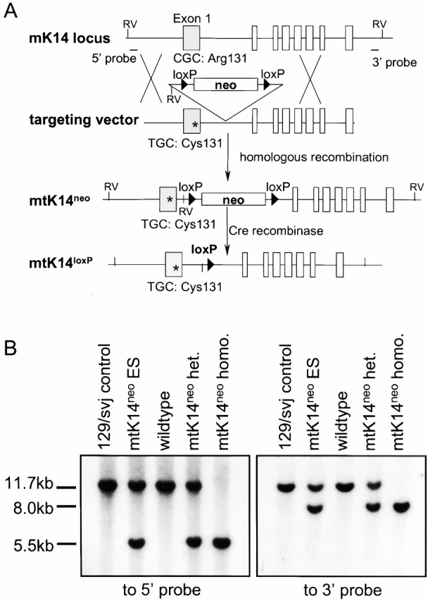
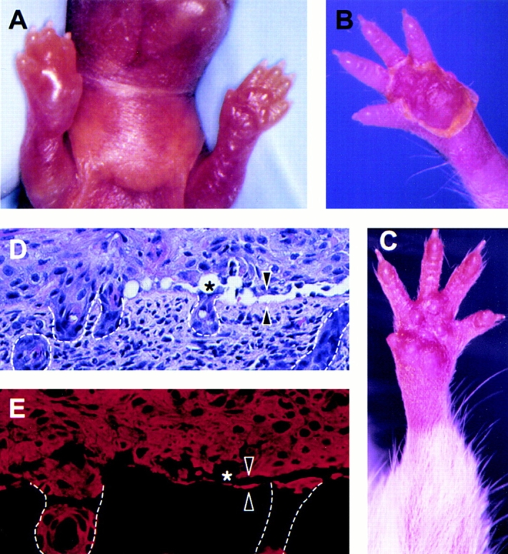
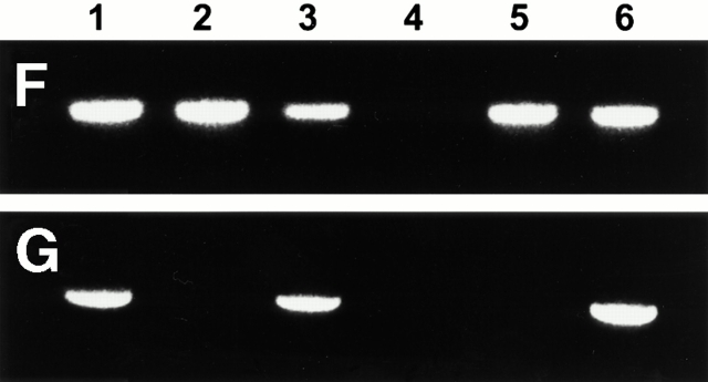
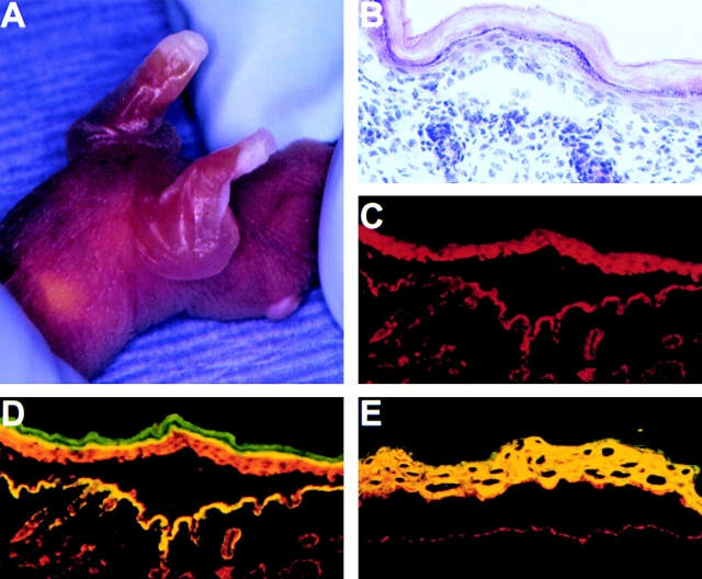
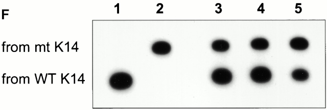
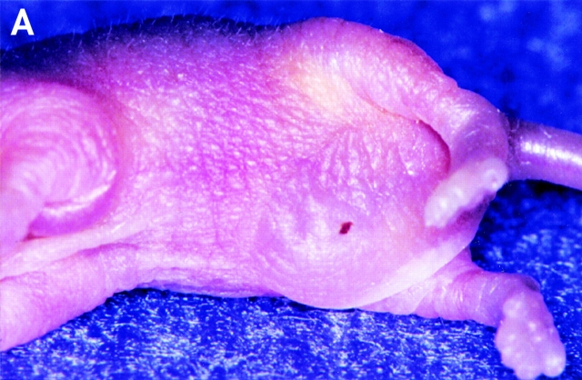
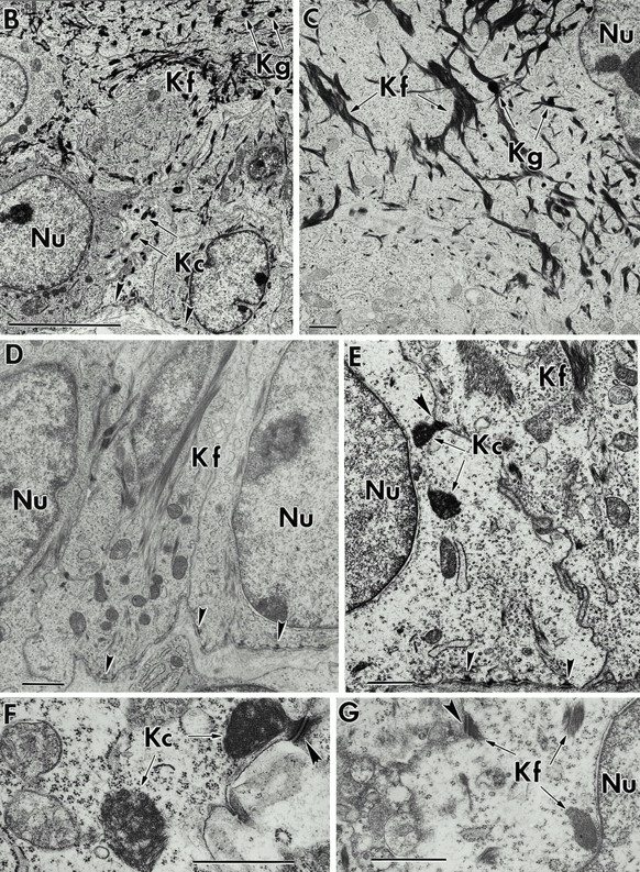
The Dowling-Meara variant of epidermolysis bullosa simplex (EBS-DM) is a severe blistering disease inherited in an autosomal-dominant fashion. Here we report the generation of a mouse model that allows focal activation of a mutant keratin 14 allele in epidermal stem cells upon topical administration of an inducer, resulting in EBS phenotypes in treated areas. Using laser capture microdissection, we show that induced blisters healed by migration of surrounding nonphenotypic stem cells into the wound bed. This observation provides an explanation for the lack of mosaic forms of EBS-DM. In addition, we show that decreased mutant keratin 14 expression resulted in normal morphology and functions of the skin. Our results have important implications for gene therapy of EBS and other dominantly inherited diseases.


Figures

Figure 1
Figure 1
Targeting strategy and Southern blot analysis. (A) Targeting strategy. Open boxes represent the exons. The asterisk denotes the C→T point mutation, and neo denotes the neomycin-resistance selection cassette. (B) Southern blot analysis of EcoRV-digested genomic DNA. The 5′ external probe (A) hybridized to an 11.7-kb fragment from the wild-type K14 locus, and a 5.5-kb fragment from the mtK14neo locus. The 3′ external probe (A) hybridized to the same 11.7-kb fragment from the wild-type K14 locus, and an 8.0-kb fragment from the mtK14neo locus. het., heterozygote; homo., homozygote.
Figure 4
Figure 4
Induction and characterization of RU486 induced blisters. (A) Gross phenotype of an induced blister on a mtK14neo/CrePR1 pup after two RU486 treatments on the right front leg. No blisters developed on the untreated leg (A) or in +/mtK14neo or CrePR1 control pups treated with RU486 (data not shown). (B) The right front paw of a mtK14neo/CrePR1 pup 10 d after blister formation upon treatment. (C) The left front paw and leg of a mtK14neo/CrePR1 mouse 6 mo after blister formation and cessation of RU486 treatment. The blistered area, including the palm and leg, healed without scarring, and there is normal hair growth on the leg. No additional blisters formed without further RU486 treatment. (D and E) H&E staining (D) and immunofluorescence microscopy with an anti-K14 antibody (Texas red, E) of an induced blister edge. Blistering occurred in the basal keratinocytes (arrowheads) on the chest (D and E), front leg, and paw (data not shown). The asterisk denotes cytolysis, and dashed lines outline the hair follicles. (F and G) PCR analysis of DNA for the presence of the loxP site (F) and neo cassette (G). (1–3) Epidermal DNA samples from different areas of a treated mtK14neo/CrePR1 pup. (1) Untreated area; (2) blister roof; (3) healed blistered area 1 wk after blister formation. (4–6) DNA samples from control mice. (4) Wild type; (5) +/mtK14loxP; (6) +/mtK14neo.
Figure 4
Figure 4
Induction and characterization of RU486 induced blisters. (A) Gross phenotype of an induced blister on a mtK14neo/CrePR1 pup after two RU486 treatments on the right front leg. No blisters developed on the untreated leg (A) or in +/mtK14neo or CrePR1 control pups treated with RU486 (data not shown). (B) The right front paw of a mtK14neo/CrePR1 pup 10 d after blister formation upon treatment. (C) The left front paw and leg of a mtK14neo/CrePR1 mouse 6 mo after blister formation and cessation of RU486 treatment. The blistered area, including the palm and leg, healed without scarring, and there is normal hair growth on the leg. No additional blisters formed without further RU486 treatment. (D and E) H&E staining (D) and immunofluorescence microscopy with an anti-K14 antibody (Texas red, E) of an induced blister edge. Blistering occurred in the basal keratinocytes (arrowheads) on the chest (D and E), front leg, and paw (data not shown). The asterisk denotes cytolysis, and dashed lines outline the hair follicles. (F and G) PCR analysis of DNA for the presence of the loxP site (F) and neo cassette (G). (1–3) Epidermal DNA samples from different areas of a treated mtK14neo/CrePR1 pup. (1) Untreated area; (2) blister roof; (3) healed blistered area 1 wk after blister formation. (4–6) DNA samples from control mice. (4) Wild type; (5) +/mtK14loxP; (6) +/mtK14neo.
Figure 2
Figure 2
Phenotype of homozygous mtK14neo pups. (A) Gross phenotype of a 1-d-old pup, showing severe blisters on the front legs, paws, and chest. (B) H&E staining of skin from a homozygous mtK14neo pup. The separation occurred in the basal cell layer within the epidermis. (C–E) Immunofluorescence microscopy of pup skin. The sections were stained for mutant K14 (Texas red, C–E), integrin α6 (FITC, D), and keratin 10 (FITC, E). Overlapping of K14 staining with either integrin α6 (D) or keratin 10 (E) results in yellow fluorescence. (F) AciI digestion of semi-quantitative RT-PCR products from mouse RNA. (1) Wild type; (2) homozygous mtK14neo; (3 and 4) +/mtK14neo; (5) +/mtK14loxP. Note the lower expression levels of mtK14neo allele in +/mtK14neo mice shown in lanes 3 and 4.
Figure 2
Figure 2
Phenotype of homozygous mtK14neo pups. (A) Gross phenotype of a 1-d-old pup, showing severe blisters on the front legs, paws, and chest. (B) H&E staining of skin from a homozygous mtK14neo pup. The separation occurred in the basal cell layer within the epidermis. (C–E) Immunofluorescence microscopy of pup skin. The sections were stained for mutant K14 (Texas red, C–E), integrin α6 (FITC, D), and keratin 10 (FITC, E). Overlapping of K14 staining with either integrin α6 (D) or keratin 10 (E) results in yellow fluorescence. (F) AciI digestion of semi-quantitative RT-PCR products from mouse RNA. (1) Wild type; (2) homozygous mtK14neo; (3 and 4) +/mtK14neo; (5) +/mtK14loxP. Note the lower expression levels of mtK14neo allele in +/mtK14neo mice shown in lanes 3 and 4.
Figure 3
Figure 3
Phenotype of +/mtK14loxP pups. (A) Gross phenotype of a 2-d-old pup, showing blisters on the left front leg and abdomen. (B–G) Transmission electron microscopy of skin samples from +/mtK14loxP pups (B, C, and E–G) and normal littermate (D). The mtK14loxP pup skin showed keratin clumps and short keratin filaments in the basal keratinocytes (B, E–G), but normal keratin filaments in the suprabasal layer of the epidermis (B and C). Normal skin showed long keratin filaments in the basal keratinocytes (D). Kc, keratin clumps; Kf, keratin filaments; Kg, keratohyalin granules; Nu, nucleus. Small arrowheads denote hemidesmosomes, indicating the position of the basement membrane. Large arrowheads denote desmosomes. Bars: (B) 5 μm; (C) 100 nm; (D–G) 1 μm.
Figure 3
Figure 3
Phenotype of +/mtK14loxP pups. (A) Gross phenotype of a 2-d-old pup, showing blisters on the left front leg and abdomen. (B–G) Transmission electron microscopy of skin samples from +/mtK14loxP pups (B, C, and E–G) and normal littermate (D). The mtK14loxP pup skin showed keratin clumps and short keratin filaments in the basal keratinocytes (B, E–G), but normal keratin filaments in the suprabasal layer of the epidermis (B and C). Normal skin showed long keratin filaments in the basal keratinocytes (D). Kc, keratin clumps; Kf, keratin filaments; Kg, keratohyalin granules; Nu, nucleus. Small arrowheads denote hemidesmosomes, indicating the position of the basement membrane. Large arrowheads denote desmosomes. Bars: (B) 5 μm; (C) 100 nm; (D–G) 1 μm.

References

    1. Alexeev V., Igoucheva O., Domashenko A., Cotsarelis G., Yoon K. Localized in vivo genotypic and phenotypic correction of the albino mutation in skin by RNA-DNA oligonucleotide. Nat. Biotechnol. 2000;18:43–47. - PubMed
    1. Anton-Lamprecht I., Schnyder U.W. Epidermolysis bullosa herpetiformis Dowling-Meara. Report of a case and pathomorphogenesis. Dermatologica. 1982;164:221–235. - PubMed
    1. Arin M.J., Longley M.A., Wang X.-J., Roop D.R. Focal activation of a mutant allele defines the role of stem cells in mosaic skin disorders. J. Cell Biol. 2001;152:645–649. - PMC - PubMed
    1. Berton T.R., Wang X.J., Zhou Z., Kellendonk C., Schutz G., Tsai S., Roop D.R. Characterization of an inducible, epidermal-specific knockout systemdifferential expression of lacZ in different Cre reporter mouse strains. Genesis. 2000;26:160–161. - PubMed
    1. Chan Y., Anton-Lamprecht I., Yu Q.C., Jackel A., Zabel B., Ernst J.P., Fuchs E. A human keratin 14 “knockout”the absence of K14 leads to severe epidermolysis bullosa simplex and a function for an intermediate filament protein. Genes Dev. 1994;8:2574–2587. - PubMed

Publication types

MeSH terms