

Immunological and PCR analyses for Borna disease virus in psychiatric patients and blood donors in Japan
- PMID: 11158085
- PMCID: PMC87754
- DOI: 10.1128/JCM.39.2.419-429.2001
Immunological and PCR analyses for Borna disease virus in psychiatric patients and blood donors in Japan
Abstract
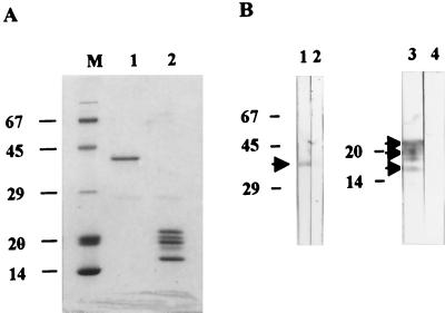
The involvement of Borna disease virus (BDV) in psychiatric diseases in humans remains controversial. T-cell memory response and seroprevalence of BDV in patients with psychiatric disorders and blood donors in Japan were evaluated collectively by Western blot (WB) analysis with inhibition test, electrochemiluminescence immunoassay, immunofluorescence assay, and T-cell proliferative response as well as detection of BDV p24 RNA in peripheral blood mononuclear cells (PBMCs). Positive proliferative responses to both BDV p40 and p24 proteins were detected in 9% of patients with mood disorders (4 of 45), 4% of schizophrenic patients (2 of 45), and 2% of blood donors (1 of 45). By WB analysis, the antibody to BDV p40 was detected only in 2% of patients with mood disorders (1 of 45). The BDV p24 antibody was detected in 2% of patients with mood disorders (1 of 45) and 9% of schizophrenic patients. (4 of 45) No plasma reacted with both BDV proteins. The finding of a lower seroprevalence than previously reported suggests the presence of false-positive cases in the previous report. BDV RNA was detected only in 2% of patients with mood disorders (1 of 45). In these three serological assays, T-cell responses, and PCR analysis, there was no significant difference in the prevalence among the three groups. However, we found three psychiatric patients who were positive for both BDV antibodies and T-cell proliferative responses and one patient who was positive for BDV RNA in PBMCs. These findings suggest the usefulness of the proliferative T-cell response and that certain individuals are infected with BDV or a BDV-related virus.
Figures


Similar articles
-
Detection of borna disease virus-reactive antibodies from patients with psychiatric disorders and from horses by electrochemiluminescence immunoassay.Clin Diagn Lab Immunol. 1999 Sep;6(5):696-700. doi: 10.1128/CDLI.6.5.696-700.1999. Clin Diagn Lab Immunol. 1999. PMID: 10473520 Free PMC article.
-
Detection and sequence analysis of borna disease virus p24 RNA from peripheral blood mononuclear cells of patients with mood disorders or schizophrenia and of blood donors.J Virol. 1998 Dec;72(12):10044-9. doi: 10.1128/JVI.72.12.10044-10049.1998. J Virol. 1998. PMID: 9811743 Free PMC article.
-
Detection of Borna disease virus (BDV) antibodies and BDV RNA in psychiatric patients: evidence for high sequence conservation of human blood-derived BDV RNA.J Virol. 1996 Nov;70(11):7713-24. doi: 10.1128/JVI.70.11.7713-7724.1996. J Virol. 1996. PMID: 8892892 Free PMC article.
-
Borna disease virus infection in psychiatric patients: are we on the right track?Lancet Infect Dis. 2001 Aug;1(1):46-52. doi: 10.1016/S1473-3099(01)00021-4. Lancet Infect Dis. 2001. PMID: 11871411 Review.
-
Borna disease virus infection in animals and humans.Emerg Infect Dis. 1997 Jul-Sep;3(3):343-52. doi: 10.3201/eid0303.970311. Emerg Infect Dis. 1997. PMID: 9284379 Free PMC article. Review.
Cited by
-
Borna disease virus.J Neurovirol. 2003 Apr;9(2):259-73. doi: 10.1080/13550280390194064. J Neurovirol. 2003. PMID: 12707857 Review.
-
Detection by radioligand assay of antibodies against Borna disease virus in patients with various psychiatric disorders.Clin Diagn Lab Immunol. 2005 May;12(5):671-6. doi: 10.1128/CDLI.12.5.671-676.2005. Clin Diagn Lab Immunol. 2005. PMID: 15879032 Free PMC article.
-
No molecular evidence of Borna disease virus among schizophrenia and bipolar disorder patients in Iran.Iran J Microbiol. 2017 Apr;9(2):112-118. Iran J Microbiol. 2017. PMID: 29214003 Free PMC article.
-
Borna disease virus (BDV) circulating immunocomplex positivity in addicted patients in the Czech Republic: a prospective cohort analysis.BMC Psychiatry. 2010 Sep 8;10:70. doi: 10.1186/1471-244X-10-70. BMC Psychiatry. 2010. PMID: 20825673 Free PMC article.
-
Failure to detect borna disease virus antibody and RNA from peripheral blood mononuclear cells of psychiatric patients.Psychiatry Investig. 2009 Dec;6(4):306-12. doi: 10.4306/pi.2009.6.4.306. Epub 2009 Nov 5. Psychiatry Investig. 2009. PMID: 20140130 Free PMC article.
References
-
- American Psychiatric Association. Diagnostic and statistical manual of mental disorders IV. Washington, D.C.: American Psychiatric Association; 1994.
-
- Berg A L, Johannisson A, Johansson M, Hein A, Berg M, Dorries R L. Peripheral and intracerebral T cell immune response in cats naturally infected with Borna disease virus. Vet Immunol Immunopathol. 1999;68:241–253. - PubMed
-
- Bilzer T, Planz O, Lipkin W I, Stitz L. Presence of CD4+ and CD8+ T cells and expression of MHC class I and MHC class II antigen in horses with Borna disease virus-induced encephalitis. Brain Pathol. 1995;5:223–230. - PubMed
-
- Bilzer T, Stitz L. Immunopathogenesis of virus diseases affecting the central nerve system. Crit Rev Immunol. 1996;16:145–222. - PubMed
-
- Bode L. Human infections with Borna disease virus (BDV) and potential pathogenic implications. Curr Top Microbiol Immunol. 1995;190:103–130. - PubMed
MeSH terms
Substances
LinkOut - more resources
Full Text Sources
Medical
