. 2001 Feb 1;530(Pt 3):431-42.
doi: 10.1111/j.1469-7793.2001.0431k.x.

Intestinal serotonin acts as a paracrine substance to mediate vagal signal transmission evoked by luminal factors in the rat

Affiliations

Intestinal serotonin acts as a paracrine substance to mediate vagal signal transmission evoked by luminal factors in the rat

J X Zhu et al. J Physiol. .

Abstract

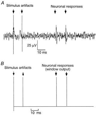
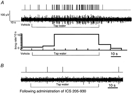
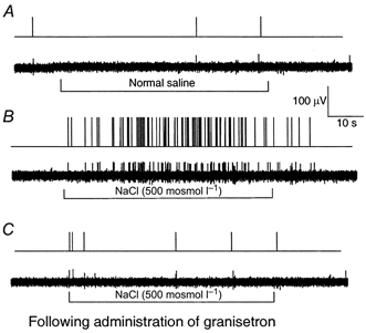
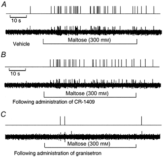
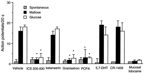
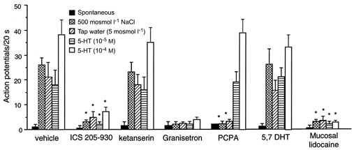
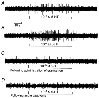
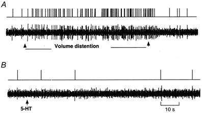
The vagus nerve conveys primary afferent information produced by a meal to the brainstem. Serotonin (5-HT), which abounds in intestinal enterochromaffin cells, is released in response to various stimuli. We have recently demonstrated that 5-HT released from intestinal enterochromaffin cells activates 5-HT3 receptors on vagal afferent fibres to mediate luminal non-cholecystokinin-stimulated pancreatic secretion. The present study was designed to evaluate the responses of vagal sensory neurons to intraluminal osmotic stimulation and luminal infusion of maltose, glucose or 5-HT. We investigated the role of endogenous 5-HT in signal transmission evoked by luminal stimuli to activate vagal sensory neurons. The discharges of vagal primary afferent neurons innervating the intestine were recorded from rat nodose ganglia. Luminal factors such as intestinal osmotic stimuli and perfusion of carbohydrates elicited powerful vagal nodose responses. Electrical subdiaphragmatic vagal stimulation activated 364 single units; 40 of these responded to intestinal mucosal stimuli. Of these 40, 30 responded to intraduodenal perfusion of hyperosmolar NaCl (500 mosmol l(-1)), 27 responded to tap water (5 mosmol l(-1)) and 20 and 19 responded to maltose (300 mM) and glucose (277.5 mM), respectively. The 5-HT3/4 antagonist tropisetron (ICS 205-930) or 5-HT3 antagonist granisetron abolished luminal stimuli-evoked nodose neuronal responses. Intraluminal infusion of 10(-5) and 10(-4) M 5-HT elicited increases in vagal afferent discharge in 25 and 31 units, respectively, by activating the 5-HT3 receptors. Acute subdiaphragmatic vagotomy, intestinal mucosal application of the local anaesthetic lidocaine (lignocaine) or administration of 5-HT3 antagonist each abolished the luminal 5-HT-induced nodose neuronal responses. In contrast, distension-sensitive neurons did not respond to duodenal infusion of 5-HT. Pharmacological depletion of 5-HT stores using p-chlorophenylalanine (PCPA), a 5-HT-synthesis inhibitor, abolished luminal factor-stimulated nodose neuronal responses. In contrast, pretreatment with 5,7-dihydroxytryptamine (5,7-DHT), a specific 5-HT neurotoxin that destroys 5-HT-containing neurons without affecting 5-HT-containing mucosal cells, had no effect on these responses. These results suggested that the nodose neuronal responses to luminal osmolarity and to the digestion products of carbohydrates are dependent on the release of endogenous 5-HT from the mucosal enterochromaffin cells, which acts on the 5-HT3 receptors on vagal afferent fibres to stimulate vagal sensory neurons.


Figures

Figure 1
Figure 1
A, neurography of the action potential elicited from a nodose ganglion cell in response to 2 consecutive electrical subdiaphragmatic vagal stimulations. Conductive distance = 0.06 m, latency = 70 ms, conductive velocity = 0.86 m s−1. B, the action potential triggered the window discriminator to generate a standard pulse.
Figure 2
Figure 2
Waveforms of a unitary action potential evoked by electrical vagal stimulation (I), and by intraduodenal infusion of 5-HT (II), 500 mosmol l−1 NaCl (III), and 300 mm maltose (IV). Note the similarity of all 4 firing patterns.
Figure 3
Figure 3
A, response of nodose ganglia neurons to intraduodenal infusion of tap water (5 mosmol l−1) (unit 20). Intraduodenal infusion of tap water (2 ml min−1) resulted in a marked increase of vagal afferent discharges, reaching a peak within 20 s and returning to basal levels after rinsing the lumen with isotonic saline. The action potential was converted to a standard pulse (top), and presented as an original tracing (middle), or a histogram (bottom, firing rate, spikes bin−1, versus time, bin width = 10 s). B, administration of the 5-HT3/4 receptor antagonist ICS 205-930 abolished nodose neuronal response evoked by tap water.
Figure 4
Figure 4
Recording of the action potentials of nodose ganglia neurons innervating the duodenal mucosa in response to intraduodenal infusion of hyperosmolar NaCl (unit 23) A, control. The nodose neuron innervating the duodenal mucosa has a very low basal level of activity. Duodenal infusion of normal saline did not affect the vagal afferent discharges. B, duodenal infusion of hypertonic NaCl (500 mosmol l−1, 2 ml min−1) produced a marked increase of vagal afferent discharges, peaking at 26 impulses (20 s)−1 within 20 s, and returning to basal levels 20 s after rinsing of the lumen with isotonic saline. C, administration of the 5-HT3 receptor antagonist granisetron abolished the nodose neuronal response evoked by hyperosmolar NaCl. The action potential was converted to a standard pulse (top) and presented as an original tracing (bottom).
Figure 5
Figure 5. Response of nodose ganglia neurons to intraduodenal infusion of maltose
A, the neurons responded to maltose after a latency of 10 s. Activity was maintained for the duration of the stimulus and ceased after rinsing the lumen with isotonic saline. B, administration of the CCK-8 antagonist CR-1409 had no effect on the nodose neuronal response to maltose (unit 15). C, administration of the 5-HT3 receptor antagonist granisetron abolished the nodose neuronal response evoked by maltose (unit 36).
Figure 8
Figure 8. Discharge of nodose ganglia neurons in response to intraluminal infusion of maltose and glucose
Values are means +s.e.m. Of 364 units activated by electrical vagal stimulation, 20 and 19 units responded to maltose and glucose, respectively. Administration of the 5-HT3/4 receptor antagonist ICS 205-930 (units 20-22), the 5-HT3 antagonist granisetron (units 35-39), or the mucosal application of lidocaine (units 10, 18, 19, 29 and 30) each abolished nodose responses to intraduodenal infusion of maltose and glucose. In contrast, administration of the 5-HT2A antagonist ketanserin (units 25-28) or the CCK-A receptor antagonist CR-1409 (units 5, 6, 15, 31 and 40) had no effect on the nodose responses induced by maltose or glucose. In a separate study, none of the 20 units tested after PCPA treatment responded to maltose and glucose. In contrast, after administration of 5,7-DHT, 5 of 22 units tested showed normal responses to intraduodenal administration of maltose and glucose. *P < 0.01 (compared with Vehicle).
Figure 9
Figure 9. Discharge of nodose ganglia neurons in response to intraluminal osmotic stimulation and to 5-HT
Values are means +s.e.m. Of 364 units activated by electrical vagal stimulation, 30 units responded to intraluminal infusion of hypertonic saline, 27 units responded to tap water, and 25 and 31 units responded to intraluminal infusion of 5-HT (10−5 or 10−4m, respectively). Administration of the 5-HT3/4 receptor antagonist ICS 205-930 (units 17 and 20-22), the 5-HT3 antagonist granisetron (units 23 and 35-39) or mucosal application of lidocaine (units 10, 12, 16, 18, 19, 29 and 30) but not the 5-HT2A receptor antagonist ketanserin (units 24-28) each abolished nodose neuronal responses evoked by osmotic or 5-HT stimulation. In separate groups of rats treated with PCPA, 20 units were tested. None responded to luminal osmotic stimuli. In contrast, 6 of these 20 units showed an increase in discharge frequency in response to intraluminal infusion of 5-HT. In another study, 22 units were tested following administration of 5,7-DHT. Five of 22 units responded to luminal osmotic stimuli and exogenous infusion of 5-HT. *P < 0.01 (compared with vehicle).
Figure 6
Figure 6. Response of nodose ganglia neurons to intraduodenal infusion of 5-HT
In the rats with intact vagus nerves, intraduodenal infusion of 10−5m 5-HT (unit 37) (A) and 10−4m 5-HT (unit 14) (B) increased the discharge frequency in a dose-dependent manner. C, following administration of granisetron, the same unit shown in A failed to respond to intraduodenal infusion of 5-HT. D, acute vagotomy abolished responses to intraduodenal infusion of 5-HT in the same unit shown in B.
Figure 7
Figure 7. Response of nodose ganglia neurons to intestinal distension and intraduodenal infusion of 5-HT
A, intestinal volume distension elicited a powerful increase in discharge frequency; latency was much shorter and the activity was maintained throughout the period of distension. B, the same unit shown in A failed to respond to 5-HT.

Comment in

References

    1. Andrews PL, Davis CJ, Bingham S, Davidson HI, Hawthorn J, Maskell L. The abdominal visceral innervation and the emetic reflex: pathways, pharmacology, and plasticity. Canadian Journal of Physiology and Pharmacology. 1990;68:325–345. - PubMed
    1. Andrews PLR, Davison JS. Activation of vagal afferent terminals by 5-HT is mediated by 5-HT3 receptors in the anaesthetized ferret. Journal of Physiology. 1990;422:92.
    1. Berthoud HR, Kressel M, Raybould HE, Nuehuber WL. Vagal sensors in the rat duodenal mucosa: distribution and structure as revealed by in vivo Dil-tracing. Anatomy and Embryology. 1995;191:203–212. - PubMed
    1. Bjorkland A, Baumgarten HG, Rensch A. 5,7-Dihydroxytryptamine: improvement of its selectivity for serotonin neurons in the CNS by pretreatment with desipramine. Journal of Neurochemistry. 1975;24:833–835. - PubMed
    1. Blackshaw LA, Grundy D. Effects of 5-hydroxy-tryptamine on discharge of vagal mucosal receptors from the upper gastrointestinal tract of the ferret. Journal of the Autonomic Nervous System. 1993;45:41–50. - PubMed

Publication types

MeSH terms