

Effect of antenatal betamethasone treatment on microtubule-associated proteins MAP1B and MAP2 in fetal sheep
- PMID: 11158279
- PMCID: PMC2278421
- DOI: 10.1111/j.1469-7793.2001.0497k.x
Effect of antenatal betamethasone treatment on microtubule-associated proteins MAP1B and MAP2 in fetal sheep
Abstract
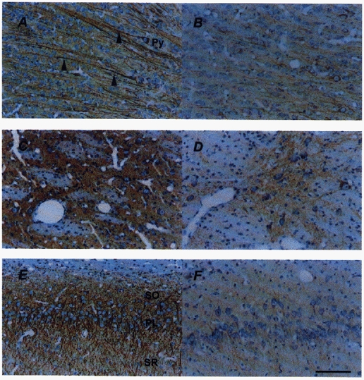
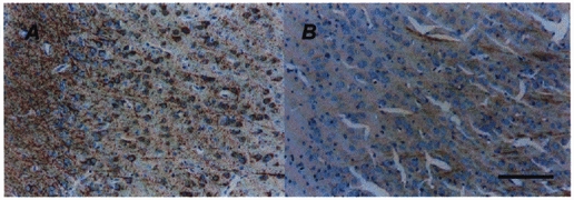
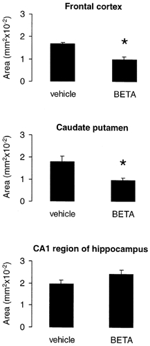
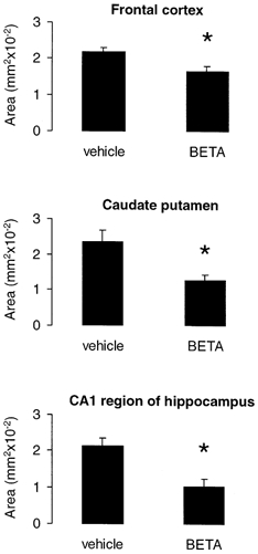
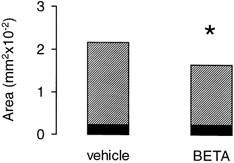
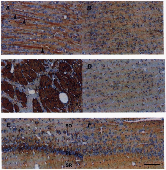
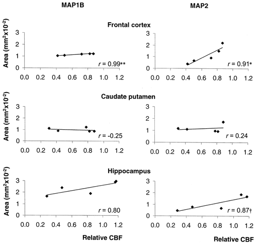
Betamethasone has been used extensively to accelerate fetal lung maturation, yet little is known of its effects on neuronal morphogenesis in the developing fetus. Microtubule-associated proteins (MAPs) are a diverse family of cytoskeletal proteins that are important for brain development and the maintenance of neuroarchitecture. Vehicle (n = 7) or betamethasone (10 ug h-1, n = 7) was infused I.V. to fetal sheep over 48 h beginning at 0.87 of gestation (128 days of gestation), producing fetal plasma betamethasone concentrations resembling those to which the human fetus is exposed during antenatal glucocorticoid therapy. Paraffin sections of the left hemisphere were stained with monoclonal antibodies against MAP1B and the MAP2 isoforms MAP2a,b,c and MAP2a,b. The level of the juvenile isoform MAP2c was determined by comparison of the two MAP2 immunostainings. We were able to detect MAP1B and MAP2 immunoreactivity (IR) in the fetal sheep brain. MAP2c was the major MAP2, constituting 90.2 % of the total MAPBetamethasone exposure diminished MAP1B IR in the frontal cortex and caudate putamen (P < 0.05) but not in the hippocampus. A decrease of MAP2 IR was found in the frontal cortex, hippocampus and caudate putamen (P < 0.05). Loss of MAP2 IR was mainly due to the loss of MAP2c IR. Haematoxylin-eosin staining did not demonstrate irreversible neuronal damage. Regional cerebral blood flow determined using coloured microspheres was significantly decreased by 28 % in the frontal cortex and by 36 % in the caudate putamen but not in the hippocampus 24 h after the onset of betamethasone exposure (P < 0.05). The loss of MAP1B and MAP2a,b,c IR showed a significant correlation to the cerebral blood flow decrease only in the frontal cortex (P < 0.05). These data suggest that mechanisms other than metabolic insufficiency caused by the decreased cerebral blood flow may contribute to the loss of MAPs. The results suggest that clinical doses of betamethasone may have acute effects on cytoskeletal proteins in the fetal brain.
Figures
References
-
- Ahima RS, Krozowski Z, Harlan RE. Type I corticosteroid receptor-like immunoreactivity in the rat CNS: distribution and regulation by corticosteroids. Journal of Comparative Neurology. 1991;313:522–538. - PubMed
-
- Albala JS, Kress Y, Liu WK, Weidenheim K, Yen SH, Shafit Zagardo B. Human microtubule-associated protein-2c localizes to dendrites and axons in fetal spinal motor neurons. Journal of Neurochemistry. 1995;64:2480–2490. - PubMed
-
- Arnold SE, Trojanowski JQ. Human fetal hippocampal development: II. The neuronal cytoskeleton. Journal of Comparative Neurology. 1996;367:293–307. - PubMed
-
- Ballard PL, Ballard RA. Scientific basis and therapeutic regimens for use of antenatal glucocorticoids. American Journal of Obstetrics and Gynecology. 1995;173:254–262. - PubMed
-
- Ballough GP, Martin LJ, Cann FJ, Graham JS, Smith CD, Kling CE, Forster JS, Phann S, Filbert MG. Microtubule-associated protein 2 (MAP-2): a sensitive marker of seizure-related brain damage. Journal of Neuroscience Methods. 1995;61:23–32. - PubMed
Publication types
MeSH terms
Substances
Grants and funding
LinkOut - more resources
Full Text Sources