Comparative Study
. 2001 Feb;21(4):1345-59.
doi: 10.1128/MCB.21.4.1345-1359.2001.

Distinctive features of Drosophila alternative splicing factor RS domain: implication for specific phosphorylation, shuttling, and splicing activation

Affiliations
Comparative Study

Distinctive features of Drosophila alternative splicing factor RS domain: implication for specific phosphorylation, shuttling, and splicing activation

E Allemand et al. Mol Cell Biol. 2001 Feb.

Abstract

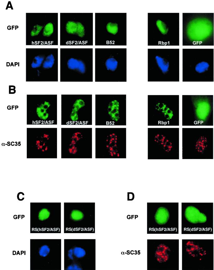
The human splicing factor 2, also called human alternative splicing factor (hASF), is the prototype of the highly conserved SR protein family involved in constitutive and regulated splicing of metazoan mRNA precursors. Here we report that the Drosophila homologue of hASF (dASF) lacks eight repeating arginine-serine dipeptides at its carboxyl-terminal region (RS domain), previously shown to be important for both localization and splicing activity of hASF. While this difference has no effect on dASF localization, it impedes its capacity to shuttle between the nucleus and cytoplasm and abolishes its phosphorylation by SR protein kinase 1 (SRPK1). dASF also has an altered splicing activity. While being competent for the regulation of 5' alternative splice site choice and activation of specific splicing enhancers, dASF fails to complement S100-cytoplasmic splicing-deficient extracts. Moreover, targeted overexpression of dASF in transgenic flies leads to higher deleterious developmental defects than hASF overexpression, supporting the notion that the distinctive structural features at the RS domain between the two proteins are likely to be functionally relevant in vivo.


Figures

FIG. 1
FIG. 1
Amino acid sequence alignments of dASF (dmSF2/p28) with hASF (hsSF2/ASF), dSC35 (dmSC35) with human SC35 (hsSC35), and d9G8 (dm9G8) with human 9G8 (hs9G8) as obtained by the ClustalW program. Identical amino acid residues are on a black background, and conservative substitutions such as RKH, IVLM, ED, FY, and ST are marked in grey. The glycine-rich region between the RNA-binding domains and the RS domains are underlined. The positions of the conserved RNP-1 and RNP-2 submotifs are indicated. The amino acids belonging to the consensus C(X)2C(X)4H(X)4C forming the zinc knuckles of human and Drosophila 9G8 are represented in red.
FIG. 2
FIG. 2
(A) Phosphorylation of purified E. coli-expressed recombinant ehASF and edASF proteins by dSRPK1 (A), human SRPK1 (B), Clk/Sty (C), and human topo I (D). Kinase assays were performed with equivalent activities of recombinant GST-Clk/Sty, SRPK1, and topo I to phosphorylate ehASF (lanes 1), edASF (lanes 2), or no substrate added other than the RS domain of hASF (lanes 3) as described in Materials and Methods. The RS domain of hASF was used as an internal control.
FIG. 3
FIG. 3
Cellular localization of GFP fusion proteins in Drosophila S2 (A) and HeLa (B) cells. Direct fluorescence of GFP (GFP), GFP-hASF (hSF2/ASF), GFP-dASF (dSF2/ASF), GFP-B52 (B52), and GFP-RBP1 (Rbp1) fusion proteins was analyzed 20 h after transfection. Expression of fusion proteins was confirmed by immunoblot analysis using anti-GFP antibody (data not shown). (C and D) Cellular localization of the GFP-RS domain of either hASF [RS(hSF2/ASF)] or dASF [RS(dSF2/ASF)] fusion proteins in S2 (C) and HeLa (D) cells. The position of nuclei was confirmed either by DAPI staining of S2 cells (DAPI) or by indirect immunofluorescent staining of HeLa cells with α-SC35, which showed the cellular localization of endogenous SR protein SC35. DAPI and α-SC35 staining were performed in the same cells transfected with GFP fusion proteins.
FIG. 4
FIG. 4
Analysis of nucleocytoplasmic shuttling of GFP-dASF, GFP-RBP1, GFP-hASF, and GFP-hASF Δ197–216 (a mutant of hASF lacking the RS repeats at the RS domain) fusion proteins by transient expression in interspecies heterokaryons (see Materials and Methods). Phase-contrast images of the heterokaryons are shown (left column). Localization of the expressed proteins was determined by direct fluorescence of GFP (middle column). The cells were simultaneously incubated with DAPI for differential staining of human and mouse nuclei within heterokaryons (right column). Arrows indicate the mouse nuclei within human-mouse heterokaryons.
FIG. 5
FIG. 5
Effects of dASF on splicing activation and alternative splicing in vitro. (A) Aliquots of 50 fmol of 32P-labeled SV40 derivative (left), E1A derivative (middle), and β-globin derivative (right) pre-mRNAs were incubated in HeLa cell NE under splicing conditions without complementation (lanes 1) or complemented with 8 or 16 pmol of bhASF (lanes 2 and 3), 8 or 16 pmol of bdASF (lanes 4 and 5), or 16 pmol of baculovirus-purified 9G8 (lanes 6). (B) Splicing-complementation activity of recombinant bdASF in S100-cytoplasmic splicing-deficient extracts. 32P-labeled Minx pre-mRNA (left) was incubated under splicing conditions (see Materials and Methods) either in HeLa S100 extracts without (lane 1) or with 4, 8, or 16 pmol of the indicated recombinant proteins (lanes 2 to 7) or in HeLa S100 extracts supplemented with 1/10 of HeLa NE in the absence (lane 9) or presence of added 8 or 16 pmol of the indicated recombinant proteins (lanes 10 to 13). Lane 8 represents a standard splicing reaction in HeLa NE. Right, splicing reactions performed as at the left, using an ftz pre-mRNA.
FIG. 6
FIG. 6
(A) Effect of dASF on U1 snRNP binding to the 5′ splice site. U1 snRNP–ASF–pre-mRNA complex formation assays were performed as previously described (22). Reactions in 10 ml contained 1.7 pmol of U1 snRNP (lanes 2, 3, 5 to 8, and 10 to 13), 40 (lanes 4, 5, and 8), 20 (lane 6), and 10 (lane 7) pmol of bhASF, 40 (lanes 9, 10, and 13), 20 (lane 11), and 10 (lane 12) pmol of bdASF, and 1.5 fmol of either 32P-labeled PIP7.A (lanes 1, 2, 4 to 7, and 9 to 12) or PIP75′AU (lanes 3, 8, and 13) pre-mRNA. (B) Physical interaction between the hASF and dASF by far-Western analysis (22). The indicated proteins (lanes 1 to 3), purified U1 snRNP (lane 4), and purified SR proteins treated with calf alkaline phosphatase (lane 5) were separated by SDS-PAGE on a 12% gel, transferred to nitrocellulose, renatured, and probed with 32P-labeled ehASF. SR proteins were purified as described by Zahler et al. (61). (C) dASF interacts with both hASF and itself, as revealed by yeast two-hybrid system.
FIG. 7
FIG. 7
dASF specifically activates enhancer-dependent splicing. 32P-labeled Sp1 transcripts containing hASF (left), 9G8 (middle), and SRp20 (right) high-affinity binding sites were incubated in a mixture of S100 cytoplasmic extracts and NF20–40 (7). Assays were supplemented with no SR protein (lanes 2, 7, and 12) or with 16 pmol of bhASF (lanes 3, 8, and 14), 16 pmol of bdASF (lanes 4, 10, and 15), 16 pmol of baculovirus-purified 9G8 (lanes 5 and 9), or 16 pmol of SRp20 (lane 13). Lanes 1, 6, and 11 represent control splicing assays using NE. ESE, exonic splicing element.
FIG. 8
FIG. 8
High levels of dASF overexpression in differentiating photoreceptor cells lead to more severe adult eye defects than hASF overexpression. (A) Virgin female flies carrying a P-element insert in which the GAL4 coding sequence was placed under the control of five glass-binding sites (GMR enhancer) were mated to male flies carrying a single UAS-hASF line 5, 1, or 6 or UAS-dASF line 7, 1, or 2 element. (B) Stereomicroscope views of adult compound eyes. Genotypes: a, GMR-GAL4/+; UAS-lacZ/+; b, GMR-GAL4/UAS-ASF5; c, GMR-GAL4/+; UAS-ASF1/+; d, GMR-GAL4/+; UAS-ASF6/+; e, GMR-GAL4/UAS-dASF7; f, GMR-GAL4/+; UAS-dASF1/+; g, GMR-GAL4/+; UAS-dASF1/+. All progeny were raised at 25°C. Overexpression of dASF in the developing retina results in necrosis of ommatidia that can be moderate (e) or severe (f and g), depending on the site of chromosomal integration of the transgene.

References

    1. Biencowe B J. Exonic splicing enhancers: mechanism of action, diversity and role in human genetic diseases. Trends Biochem. 2000;25:106–110. - PubMed
    1. Brand A H, Manoukian A S, Perrimon N. Ectopic expression in Drosophila. Methods Cell Biol. 1994;44:635–654. - PubMed
    1. Cáceres J F, Krainer A R. Functional analysis of pre-mRNA splicing factor SF2/ASF structural domains. EMBO J. 1993;12:4715–4726. - PMC - PubMed
    1. Cáceres J F, Misteli T, Screaton G R, Spector D L, Krainer A R. Role of the modular domains of SR proteins in subnuclear localization and alternative splicing specificity. J Cell Biol. 1997;138:225–238. - PMC - PubMed
    1. Cáceres J F, Screaton G R, Krainer A R. A specific subset of SR proteins shuttles continuously between the nucleus and the cytoplasm. Genes Dev. 1998;12:55–66. - PMC - PubMed

Publication types

MeSH terms