. 2001 Jan;13(1):47-60.
doi: 10.1105/tpc.13.1.47.

The control of single-celled cotton fiber elongation by developmentally reversible gating of plasmodesmata and coordinated expression of sucrose and K+ transporters and expansin

Affiliations

The control of single-celled cotton fiber elongation by developmentally reversible gating of plasmodesmata and coordinated expression of sucrose and K+ transporters and expansin

Y L Ruan et al. Plant Cell. 2001 Jan.

Abstract

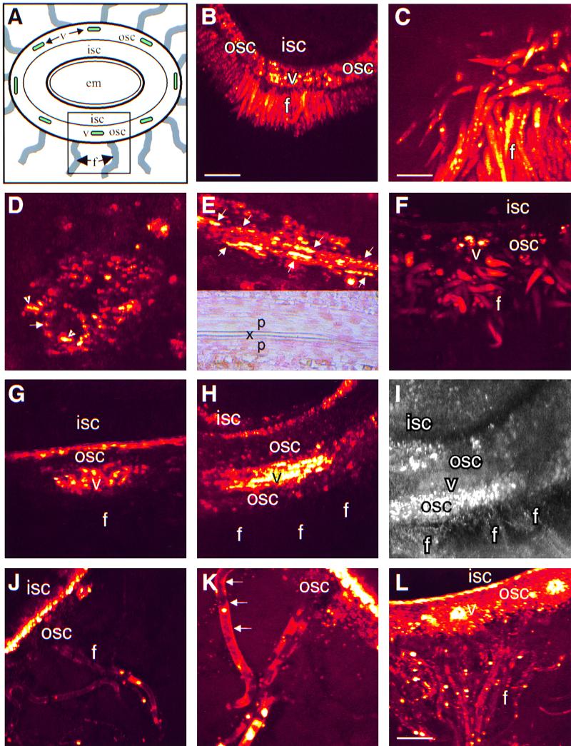
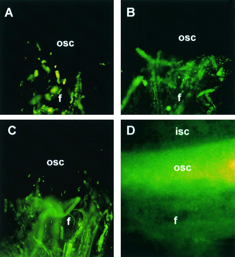
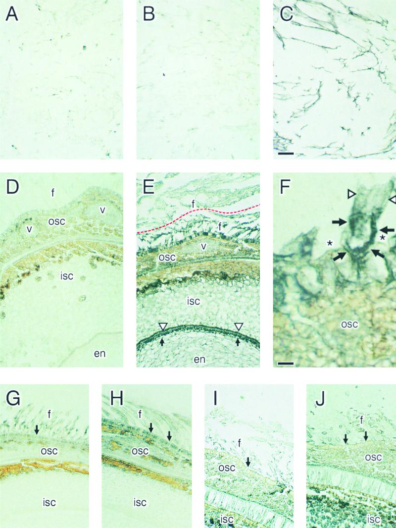
Each cotton fiber is a single cell that elongates to 2.5 to 3.0 cm from the seed coat epidermis within approximately 16 days after anthesis (DAA). To elucidate the mechanisms controlling this rapid elongation, we studied the gating of fiber plasmodesmata and the expression of the cell wall-loosening gene expansin and plasma membrane transporters for sucrose and K(+), the major osmotic solutes imported into fibers. Confocal imaging of the membrane-impermeant fluorescent solute carboxyfluorescein (CF) revealed that the fiber plasmodesmata were initially permeable to CF (0 to 9 DAA), but closed at approximately 10 DAA and re-opened at 16 DAA. A developmental switch from simple to branched plasmodesmata was also observed in fibers at 10 DAA. Coincident with the transient closure of the plasmodesmata, the sucrose and K(+) transporter genes were expressed maximally in fibers at 10 DAA with sucrose transporter proteins predominately localized at the fiber base. Consequently, fiber osmotic and turgor potentials were elevated, driving the rapid phase of elongation. The level of expansin mRNA, however, was high at the early phase of elongation (6 to 8 DAA) and decreased rapidly afterwards. The fiber turgor was similar to the underlying seed coat cells at 6 to 10 DAA and after 16 DAA. These results suggest that fiber elongation is initially achieved largely by cell wall loosening and finally terminated by increased wall rigidity and loss of higher turgor. To our knowledge, this study provides an unprecedented demonstration that the gating of plasmodesmata in a given cell is developmentally reversible and is coordinated with the expression of solute transporters and the cell wall-loosening gene. This integration of plasmodesmatal gating and gene expression appears to control fiber cell elongation.


Figures

Figure 1.
Figure 1.
Confocal Imaging of CF Transport from Phloem in Seed Coat into Elongating Cotton Fibers at 2, 6, 10, and 16 DAA. (A) A schematic representation of a developing cotton seed. The boxed area corresponds to the following confocal images of CF movement from the vascular bundle in the outer seed coat into fibers. (B) Optical cross-section of seed at 2 DAA from shoot fed with CF for 24 hr, showing CF movement from the vascular bundle into fibers. (C) Imaging of the surface of the intact seed shown in (B). Note strong CF signals in fibers. (D) Cross-section of a major vascular bundle from seed at 6 DAA after feeding CF for 16 hr. Note that CF signals were initially detected in the sieve element (arrow) and sieve element–companion cell complexes (arrowhead), but not in xylem between them. (E) Longitudinual section of a vascular bundle at 6 DAA, showing CF fluorescence in phloem (arrows), flanked by nonfluorescence xylem, which is shown in the inset. Note the thicker cell wall of the xylem in the inset. (F) Preferential transport and accumulation of CF from unloading area to fibers at 6 DAA after 24-hr feeding. (G) Blockage of CF movement into fibers at 10 DAA after 24-hr feeding. Note the stronger and wider spread CF signals in the vascular region than that at 6 DAA (F), suggesting that sufficient CF has been unloaded. Also, the dye preferentially accumulated at the inner side of the outer seed coat, in contrast to that at 6 DAA (F). (H) Optical section at 10 DAA after extended feeding of CF for 48 hr. The dye spread throughout the outer seed coat but was not present in fibers. (I) Autofluorescense image of (H) at 514 nm, showing the position of fiber and other tissues. (J) CF signals were detected again in fibers at 16 DAA after 24-hr feeding. (K) Enlarged view of fiber shown in (J). Note CF signals in cytosol lining to plasma membranes (arrows) and appeared patchy in some areas. (L) A montaged image of seed coat at 16 DAA after extended feeding for 48 hr. CF moved extensively into fibers. f, fiber; isc, inner seed coat; osc, outer seed coat; p, phloem; v, vascular bundle; x, xylem. formula image (B) formula image for (B) and (F) to (J); bar in (C) formula image 200 μm for (C) to (E) and (K); bar in (L) formula image 1200 μm.
Figure 2.
Figure 2.
Light-Fluorescent Micrographs of CF in Hand-Cut Transverse Sections of Developing Cotton Seed Coats at 10 ([A] and [B]) and 16 ([C] and [D]) DAA. (A) and (C) Fluorescent signals were confined in fibers after 20-min local loading into fibers from an attached fruit. (B) and (D) After 2-hr incubation in buffer, CF failed to move into the outer seed coat from fiber at 10 DAA (B). In contrast, at 16 DAA, the dye readily moved into the seed coat (D). f, fiber; isc, inner seed coat; osc, outer seed coat.
Figure 3.
Figure 3.
Plasmodesmata of Elongating Cotton Fibers. (A) Simple plasmodesmata (arrows) at 6 DAA in longitudinal orientation. (B) Branched (arrowheads) and “swollen” simple (arrow) plasmodesmata at 10 DAA. Branching occurred at the fiber side. cw, cell wall; epc, epidermal cell; f, fiber cell. formula image (A) formula image for (A) and (B).
Figure 4.
Figure 4.
Transcript Expression of GhSUT1, GhKT1, GhEXP1, and GhSuSy in Elongating Cotton Fibers and Other Sink Tissues. RNA gel blot with 25 μg total RNA in each lane was sequentially hybridized with GhSUT1, GhKT1, GhEXP1, and GhSuSy cDNA probes. The blot was finally hybridized with a maize rRNA cDNA probe to show equal loading and transfer of RNA in each lane. Number on fiber samples indicates days after anthesis.
Figure 5.
Figure 5.
Immunogold Localization of SUT Proteins in Developing Cotton Seed at 10, 5, and 18 DAA. For samples at 10 and 18 DAA, fibers were excised from the seed surface at their base region and treated separately. The black signal represents SUT proteins. (A) Fibers at 10 DAA treated with preimmune serum. (B) Fibers at 10 DAA treated with antiserum against SUT. No signals were detected compared with (A). (C) Fibers at 10 DAA treated with antibody against cotton SuSy, showing strong signals of SuSy protein. (D) Cross-section of seed at 10 DAA treated with preimmune serum. (E) A consecutive section of (D) but treated with antiserum against SUT. Note strong SUT signals at the fiber base region interconnecting the outer seed coat (below the red line). The signals became much weaker beyond the base region (above the red line). Also note specific strong signals at the innermost of inner seed coat (triangles) and at the outermost of the endosperm (arrows). (F) Magnified view of a base region of fibers shown in (E). Note strong SUT signals confined at the base region of fibers (arrows) with much de
Figure 6.
Figure 6.
Developmental Changes of Osmotic and Turgor Potentials in Elongating Cotton Fibers and Seed Coats. (A) Osmotic potentials. (B) Turgor potentials. Bars represent ±sem (four replicates).
Figure 7.
Figure 7.
An Integrated Model of the Control of Cotton Fiber Elongation by Reversible Gating of Plasmodesmata and Coordinated Expression of Plasma Membrane Sucrose and K+ Transporters and Expansin. The thicker the curve representing fiber cell wall, the less the expansin expressed and the more rigid the wall is.

References

    1. Basra, A., and Malik, C.P. (1984). Development of the cotton fiber. Int. Rev. Cytol. 89 65–113.
    1. Beffagna, N., and Romani, G. (1988). Effects of two plasmalemma ATPase inhibitors on H+ extrusion and intracellular pH in Elodea densa leaves. J. Exp. Bot. 39 1039–1043.
    1. Buchala, A.J. (1987). Acid β-fructofuranoside fructohydrolase (invertase) in developing cotton (Gossypium arboreum L.) fibers and its relationship to β-glucan synthesis from sucrose fed to the fiber apoplast. J. Plant Physiol. 127 219–230.
    1. Cassman, K.G., Kerby, T.A., Roberts, B.A., Bryant, D.C., and Higashi, S.L. (1990). Potassium nutrition effects on lint yield and fiber quality of Acala cotton. Crop Sci. 30 672–677.
    1. Cosgrove, D.J. (1997). Relaxation in a high-stress environment: The molecular bases of extensible cell walls and cell enlargement. Plant Cell 9 1031–1041. - PMC - PubMed

MeSH terms