. 2001 Jan 30;98(3):992-9.
doi: 10.1073/pnas.98.3.992.

Defective repression of c-myc in breast cancer cells: A loss at the core of the transforming growth factor beta growth arrest program

Affiliations

Defective repression of c-myc in breast cancer cells: A loss at the core of the transforming growth factor beta growth arrest program

C R Chen et al. Proc Natl Acad Sci U S A. .

Abstract

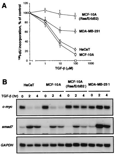
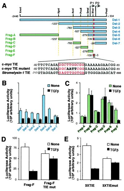
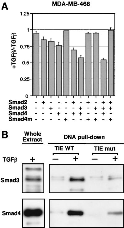
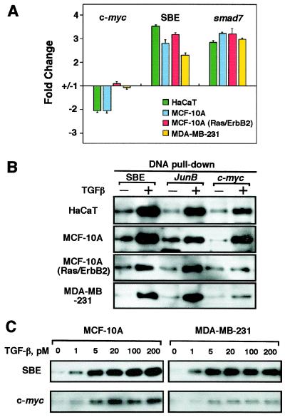
Loss of growth inhibitory responses to the cytokine transforming growth factor beta (TGF-beta) in cancer cells may result from mutational inactivation of TGF-beta receptors or their signal transducers, the Smad transcription factors. In breast cancer, however, loss of TGF-beta growth inhibition often occurs without a loss of these signaling components. A genome-wide analysis of rapid TGF-beta gene responses in MCF-10A human mammary epithelial cells and MDA-MB-231 breast cancer cells shows that c-myc repression, a response that is key to the TGF-beta program of cell cycle arrest, is selectively lost in the cancer cell line. Transformation of MCF-10A cells with c-Ha-ras and c-erbB2 oncogenes also led to a selective loss of c-myc repression and cell cycle arrest response. TGF-beta stimulation of epithelial cells rapidly induces the formation of a Smad complex that specifically recognizes a TGF-beta inhibitory element in the c-myc promoter. Formation of this complex is deficient in the oncogenically transformed breast cells. These results suggest that a Smad complex that specifically mediates c-myc repression is a target of oncogenic signals in breast cancer.


Figures

Figure 1
Figure 1
Loss of c-Myc down-regulation in breast cancer cells resistant to TGF-β growth inhibition. (A) Inhibition of [125I]deoxyuridine incorporation into DNA by TGF-β in three cell lines used in DNA microarray analysis and in HaCaT cells. Exponentially growing cultures were incubated for 20 h with the indicated concentrations of TGF-β. Data are the average of triplicate determinations ± SD. (B) Endogenous gene responses in various human cell lines. Exponentially growing cultures of HaCaT, MCF-10A, MCF-10A(Ras/ErbB2), or MDA-MB-231 cells were incubated with 100 pM TGF-β for the indicated time. Total RNA was isolated and subjected to Northern analysis. Blots were probed with human c-myc, smad7, and glyceraldehyde-3-phosphate dehydrogenase (GAPDH) cDNA probes.
Figure 2
Figure 2
A c-myc TIE-like element mediates TGF-β repression. (A) Diagram of the human c-myc promoter/luciferase constructs and sequence of TIE in c-myc and stromelysin-1. The bars represent the inserted c-myc promoter sequence in the reporter plasmid. P1 and P2 are two transcriptional start sites at the c-myc promoter. Annotations of promoter nucleotide sequence are relative to P2 transcription starting site. TIE-like elements are highlighted by red bars. Thirty-base pair sequences encompassing the TIE element are shown, with the TIE element highlighted in red letters and boxed. The mutations introduced in the c-myc TIE are shown in bold letters. (BE) Transient expression analysis of c-myc reporter constructs in proliferating HaCaT cells. Cells were transfected with the indicated plasmids and were left untreated or treated with TGF-β for 16 h before luciferase activities were determined. Data are mean ± SD of triplicate experiments.
Figure 3
Figure 3
Recognition of the c-myc TIE by TGF-β-dependent Smad complex. (A) MDA-MB-468 cells were cotransfected with c-myc-luciferase reporter (Del-2, 1 μg) and the indicated combinations of constructs [200 ng of pCMV5-Smad2, 30 ng of pCMV5-Smad3, 60 ng of pCMV5-Smad4, or pCMV5 vector encoding the Smad4(1–514) mutant]. Transfected cells were left untreated (−) or treated (+) with TGF-β for 16 h. Luciferase activity was determined and is represented as the ratio relative to the values without TGF-β treatment. The amount of DNA used in the transfections was kept equal by using empty pCMV5 plasmid. (B) Binding of a TGF-β-induced endogenous Smad complex to the TIE. Cell extracts from HaCaT cells untreated or pretreated with 100 pM TGF-β for 1 h were incubated with biotinylated wild-type TIE or mutant TIE oligos and streptavidin-agarose beads. Protein-DNA complexes were subjected to Western blotting analysis and probed with anti-Smad3 and anti-Smad4 polyclonal antibodies.
Figure 4
Figure 4
Attenuation of Smad binding to the c-myc TIE in cells resistant to growth inhibition by TGF-β. (A) Reporter responses in various cell lines. HaCaT, MCF-10A, MCF-10A(Ras/ErbB2), and MDA-MB-231 cells were transfected with the indicated reporter constructs with (+) or without (−) TGF-β treatment for 16 h before luciferase activity was determined, and the change relative to the basal activity was calculated and plotted. (B and C) Binding of TGF-β-induced Smad complexes to the c-myc TIE element. Cell extracts prepared from HaCaT, MCF-10A, MCF-10A(Ras/ErbB2), and MDA-MB-231 untreated (−) or pretreated with indicated concentration of TGF-β for 1 h were incubated with biotinylated oligonucleotides corresponding to the TGF-β response elements of c-myc or junB, or a multimer of the Smad binding element GTCT (SBE). Precipitates were subjected to Smad4 Western immunoblotting analysis.

References

    1. Roberts A B, Sporn M B. In: Peptide Growth Factors and Their Receptors. Sporn M B, Roberts A B, editors. Heidelberg: Springer; 1990. pp. 419–472.
    1. Alexandrow M G, Moses H L. Cancer Res. 1995;55:1452–1457. - PubMed
    1. Massagué J. Nat Rev Mol Cell Biol. 2000;1:169–181. - PubMed
    1. Massagué J, Blain S W, Lo R S. Cell. 2000;103:295–309. - PubMed
    1. Derynck R, Zhang Y, Feng X H. Cell. 1998;95:737–740. - PubMed

Publication types

MeSH terms

Substances