. 2001 Jan 30;98(3):1065-70.
doi: 10.1073/pnas.98.3.1065. Epub 2001 Jan 16.

PDX:PBX complexes are required for normal proliferation of pancreatic cells during development

Affiliations

PDX:PBX complexes are required for normal proliferation of pancreatic cells during development

S Dutta et al. Proc Natl Acad Sci U S A. .

Abstract

The homeobox factor PDX-1 is a key regulator of pancreatic morphogenesis and glucose homeostasis; targeted disruption of the PDX-1 gene leads to pancreatic agenesis in pdx-1(-/-) homozygotes. Pdx-1 heterozygotes develop normally, but they display glucose intolerance in adulthood. Like certain other homeobox proteins, PDX-1 contains a consensus FPWMK motif that promotes heterodimer formation with the ubiquitous homeodomain protein PBX. To evaluate the importance of PDX-1:PBX complexes in pancreatic morphogenesis and glucose homeostasis, we expressed either wild-type or PBX interaction defective PDX-1 transgenes under control of the PDX-1 promoter. Both wild-type and mutant PDX-1 transgenes corrected glucose intolerance in pdx-1 heterozygotes. The wild-type PDX-1 transgene rescued the development of all pancreatic lineages in pdx-1(-/-) animals, and these mice survived to adulthood. In contrast, pancreata from pdx-1(-/-) mice expressing the mutant PDX-1 transgene were hypoplastic, and these mice died within 3 weeks of birth from pancreatic insufficiency. All pancreatic cell types were observed in pdx-1(-/-) mice expressing the mutant PDX-1 transgene; but the islets were smaller, and increased numbers of islet hormone-positive cells were noted within the ductal epithelium. These results indicate that PDX-1:PBX complexes are dispensable for glucose homeostasis and for differentiation of stem cells into ductal, endocrine, and acinar lineages; but they are essential for expansion of these populations during development.


Figures

Figure 1
Figure 1
Expression of wild-type or PBX interaction defective PDX-1 transgenes rescues glucose homeostasis in pdx-1 (+/−) mice. (A) Transient transfection assay of wild-type and PBX interaction defective (Pdxμ) PDX-1 constructs in 293T cells, with a TSEII luciferase reporter containing two PDX:PBX heterodimer recognition sites. Cells were transfected with wild-type or mutant PDX plus E2A-PBX expression vector where indicated. The first bar on the left represents control transfection with empty vector alone plus TSEII reporter. Luciferase activity shown after normalizing to β-galactosidase activity from cotransfected RSV-βgal plasmid. (B) Genotypic analysis of mice expressing PDX-1 transgene. (Left) Inheritance of PDX-1 transgene was evaluated by PCR assay with primers that are selective for rat PDX-1. (Right) Mice heterozygous or homozygous for targeted disruption of the murine PDX-1 gene were identified by genomic Southern blot analysis with a 32P-labeled PDX-1 cDNA probe. Insertion of the inactivating β-galactosidase gene is indicated by a 3.8-kb vs. a 3.0-kb EcoRI fragment. (C) Wild-type (Lane 1) and mutant (Lane 2) PDX-1 transgenes are comparably expressed in transgenic mice. (Left) RNase protection assay of total RNA from whole pancreas of adult mice, with the use of 32P-labeled wild-type PDX-1 antisense RNA probe. The shorter fragment in mutant mice results from RNase digestion in sequences encoding mutant FPWMK/AAGGQ motif. Lane 3, control nontransgenic littermate RNA; the rat PDX-1 probe does not recognize murine PDX-1. Lane 4, position of undigested PDX-1 antisense probe. Lane 5, molecular weight marker. (Right) Western blot assay of whole pancreas extract, showing levels of PDX-1 protein in wild-type, pdx-1(+/−), pdx-1(+/−);mTg, and pdx-1(−/−);wtTg mice. (D) Line graph of blood glucose levels in wild-type (—■—), pdx-1(+/−) heterozygous (– –⧫– –), pdx-1(+/−); wtTg, and pdx-1 (+/−);mTg mice (⋅⋅⋅●⋅⋅⋅) after i.p. glucose injection. Results for pdx-1(+/−); wtTg and pdx-1(+/−);mTg mice were indistinguishable.
Figure 2
Figure 2
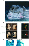
Islet hypoplasia in pdx-1(−/−);mTg neonates. (A) Comparison of pdx-1(−/−);mTg (smaller) and wild-type (larger) littermates 16 days after birth. (B) Whole mount of pdx-1(−/−); wtTg and pdx-1(−/−); mTg mice. Ventral (Left) and dorsal (Right) views are included. p, pancreas; d, duodenum; sp, spleen; s, stomach. (C) Immunocytochemical analysis of insulin (red, arrows) and glucagon + somatostatin (green, asterisks) cells in sections from pdx-1(−/−); wtTg (Upper) and pdx-1(−/−):mTg (Lower). (D) Whole mount showing smaller developing islets (dark blue; arrowheads) in pdx-1(−/−) mTg compared with pdx-1(+/−) mTgt mice. Blue color indicates expression of β-galactosidase from the endogenous pdx-1 allele pdx-1lacZKO (1).
Figure 3
Figure 3
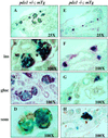
Pancreatic islets of neonatal pdx-1(−/−);mTg mice are smaller and have disrupted architecture. (A–D) Sections of pdx-1(+/−); mTg islets. (E–H) Sections of pdx-1(−/−); mTg islets. pdx-1(−/−); mTg islets are much smaller and are located in close proximity to the ducts. Immunohistochemistry (brown) shows insulin-positive (F), glucagon-positive (G), and somatostatin-positive (H) cells in pdx-1(−/−); mTg islets. Glucagon- and somatostatin-positive cells (G and H) are not localized at the islet periphery as in wild-type islets (compare with C and D).
Figure 4
Figure 4
Pancreata from pdx-1 (−/−);mTgt mice show a defect in proliferation and contain increased numbers of hormone-positive cells in the ductal epithelium. (A) Immunocytochemical staining of whole-pancreas sections from pdx-1 (−/−);wtTg and pdx-1 (−/−);mTg neonates, with Ki-67 antiserum as the marker of cell proliferation. (B) Immunocytochemistry of pancreas sections from neonatal wild-type mice, showing the highest levels of PBX-1 (green) in ductal cells (D), followed by islet cells. Acinar cells stain weakly for PBX-1. Insulin immunostaining (red) is overlaid to indicate the islet. (C) Immunohistochemical detection of insulin (Left), glucagon (Middle), and somatostatin (Right) cells (arrowheads) within the pancreatic ductal epithelium of neonatal pdx-1 (−/−);mTg mice. Mice of all other genotypes did not show hormone-positive cells within ducts in any sections examined.

Similar articles

Cited by

References

    1. Offield M, Jetton T, Labosky P, Ray M, Stein R, Magnuson M, Hogan B, Wright C. Development (Cambridge, UK) 1996;122:983–995. - PubMed
    1. Jonsson J, Carlsson L, Edlund T, Edlund H. Nature (London) 1994;13:606–609. - PubMed
    1. Stoffers D, Zinkin N, Stanojevic V, Clarke W L, Habener J F. Nat Genet. 1997;15:106–110. - PubMed
    1. Dutta S, Bonner-Wier S, Wright C, Montminy M. Nature (London) 1998;392:560. - PubMed
    1. Ahlgren U, Jonsson J, Jonsson L, Simu K, Edlund H. Genes Dev. 1998;12:1763–1768. - PMC - PubMed

Publication types

MeSH terms