. 2001 Jan 30;98(3):1118-23.
doi: 10.1073/pnas.98.3.1118.

A mouse model of multiple endocrine neoplasia, type 1, develops multiple endocrine tumors

Affiliations

A mouse model of multiple endocrine neoplasia, type 1, develops multiple endocrine tumors

J S Crabtree et al. Proc Natl Acad Sci U S A. .

Abstract

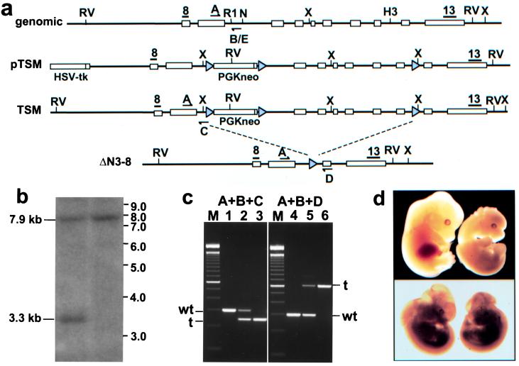
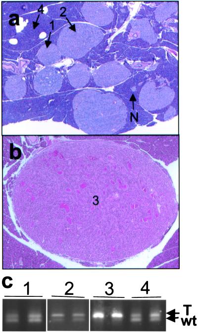
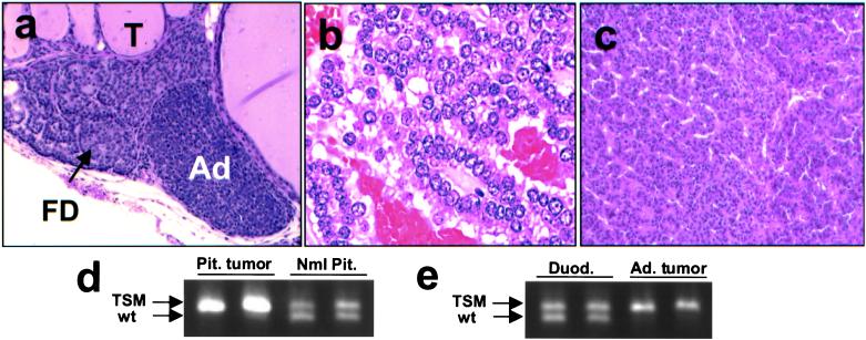
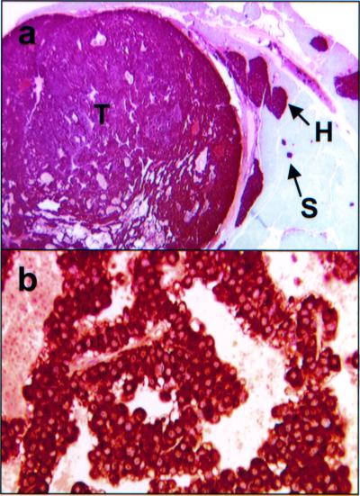
Multiple endocrine neoplasia type 1 (MEN1) is an autosomal dominant cancer syndrome, characterized primarily by multiple tumors in the parathyroid glands, endocrine pancreas, and anterior pituitary. Other tumors, including gastrinoma, carcinoid, adrenal cortical tumors, angiofibroma, collagenoma, and lipoma, also occur in some patients. Individuals with MEN1 almost always have loss-of-function mutations in the MEN1 gene on chromosome 11, and endocrine tumors arising in these patients usually show somatic loss of the remaining wild-type allele. To examine the role of MEN1 in tumor formation, a mouse model was generated through homologous recombination of the mouse homolog Men1. Homozygous mice die in utero at embryonic days 11.5-12.5, whereas heterozygous mice develop features remarkably similar to those of the human disorder. As early as 9 months, pancreatic islets show a range of lesions from hyperplasia to insulin-producing islet cell tumors, and parathyroid adenomas are also frequently observed. Larger, more numerous tumors involving pancreatic islets, parathyroids, thyroid, adrenal cortex, and pituitary are seen by 16 months. All of the tumors tested to date show loss of the wild-type Men1 allele, further supporting its role as a tumor suppressor gene.


Figures

Figure 1
Figure 1
(a) Targeting strategy for the Men1 knockouts. Restriction sites: RV, EcoRV; X, XbaI; R1, EcoRI; n, NdeI; H3, HindIII. Gray triangles, LoxP sites; black bars, probes 8 and 13; arrows, genotyping primers. (b) Southern blot of ES cell genomic DNA digested with EcoRV and probed with probe 8. Wild-type allele (7.9 kb) and targeted allele (3.3 kb) are indicated. (c) Agarose gel electrophoresis of genotype PCRs. Lanes 1–3 are wild-type, heterozygote, and homozygote TSM mice, respectively. Lanes 4–6 are wild-type, heterozygote, and homozygote ΔN3–8 mice, respectively. (d) Lethality in homozygote embryos. (Upper) Wild-type E12.5 embryo on left, TSM homozygote E12.5 embryo on right. (Lower) Wild-type E11.5 embryo on left, ΔN3–8 E11.5 embryo on right.
Figure 2
Figure 2
Pancreatic lesions in Men1TSM/+ mice. (a) H&E-stained section (×15) of pancreas from a 12-month-old mouse, showing normal islets (N), hyperplastic islets (1), hyperplastic islets with dysplasia (2), and acinar pancreas (4). (b) H&E-stained section (×15) of a pancreatic islet cell tumor (3) from a 15-month-old mouse, showing increased vascularization and ribbon-like nuclei formation around capillaries. (c) Agarose gel electrophoresis of PCR from microdissected samples. Numbers correlate with the type of tissue indicated in a and b: 1 is hyperplastic islet, 2 is hyperplastic islet with dysplasia, 3 is pancreatic islet cell tumor, and 4 is acinar tissue. The upper band arises from the TSM allele (T), and the lower band from the wild-type allele (wt).
Figure 3
Figure 3
Other lesions in Men1TSM/+ mice. (a) H&E-stained section (×40) of parathyroid from a 12-month-old mouse with thyroid follicles (T), focal parathyroid dysplasia (FD), and parathyroid adenoma (Ad). (b) H&E-stained section (×120) of pituitary adenoma from a 21-month-old mouse. (c) H&E-stained section (×40) of adrenal cortical tumor from a 15-month-old mouse. (d) Agarose gel electrophoresis, showing loss of the wild-type allele in pituitary adenoma and retention of both alleles in normal pituitary tissue. (e) Agarose gel electrophoresis, showing loss of the wild-type allele in adrenal cortical tumor and retention of both alleles in normal duodenum tissue from the same animal. In LOH the upper band arises from the TSM allele, and the lower band, from the wild-type allele.
Figure 4
Figure 4
Immunohistochemistry of Men1TSM/+ mouse lesions. (a) Insulin immunohistochemical staining of a pancreatic section from a 20-month-old mouse. This section (×10) shows small islets (S), hyperplastic islets (H), and tumor (T), all containing insulin at appreciable levels. (b) Prolactin immunohistochemical staining of a pituitary adenoma section (×120) from a 21-month-old mouse.

References

    1. Trump D, Farren B, Wooding C, Pang J T, Besser G M, Buchanan K D, Edwards C R, Heath D A, Jackson C E, Jansen S, et al. Q J Med. 1996;89:653–669. - PubMed
    1. Marx S. In: Genetic Basis of Human Cancer. Vogelstein B, Kinzler K, editors. New York: McGraw–Hill; 1998. pp. 489–506.
    1. Larsson C, Skogseid B, Oberg K, Nakamura Y, Nordenskjold M. Nature (London) 1988;332:85–87. - PubMed
    1. Chandrasekharappa S C, Guru S C, Manickam P, Olufemi S E, Collins F S, Emmert-Buck M R, Debelenko L V, Zhuang Z, Lubensky I A, Liotta L A, et al. Science. 1997;276:404–407. - PubMed
    1. Agarwal S K, Kester M B, Debelenko L V, Heppner C, Emmert-Buck M R, Skarulis M C, Doppman J L, Kim Y S, Lubensky I A, Zhuang Z, et al. Hum Mol Genet. 1997;6:1169–1175. - PubMed

MeSH terms