. 2001 Jan 30;98(3):1142-7.
doi: 10.1073/pnas.98.3.1142.

Genetic remodeling of protein glycosylation in vivo induces autoimmune disease

Affiliations

Genetic remodeling of protein glycosylation in vivo induces autoimmune disease

D Chui et al. Proc Natl Acad Sci U S A. .

Abstract

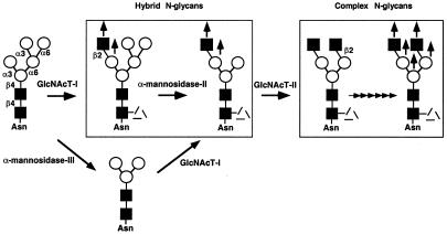
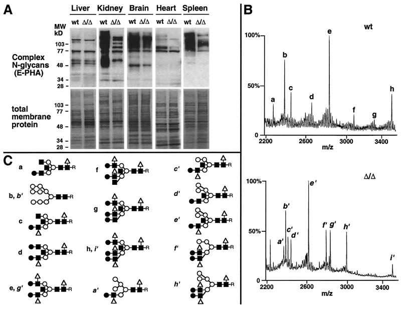
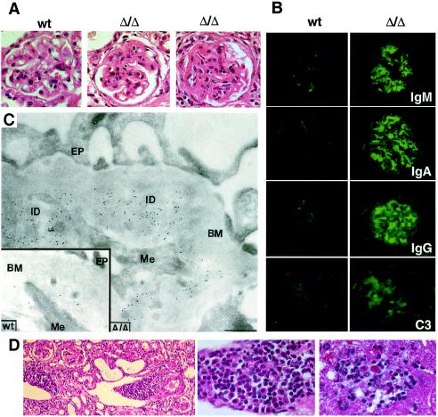
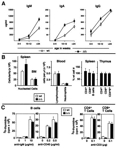
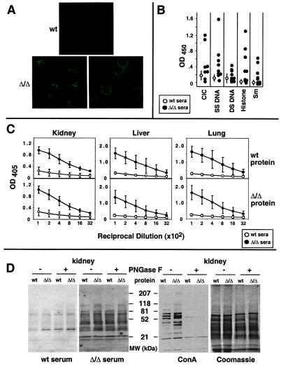
Autoimmune diseases are among the most prevalent of afflictions, yet the genetic factors responsible are largely undefined. Protein glycosylation in the Golgi apparatus produces structural variation at the cell surface and contributes to immune self-recognition. Altered protein glycosylation and antibodies that recognize endogenous glycans have been associated with various autoimmune syndromes, with the possibility that such abnormalities may reflect genetic defects in glycan formation. We show that mutation of a single gene, encoding alpha-mannosidase II, which regulates the hybrid to complex branching pattern of extracellular asparagine (N)-linked oligosaccharide chains (N-glycans), results in a systemic autoimmune disease similar to human systemic lupus erythematosus. alpha-Mannosidase II-deficient autoimmune disease is due to an incomplete overlap of two conjoined pathways in complex-type N-glycan production. Lymphocyte development, abundance, and activation parameters are normal; however, serum immunoglobulins are increased and kidney function progressively falters as a disorder consistent with lupus nephritis develops. Autoantibody reactivity and circulating immune complexes are induced, and anti-nuclear antibodies exhibit reactivity toward histone, Sm antigen, and DNA. These findings reveal a genetic cause of autoimmune disease provoked by a defect in the pathway of protein N-glycosylation.


Figures

Figure 1
Figure 1
Two pathways to complex protein N-glycosylation in mammals. Complex-type N-glycans are produced in the Golgi apparatus and are the predominant forms among extracellular compartments. Each pathway depends on a separate α-mannosidase activity to produce the hybrid N-glycan substrate for the GlcNAcT-II glycosyltransferase. Differential use of each pathway among glycoprotein substrates indicates additional controls in N-glycan repertoire expression. Black square, N-acetylglucosamine; open triangle, fucose; black circles, galactose; open circles, mannose. Anomeric linkage states are denoted. The α1–6 linkage of fucose to the asparagine-proximal N-acetylglucosamine (dashed lines) can be found on both hybrid and complex N-glycans.
Figure 2
Figure 2
Reduction in complex N-glycans and increased hybrid N-glycan structures in the absence of α-mannosidase II. (A) Complex N-glycans are deficient on glycoproteins from some tissues in mice homozygous for a deletion in the α-mannosidase II gene (Δ/Δ). Membrane protein was isolated from various tissues, and complex N-glycans were visualized by binding to E-phytohemagglutinin (Upper) as previously described (14). Equivalent amounts of membrane protein were used in the analyses (Lower). (B) Mass spectrometry of N-glycans from various tissues (kidney shown) was accomplished after isolation from glycoproteins by PNGase F. Subsequent treatment with various glycosidases (not shown) provided additional information on specific saccharide linkages (16). (C) N-Glycan structures (desialylated) defined by mass spectrometry from wild-type tissues were mostly complex types with fully modified mannose termini bearing N-acetylglucosamine linkages (a, c–h), whereas structures in the absence of α-mannosidase II contained hybrid N-glycans noted by terminal mannose residues (a′, c′, d′, e′, f′, h′). The anomeric glycosidic linkages among the core regions are indicated (Fig. 1). Antennary extensions are with β1–2-linked glucosamine, β1–4-linked galactose, and α1–6-linked fucose and are as described for the relevant Lewis antigens (38). R indicates the position of the asparagine residue before release of N-glycans from glycoproteins by PNGase F. For monosaccharide symbols, see Fig. 1 legend.
Figure 3
Figure 3
Immune complex glomerulonephritis in the absence of α-mannosidase II. (A) In comparison with glomeruli from wild-type mice (Left), thickening of the mesangium is observed with capillary lumen obstruction in mutant mice (Center and Right). (Hematoxylin/eosin stain of glomeruli at ×100 magnification is shown.) (B) The glomeruli of mice lacking α-mannosidase II contain high levels of Ig deposits composed of IgM, IgA, and IgG [the latter include IgG1, IgG2a, and IgG2b (not shown)], as well as complement component C3 (×400 magnification shown). Antibody deposits were also noted in other tissues, including lung and liver, but to a lesser extent (not shown). (C) Immunogold labeling of a glomerulus from a mutant mouse, showing immune deposits (ID) between the glomerular basement membrane (BM) and mesangial cell processes (Me). Gold particles indicate the presence of IgG/IgM in the ID. (Inset) Similar field from a wild-type mouse, showing few gold particles. EP, Foot processes of glomerular epithelium (bar, 0.5 μm). (D) Mononuclear leukocytic infiltrates in kidney (Left and Center) and liver of mutant mice (×400): lymphocytes, plasma cells, and neutrophils. Infiltrates in liver (Right) are found with evidence of hepatocyte degeneration and accumulated bile. Results shown are from mice that are either 8 months (A, B, and D) or 13 months (C) of age.
Figure 4
Figure 4
Hematopoietic and immune parameters in the absence of α-mannosidase II. (A) Serum Ig levels comprising IgM, IgA, and IgG were elevated by 10 weeks of age (16 mice of each genotype used). Elevations in IgG levels included IgG1, IgG2a, and IgG2b, whereas no changes were seen in IgE or IgG3 levels (not shown). (B) Lymphocyte cellularity was not altered among lymphoid organs or in circulation. Increased cellularity in the spleen was due to increased numbers of nucleated erythroblasts, as described (14). Frequencies of T cells (CD4+ and CD8+) and B cells (B220+) were also normal. (C) T and B cell proliferation after antigen receptor crosslinking was unaffected. At east five mice of each genotype were studied.
Figure 5
Figure 5
Anti-nuclear antibodies and autoantibodies are found in mice lacking α-mannosidase II. (A) Reactivity of wild-type sera (wt) or α-mannosidase II deficient sera (Δ/Δ) to HEpG2 cells visualized by fluorescent microscopy (×200). Nucleolar (Bottom left) and nuclear membrane (Bottom right) binding by immunoglobulins in the sera of different mice. Sera dilutions were 1:250. (B) Autoantibody reactivity to various nuclear antigens. Eight mice of each genotype were tested (wild type plotted as OD ± SD, Δ/Δ plotted individually). (C) Autoantibodies to kidney, lung, and liver proteins are induced in α-mannosidase II-deficient mice. Similarly increased autoreactivity is noted when wild-type protein or α-mannosidase II-deficient protein is used as the substrate. (D) Cellular proteins of the indicated genotype were subjected to SDS-PAGE with or without PNGase F pretreatment. Protein blots were incubated with indicated sera (1:2,000 dilution) and processed as described (Materials and Methods). Identical genotypes represent autologous samples in C and D.

References

    1. Steinman L. Cell. 1995;80:7–10. - PubMed
    1. Cohen P L. In: Fundamental Immunology. Paul W E, editor. Philadelphia: Lippincott; 1999. pp. 1067–1088.
    1. Gleeson P A. Biochim Biophys Acta. 1994;1197:237–255. - PubMed
    1. Delves P J. Autoimmunity. 1998;27:239–253. - PubMed
    1. Nyame A K, Debose-Boyd R, Long T D, Tsang V C W, Cummings R D. Glycobiology. 1998;8:615–624. - PubMed

Publication types

MeSH terms