. 2001 Jan 30;98(3):1188-93.
doi: 10.1073/pnas.98.3.1188. Epub 2001 Jan 23.

Regulation of p53 stability and p53-dependent apoptosis by NADH quinone oxidoreductase 1

Affiliations

Regulation of p53 stability and p53-dependent apoptosis by NADH quinone oxidoreductase 1

G Asher et al. Proc Natl Acad Sci U S A. .

Abstract

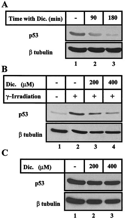
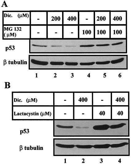
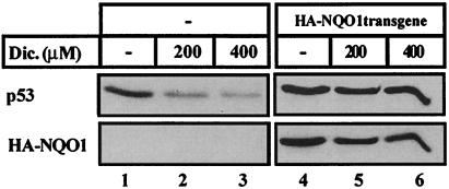
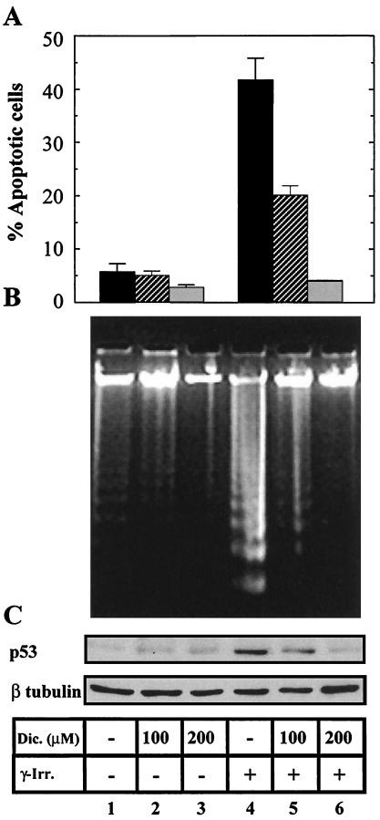
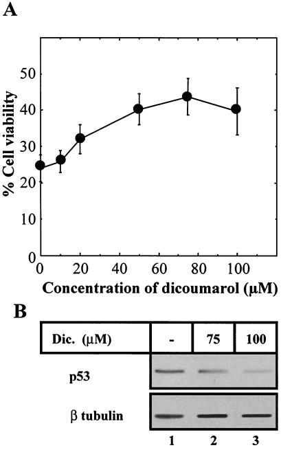
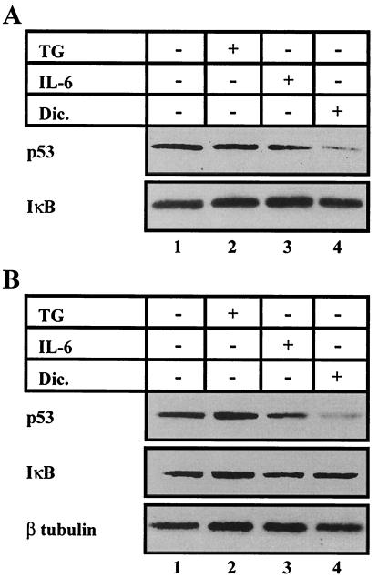
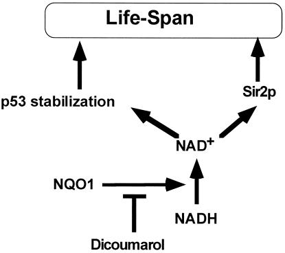
The tumor suppressor gene wild-type p53 encodes a labile protein that accumulates in cells after different stress signals and can cause either growth arrest or apoptosis. One of the p53 target genes, p53-inducible gene 3 (PIG3), encodes a protein with significant homology to oxidoreductases, enzymes involved in cellular responses to oxidative stress and irradiation. This fact raised the possibility that cellular oxidation-reduction events controlled by such enzymes also may regulate the level of p53. Here we show that NADH quinone oxidoreductase 1 (NQO1) regulates p53 stability. The NQO1 inhibitor dicoumarol caused a reduction in the level of both endogenous and gamma-irradiation-induced p53 in HCT116 human colon carcinoma cells. This reduction was prevented by the proteasome inhibitors MG132 and lactacystin, suggesting enhanced p53 degradation in the presence of dicoumarol. Dicoumarol-induced degradation of p53 also was prevented in the presence of simian virus 40 large T antigen, which is known to bind and to stabilize p53. Cells overexpressing NQO1 were resistant to dicoumarol, and this finding indicates the direct involvement of NQO1 in p53 stabilization. NQO1 inhibition induced p53 degradation and blocked wild-type p53-mediated apoptosis in gamma-irradiated normal thymocytes and in M1 myeloid leukemic cells that overexpress wild-type p53. Dicoumarol also reduced the level of p53 in its mutant form in M1 cells. The results indicate that NQO1 plays an important role in regulating p53 functions by inhibiting its degradation.


Figures

Figure 1
Figure 1
Dicoumarol affects p53 stability. (A) HCT116 cells were incubated without (−) or with 400 μM dicoumarol for 90 and 180 min. (B) HCT116 cells were γ-irradiated at 6 Gy and incubated without (−) or with 200 and 400 μM dicoumarol for 4 h. This blot was underexposed compared with A to highlight the increase in p53 protein level after irradiation. (C) COS 1 cells were γ-irradiated at 6 Gy and incubated without (−) or with 200 and 400 μM dicoumarol for 4 h. Protein extraction and immunoblot analysis were carried out as described in Materials and Methods, with the use of Pab 1801 monoclonal anti-p53 antibody. The blots were then stripped and reprobed with monoclonal anti-β-tubulin antibody as a control for equal protein loading in each lane. Dic., dicoumarol.
Figure 2
Figure 2
Dicoumarol-induced p53 decrease occurs by proteasomal degradation. (A) HCT116 cells were incubated without (−) or with 200 and 400 μM dicoumarol and without (−) or with 100 μM MG 132 for 4 h. (B) HCT116 cells were incubated without (−) or with 400 μM dicoumarol without (−) or with 40 μM lactcystin for 4 h. Dic., dicoumarol.
Figure 3
Figure 3
Dicoumarol-induced p53 degradation is inhibited by overexpression of NQO1. Parental HCT116 cells (−) and a pool of HCT116 stable clones overexpressing HA-tagged NQO1 were incubated without (−) or with 200 and 400 μM dicoumarol for 4 h. Immunoblot analysis was carried out with Pab 1801 monoclonal anti-p53 antibody, and the blots were then stripped and reprobed with monoclonal anti-HA antibody as a control for NQO1 expression.
Figure 4
Figure 4
Dicoumarol inhibits p53 accumulation and p53-dependent apoptosis in γ-irradiated thymocytes. Thymocytes that were not irradiated (−) or were γ-irradiated at 4 Gy (+) were cultured for 5 h without (−) or with 100 and 200 μM dicoumarol. (A) The percentage of apoptotic cells was determined on May–Grünwald–Giemsa-stained cytospin preparations. (B) DNA fragmentation at internucleosomal sites. (C) Immunoblot analysis was carried out with Pab 240 monoclonal anti-p53 antibody. Dic., dicoumarol; γ-irr., γ-irradiation.
Figure 5
Figure 5
Dicoumarol decreases the p53 level and p53-dependant apoptosis in M1-t-p53 cells. (A) M1-t-p53 myeloid leukemic cells were cultured at 32°C without or with different concentrations of dicoumarol, and the percentage of viable cells was determined after 23 h. Concentrations of dicoumarol above 125 μM were toxic to these cells. (B) Immunoblot analysis of the p53 level in cells cultured at 32°C for 16 h without (−) or with 75 or 100 μM dicoumarol was carried out with Pab 240 monoclonal anti-p53 antibody. Dic., dicoumarol.
Figure 6
Figure 6
Degradation of mutant and wild-type p53 by dicoumarol but not by other antiapoptotic agents such as IL-6 or thapsigargin. M1-t-p53 cells were cultured for 6 h without (−) or with 10 nM thapsigargin, 50 ng/ml IL-6, or 100 μM dicoumarol. (A) At 32°C, when p53 behaves like wild type. (B) At 37°C, when p53 behaves like a mutant. Immunoblot analysis was carried out with Pab 240 monoclonal anti-p53 antibody. The blots were then stripped and re-probed with anti-IκB and anti-β-tubulin antibody. Dic., dicoumarol.
Figure 7
Figure 7
Model of the role of NQO1 in p53 stabilization and life span. It is assumed that NQO1 determines the level of NAD+ and that this regulates the level of p53. The stabilization of p53 results in either apoptosis or growth arrest, which regulate life span. Also shown is the NAD+–Sir2p pathway that regulates life span in yeast (56).

References

    1. Oren M. FASEB J. 1992;6:3169–3176. - PubMed
    1. Levine A J. Cell. 1997;88:323–331. - PubMed
    1. Yonish-Rouach E, Resnitzky D, Lotem J, Sachs L, Kimchi A, Oren M. Nature (London) 1991;352:345–347. - PubMed
    1. Lotem J, Sachs L. Blood. 1993;82:1092–1096. - PubMed
    1. Lowe S W, Schmitt E M, Smith S W, Osborne B A, Jacks T. Nature (London) 1993;362:847–849. - PubMed

Publication types

MeSH terms