. 2001 Feb;45(2):413-9.
doi: 10.1128/AAC.45.2.413-419.2001.

Plasmid location and molecular heterogeneity of the L1 and L2 beta-lactamase genes of Stenotrophomonas maltophilia

Affiliations

Plasmid location and molecular heterogeneity of the L1 and L2 beta-lactamase genes of Stenotrophomonas maltophilia

M B Avison et al. Antimicrob Agents Chemother. 2001 Feb.

Abstract

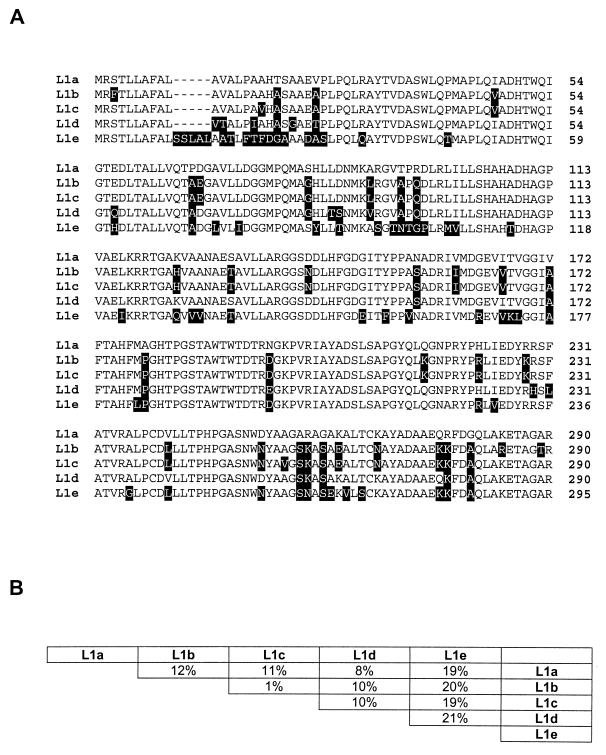
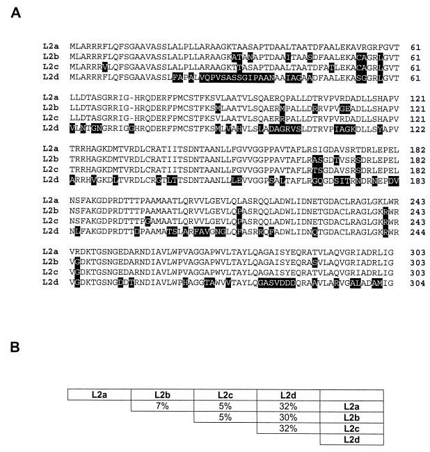
An approximately 200-kb plasmid has been purified from clinical isolates of Stenotrophomonas maltophilia. This plasmid was found in all of the 10 isolates examined and contains both the L1 and the L2 beta-lactamase genes. The location of L1 and L2 on a plasmid makes it more likely that they could spread to other gram-negative bacteria, potentially causing clinical problems. Sequence analysis of the 10 L1 genes revealed three novel genes, L1c, L1d, and L1e, with 8, 12, and 20% divergence from the published strain IID 1275 L1 (L1a), respectively. The most unusual L1 enzyme (L1e) displayed markedly different kinetic properties, with respect to hydrolysis of nitrocefin and imipenem, compared to those of L1a (250- and 100-fold lower k(cat)/K(m) ratios respectively). L1c and L1d, in contrast, displayed levels of hydrolysis very similar to that of L1a. Several nonconservative amino acid differences with respect to L1a, L1b, L1c, and L1d were observed in the substrate binding-catalytic regions of L1e, and this could explain the kinetic differences. Three novel L2 genes (L2b, L2c, and L2d) were sequenced from the same isolates, and their sequences diverge from the published sequence of strain IID 1275 L2 (L2a) by 4, 9, and 25%, respectively. Differences in L1 and L2 gene sequences were not accompanied by similar divergences in 16S rRNA gene sequences, for which differences of <1% were found. It is therefore apparent that the L1 and L2 genes have evolved relatively quickly, perhaps because of their presence on a plasmid.


Figures

FIG. 1
FIG. 1
Alignment of L1 amino acid sequences. Both strands of the L1-FULL PCR products from all 10 S. maltophilia isolates were sequenced as described in Materials and Methods. The predicted amino acid sequences of L1c, L1d, and L1e are novel, and in panel A they are aligned with the previously published L1 amino acid sequences, L1a (30) and L1b (24). All differences from L1a are shaded. (B) Percent divergence of the L1 proteins compared to each other.
FIG. 2
FIG. 2
Alignments of L2 amino acid sequences. Both strands of the L2-FULL PCR products from all 10 S. maltophilia isolates were sequenced as described in Materials and Methods. The predicted amino acid sequences of L2b, L2c, and L2d are novel, and in panel A they are aligned with the previously published L2a amino acid sequence (31). All differences from L2a are shaded. (B) Divergence among the L2 proteins.
FIG. 3
FIG. 3
PCR to show that the L1 and L2 genes of S. maltophilia are plasmid encoded. Plasmids were isolated from 1.5 ml of an overnight culture of S. maltophilia strains 1 to 10 (lanes 1 to 10, respectively, in each panel). (Table 1), (A) A total of 5 μl of the plasmid preparation was resolved by using a 0.8% agarose gel. (B and C) A total of 5 μl of the plasmid preparation was used as a template for PCR with the L1-FULL (B) or L2-FULL (C) primer sets. Primer sequences and PCR conditions are described in Materials and Methods. (B and C) A total of 10 μl of the PCR product was resolved by using a 1.2% agarose gel. DNA molecular size markers (1 kb plus ladder; Life Technologies Ltd., Paisley, United Kingdom) were run in parallel (lanes M) to check the sizes of the PCR products. The figures are photographs of the resultant ethidium bromide-stained gels under UV irradiation.
FIG. 4
FIG. 4
Assessment of chromosomal DNA contamination of plasmid preparations by multiplex PCR. (A) Multiplex PCR was performed with 5 μl of plasmid preparation (lane P) or 20 μl of genomic DNA (lane G) (see Materials and Methods) with the L1-FULL and rDNA primer sets (Materials and Methods). (B) Multiplex PCR was performed with the L2-MID and D-PEP primer sets. The L2-MID primers were used instead of the L2-FULL primers because of the similarity in size between the L2-FULL and D-PEP amplicons. The PCR was performed as described in Materials and Methods, and PCR products were separated and visualized as described in the legend to Fig. 3. Lanes M, DNA molecular size markers.

References

    1. Alonso A, Martinez J I. Multiple antimicrobial resistance in Stenotrophomonas maltophilia. Antimicrob Agents Chemother. 1997;41:1140–1142. - PMC - PubMed
    1. Avison M B, Bennett P M, Walsh T R. β-Lactamase expression in Plesiomonas shigelloides. J Antimicrob Chemother. 2000;45:877–880. - PubMed
    1. Bennett P M. The spread of drug resistance. In: Baumberg S, Young J P W, Wellington E M H, Saunders J R, editors. Population genetics of bacteria. Cambridge, United Kingdom: Cambridge University Press; 1995. pp. 317–344.
    1. Bennett P M. Integrons and gene cassettes: a genetic construction kit for bacteria. J Antimicrob Chemother. 1999;41:1–4. - PubMed
    1. Bennett P M, Chopra I P. Molecular basis of β-lactamase induction in bacteria. Antimicrob Agents Chemother. 1992;37:27–32. - PMC - PubMed

Publication types

LinkOut - more resources