Comparative Study
. 2001 Jan;22(1):27-34.

Aneurysms of the posterior cerebral artery: classification and endovascular treatment

Affiliations
Comparative Study

Aneurysms of the posterior cerebral artery: classification and endovascular treatment

E F Ciceri et al. AJNR Am J Neuroradiol. 2001 Jan.

Abstract

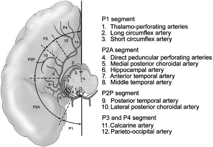
Background and purpose: We present a retrospective review of our experience in the endovascular treatment of posterior cerebral artery (PCA) aneurysms. We detail the anatomic location of these aneurysms, the technique of endovascular treatment, morphologic results, and clinical outcome. We also discuss the segmental anatomy of the PCA as it relates to the various neurologic deficits that may result from occlusion of the parent artery.

Methods: From 1993 to 1998, 20 patients (12 female, eight male; mean age, 44 yrs) harboring a PCA aneurysm were treated via an endovascular approach. One patient had two aneurysms, comprising a total of 21 lesions. Fourteen (66%) of 21 aneurysms were saccular in nature, five (24%) were giant serpentine aneurysms, and two (10%) were posttraumatic. All aneurysms were treated using Guglielmi detachable coils (GDC) either by selective obliteration of the aneurysm sac or by parent artery occlusion.

Results: Fourteen (66%) of the 21 aneurysms were successfully treated with preservation of the parent artery. In the remaining seven (33%), the parent artery was permanently occluded. The overall complication rate in this series was 15%, with a permanent morbidity rate of 10% and a 0% mortality rate.

Conclusion: Aneurysms of the PCA are rare compared with other locations in the intracranial circulation. Saccular PCA aneurysms can be treated effectively, by use of GDC, to obliterate the aneurysm yet preserve the parent artery. Fusiform and giant serpentine aneurysms of the PCA can effectively be treated by permanent occlusion of the parent artery; in these cases, thorough knowledge of the PCA segmental anatomy is crucial in order to select the site of occlusion and to avoid major neurologic deficits.


Figures

<sc>fig</sc> 1.
fig 1.
Anatomic segments of the PCA and their branches. Composite line tracing of all four anatomic segments of the PCA.
<sc>fig</sc> 2.
fig 2.
Giant serpentine aneurysm of the P2A segment of the PCA presenting with intracerebral and subarachnoid hemorrhage in a 12-year-old boy. A, Noncontrast CT scan of the head shows the large partially clotted aneurysm in the perimesencephalic cistern, associated with SAH. B, Towne's view of the vertebral angiogram shows the residual lumen of the partially clotted aneurysm (arrow), non-filling of the distal branches of the ipsilateral PCA, and avascular mass effect on the brain stem structures (arrowheads). C, The lumen of the aneurysm and the parent artery were permanently occluded with GDC. The nonsubtracted Towne's view shows the metallic cast of the coils. D, The corresponding subtracted image of the posttreatment vertebral angiogram shows complete obliteration of the aneurysm.
<sc>fig</sc> 3.
fig 3.
Successful obliteration of a ruptured saccular aneurysm of the PCA with preservation of the parent artery. A, The oblique subtracted view of a vertebral angiogram shows an irregular bilobed saccular aneurysm arising from the origin of the P3/P4 segments. The patient is a 52-year-old woman presenting with acute SAH. B, The oblique nonsubtracted view of the posttreatment vertebral angiogram shows the cast of the GDC obliterating the aneurysm with preservation of the parent artery. C, The oblique subtracted view of the follow-up vertebral angiogram, obtained 2 years after the treatment, shows persistent obliteration of the aneurysm.
<sc>fig</sc> 4.
fig 4.
Parent artery occlusion of a giant serpentine aneurysm of the PCA complicated by cerebral infarction in the distal arterial territory. A, Axial T2-weighted MR image of the head showing a giant serpentine aneurysm of the P2/P3 segments of the left PCA. B, Towne's view of a left vertebral angiogram showing the serpentine nature of the giant aneurysm, which involves the P2 and P3 segments of the left PCA. C, Subtracted Towne's view of the posttreatment vertebral angiogram shows obliteration of the aneurysm and no antegrade flow in the distal branches of the left PCA. Note the retrograde flow from the cortical branches of the ipsilateral middle cerebral artery (arrows). D, Homonymous hemianopsia and contralateral hemiparesis complicated the treatment. The posttreatment axial T2-weighted MR image shows an acute infarction in the distribution of the left PCA territory.
<sc>fig</sc> 5.
fig 5.
P1 segment aneurysm associated with a distal cortical AVM. A, Pretreatment Towne's view of a vertebral angiogram showing a P1 segment aneurysm associated with a temporooccipital AVM. Note the poor ratio of the fundus of the aneurysm to the size of its neck. B, Same view of the vertebral angiogram after the endovascular treatment of the aneurysm with GDC C, Follow-up vertebral angiogram obtained 42 months after the initial treatment showing complete obliteration of the AVM and questionable minimal residual filling of the aneurysm.
<sc>fig</sc> 6.
fig 6.
Multiple PCA associated with Moya-Moya disease, successfully treated with GDC. A, Frontal view of left internal carotid angiogram showing occlusion of the anterior and a middle cerebral arteries and Moya-Moya collateral circulation. There is increased flow in a dysplastic and enlarged ipsilateral PCA (arrow). B, Oblique view of the internal carotid angiogram showing an aneurysm originating from the P1/P2 junction of the left posterior cerebral artery (arrow) and a second aneurysm arising from the P2P segment (arrowhead). Note the presence of at least two flow-related aneurysms arising from the supraclinoid segment of the internal carotid artery (double arrows). C, Oblique view of the internal carotid angiogram, obtained 9 months after treatment, shows persistent obliteration of both PCA aneurysms (arrows).

References

    1. Drake CG. Giant posterior cerebral aneurysms: 66 patients. In: Drake CG, Peerless SJ, Hruesniemi JA, eds. Surgery of Vertebrobasilar Aneurysms: London, Ontario, Experience on 1767 Patients. New York: Springer Verlag 1996;;230-248
    1. Gerber CJ, Neil-Dwyer G. A review of the management of 15 cases of aneurysms of the posterior cerebral artery. Br J Neurosurg 1992;;6:521-527 - PubMed
    1. Pia HW, Fontana H. Aneurysms of the posterior cerebral artery. Locations and clinical pictures. Acta Neurochir (Wien) 1977;;38:13-35 - PubMed
    1. Sakata S, Fujii K, Matsushima T, et al. Aneurysms of the posterior cerebral artery: report of eleven cases- surgical approaches and procedures. Neurosurgery 1993;;32:163-167 - PubMed
    1. Guglielmi G, Vinuela F, Duckwiler G, et al. Endovascular treatment of the posterior circulation aneurysms by electrothrombosis using electrically detachable coils. J Neurosurg 1992;;77:515-524 - PubMed

Publication types