

Fetal cerebral cortex: normal gestational landmarks identified using prenatal MR imaging
- PMID: 11158907
- PMCID: PMC7975558
Fetal cerebral cortex: normal gestational landmarks identified using prenatal MR imaging
Abstract
Background and purpose: Few investigators have analyzed the MR imaging patterns of fetal gyration. Our purpose was to establish, with a large prospective series, the normal sulcation landmarks according to gestational age by using in utero MR imaging and to correlate our findings with established neuroanatomic timetables.
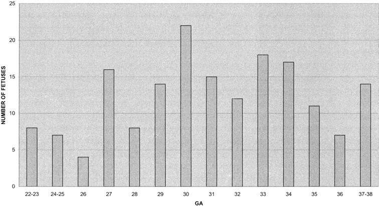
Methods: A standardized fetal cerebral MR examination was performed in 173 normal fetuses at 22 to 38 weeks' gestation. Eight T1- and T2-weighted coronal, axial, and sagittal slices were obtained for each fetus and systematically analyzed. The sequential development of the different fissures and sulci of the cerebral cortex with respect to gestational age were tabulated.
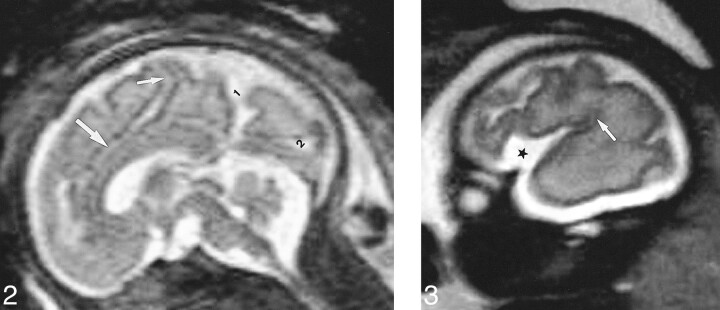
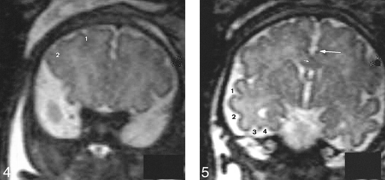
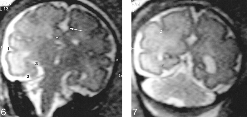
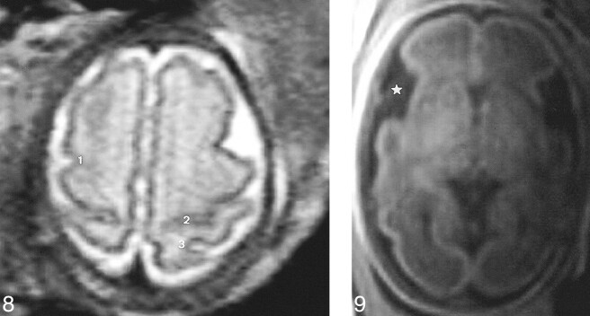
Results: Sulcation of the medial, lateral, and inferior surfaces of the brain was depicted, and a timetable for the MR depiction of the primary and secondary sulci was established for the 22- to 38-week gestational period. This timetable was in good agreement with the neuroanatomic standards of reference, with a mean lag time of 1 week.
Conclusion: This analysis of fetal brain sulcation in a large series of fetuses contributes to a better understanding of the maturation of the fetal cortex on MR imaging studies. It furthermore provides a standard of reference that can be used to assess the normality of fetal sulcation and to diagnose gyrational abnormalities with prenatal MR imaging.
Figures
References
-
- Naidich TP, Grant JL, Altman N, et al. The developing cerebral surface. Neuroimaging Clin N Am 1994;4:201-240 - PubMed
-
- Van der Knaap MS, Van Wezel-Meijler G, Barth PG, Barkhof F, Ader HJ, Valk J. Normal gyration and sulcation in preterm and term neonates: appearance on MR images. Radiology 1996;200:389-396 - PubMed
-
- Chi JG, Dooling EC, Gilles FH. Gyral development of the human brain. Ann Neurol 1977;1:86-93 - PubMed
-
- Dorovini-Zis K, Dolman CL. Gestational development of brain. Arch Pathol Lab Med 1977;101:192-195 - PubMed
-
- Larroche JC. Critères morphologiques du développement du système nerveux central du foetus humain. J Neuroradiol 1981;8:93-108 - PubMed
MeSH terms
LinkOut - more resources
Full Text Sources
Medical