. 2001 Feb;158(2):453-68.
doi: 10.1016/S0002-9440(10)63988-0.

Hippocampal apoptosis in major depression is a minor event and absent from subareas at risk for glucocorticoid overexposure

Affiliations

Hippocampal apoptosis in major depression is a minor event and absent from subareas at risk for glucocorticoid overexposure

P J Lucassen et al. Am J Pathol. 2001 Feb.

Abstract

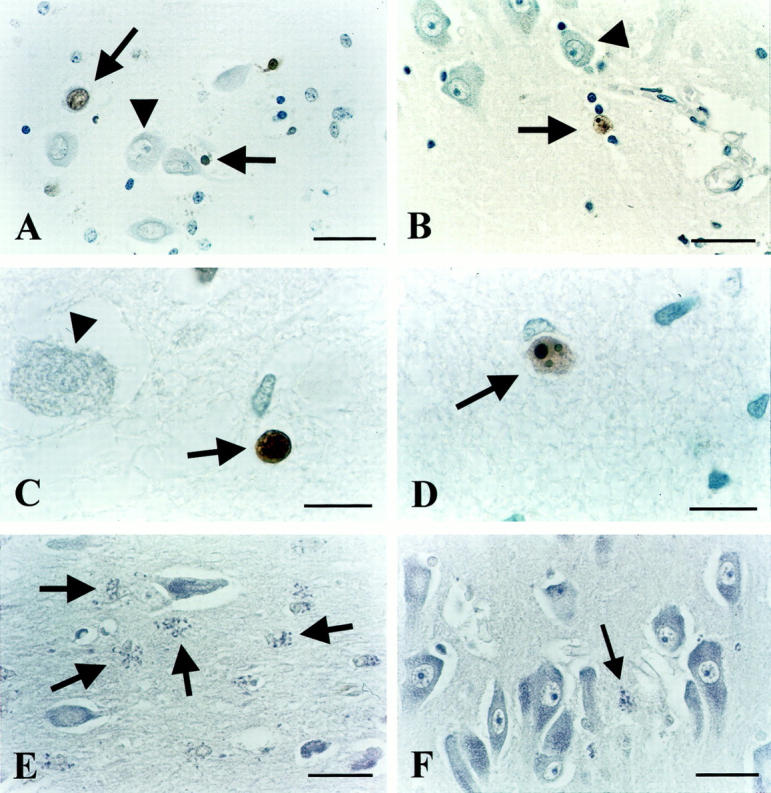
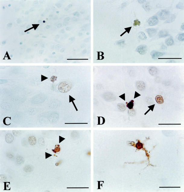
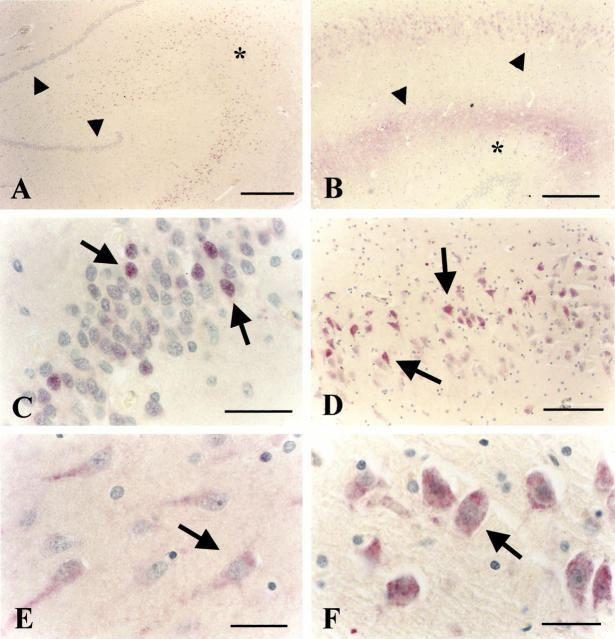
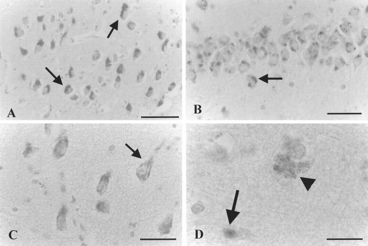
Glucocorticoid (GC) overexposure in animals has been implicated in hippocampal dysfunctioning and neuronal loss. In major depression, hypercortisolemia, hypothalamic-pituitary-adrenocortical-axis alterations, and reduced hippocampal volumes are commonly observed; hence, hippocampal neurodegeneration is also expected. To study possible GC-related pathology, we investigated hippocampal tissue of 15 major-depressed patients, 16 matched controls, and 9 steroid-treated patients, using in situ-end-labeling for DNA fragmentation and apoptosis, and heat-shock protein 70 and nuclear transcription factor kappaB immunocytochemistry for damage-related responses. No obvious massive cell loss was observed in any group. In 11 of 15 depressed patients, rare, but convincing apoptosis was found in entorhinal cortex, subiculum, dentate gyrus, CA1, and CA4. Also in three steroid-treated patients, apoptosis was found. Except for several steroid-treated patients, heat-shock protein 70 staining was generally absent, nor was nuclear transcription factor-kappaB activation found. The detection in 11 of 15 depressed patients, in three steroid-treated, and in one control patient, demonstrates for the first time that apoptosis is involved in steroid-related changes in the human hippocampus. However, in absence of major pyramidal loss, its rare occurrence, that notably was absent from areas at risk for GC damage such as CA3, indicates that apoptosis probably only contributes to a minor extent to the volume changes in depression.


Figures

Figure 1.
Figure 1.
ISEL results. A: Positive labeling in the CA4 area of depressed patient 94-112, showing necrotic morphology (upper arrow) as indicated by the comparable size as an intact, neighboring neuron (arrowhead) without chromatin re-organization or apoptotic bodies visible. Also seen is a labeled apoptotic cell as evidenced by its pycnotic appearance, strong condensation, and brown DAB precipitate (horizontal arrow). B: ISEL-positive neuron (arrow) just outside the CA1 cell layer of depressed patient 90-001 with clear apoptotic morphology, ie, a reduced size as compared to unstained, healthy-looking neurons (triangle), and apoptotic bodies clearly visible. C: ISEL-positive, apoptotic cell (arrow) with a pycnotic, condensed appearance adjacent to a nonstained large cell (arrowhead). CA1 of depressed patient 94-094. D: Apoptotic neuron (arrow) in the subiculum of depressed patient 94-032 with three clear apoptotic bodies visible. E: Frequent, granular morphology (arrows) suggestive of chromatolytic processes, adjacent to normal looking neurons in CA3 of depressed patient 90-001. F: Normal-appearing neurons in CA1 of control subject 94-123. Also, one granular, chromatolytic-like structure is visible (arrow). Scale bars: 34 μm (A, B, E, and F) and 15 μm (C and D).
Figure 2.
Figure 2.
ISEL results (continued from Figure 1 ▶ ). A: Isolated, ISEL-positive apoptotic cell with a clearly condensed size (arrow) in an otherwise ISEL-negative DG area of depressed patient 93-146. B: ISEL-positive apoptotic cell (arrow) in the DG of steroid-treated patient 95-11. C: Apoptotic cell (arrowhead) with clear membrane blebbing as well as a single apoptotic body visible, adjacent to an isolated ISEL-positive nucleus (large arrow) that appears necrotic, with a comparable size that seems even a little swollen, as compared to neighboring DG cells. Both cells are located on the inner border between the otherwise ISEL-negative DG and CA4. Depressed patient 93-090. D: Similar to C, an apoptotic cell close to a necrotic one (arrow). Clearly apoptotic bodies are indicated by arrowheads. Also, the enhanced levels of DNA fragmentation in some of the other cells is visible. Depressed patient 94-17. E: Apoptotic cell at the inner border of the DG of steroid-treated patient 83-004. Arrowheads indicate apoptotic bodies. F: Prominent ISEL labeling of a cell displaying glia morphology with DAB precipitate present throughout its protrusions. Steroid-treated patient 93-021. Scale bars: 42 μm (A), 25 μm (B), 16 μm (C–E), and 10 μm (F).
Figure 3.
Figure 3.
iHSP70 immunocytochemistry. A: Clear cellular labeling of the pyramidal CA1, CA2, CA3, and CA4 layers of 71-year-old depressed patient 94-094. Arrowheads indicate DG granule cell layer. The asterisk marks the CA3 area. No immuno-cytochemical staining is present in the DG. B: Predominant cellular (top arrowhead) and neuropil staining (between bottom arrowhead and the asterisk) in the CA1-3 area of control subject 94-118. C: Higher magnification of the DG of a 63-year-old steroid-treated patient 95-11 that shows some individual neurons showing clearly enhanced staining (arrows). D: CA1 area of depressed patient 94-112 showing prominent cellular staining (arrows) in a subpopulation of neurons in this area whereas cells without this cytoplasmic staining are also observed in the same area. E: Weak cytoplasmic staining of CA1 neurons of steroid-treated patient 95-054. F: Prominent cellular staining of CA4 neurons in steroid-treated patient 95-11. Scale bars: 400 μm (A and B), 42 μm (C), 80 μm (D), and 40 μm (E and F).
Figure 4.
Figure 4.
NF-κB immunocytochemistry. A: CA4 neurons of depressed patient 92-003 showing a cytoplasmic distribution of staining throughout the area. B: DG neurons of the same patient as in A showing a granular, punctate staining pattern for the p65 subunit only in the cytoplasm. C: Higher magnification of CA1 of control subject 94-035 with prominent staining in the cytoplasm (arrows). D: High magnification of the CA area of an Alzheimer patient that was included as a positive control, showing a similar, granular pattern (arrowhead), but also clear nuclear staining (arrow), indicative of NF-κB activation in selected cells. No such pattern was observed in depressed or steroid-treated patients. Scale bars: 90 μm (A), 38 μm (B), 50 μm (C), and 25 μm (D).

References

    1. Raadsheer FC, Sluiter AA, Ravid R, Tilders FJ, Swaab DF: Localization of corticotropin-releasing hormone (CRH) neurons in the paraventricular nucleus of the human hypothalamus; age-dependent colocalization with vasopressin. Brain Res 1993, 615:50-62 - PubMed
    1. Raadsheer FC, van Heerikhuize JJ, Lucassen PJ, Hoogendijk WJ, Tilders FJ, Swaab DF: Corticotropin-releasing hormone mRNA levels in the paraventricular nucleus of patients with Alzheimer’s disease and depression. Am J Psychiatry 1995, 152:1372-1376 - PubMed
    1. Reul JM, de Kloet ER: Two receptor systems for corticosterone in rat brain: microdistribution and differential occupation. Endocrinology 1985, 117:2505-2511 - PubMed
    1. De Kloet ER, Vreugdenhil E, Oitzl MS, Joels M: Brain corticosteroid receptor balance in health and disease. Endocr Rev 1998, 19:269-301 - PubMed
    1. Herman JP, Cullinan WE, Morano MI, Akil H, Watson SJ: Contribution of the ventral subiculum to inhibitory regulation of the hypothalamo-pituitary-adrenocortical axis. J Neuroendocrinol 1995, 7:475-482 - PubMed

Publication types

MeSH terms