

Regional distribution of amyloid-Bri deposition and its association with neurofibrillary degeneration in familial British dementia
- PMID: 11159188
- PMCID: PMC1850296
- DOI: 10.1016/S0002-9440(10)63993-4
Regional distribution of amyloid-Bri deposition and its association with neurofibrillary degeneration in familial British dementia
Abstract
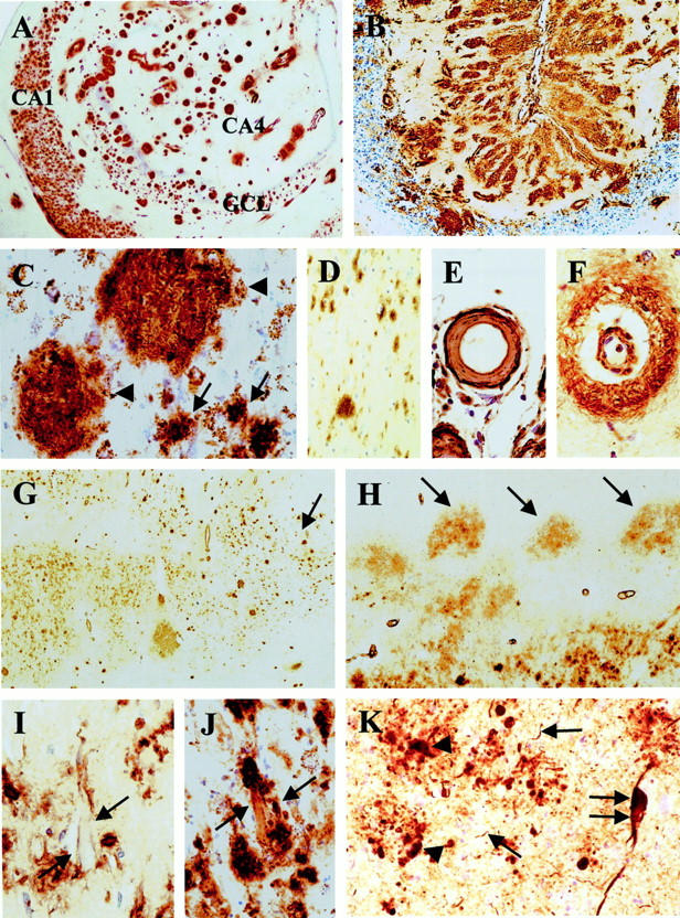
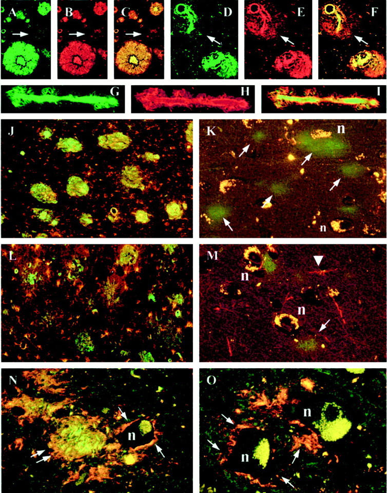
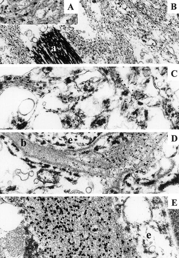
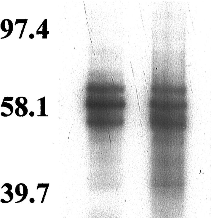
Familial British dementia (FBD), pathologically characterized by cerebral amyloid angiopathy (CAA), amyloid plaques, and neurofibrillary degeneration, is associated with a stop codon mutation in the BRI gene resulting in the production of an amyloidogenic fragment, amyloid-Bri (ABri). The aim of this study was to assess the distribution of ABri fibrillar and nonfibrillar lesions and their relationship to neurofibrillary pathology, astroglial and microglial response using immunohistochemistry, confocal microscopy, and immunoelectron microscopy in five cases of FBD. Abnormal tau was studied with immunoblotting. We present evidence that ABri is deposited throughout the central nervous system in blood vessels and parenchyma where both amyloid (fibrillar) and pre-amyloid (nonfibrillar) lesions are formed. Ultrastructurally amyloid lesions appear as bundles of fibrils recognized by an antibody raised against ABri, whereas Thioflavin S-negative diffuse deposits consist of amorphous electron-dense material with sparse, dispersed fibrils. In contrast to nonfibrillar lesions, fibrillar ABri is associated with a marked astrocytic and microglial response. Neurofibrillary tangles and neuropil threads occurring mainly in limbic structures, are found in areas affected by all types of ABri lesions whereas abnormal neurites are present around amyloid lesions. Immunoblotting for tau revealed a triplet electrophoretic migration pattern. Our observations confirm a close link between ABri deposition and neurodegeneration in FBD.
Figures
Similar articles
-
Familial Danish dementia: a novel form of cerebral amyloidosis associated with deposition of both amyloid-Dan and amyloid-beta.J Neuropathol Exp Neurol. 2002 Mar;61(3):254-67. doi: 10.1093/jnen/61.3.254. J Neuropathol Exp Neurol. 2002. PMID: 11895040
-
Effect of the disulfide bridge and the C-terminal extension on the oligomerization of the amyloid peptide ABri implicated in familial British dementia.Biochemistry. 2001 Mar 27;40(12):3449-57. doi: 10.1021/bi002287i. Biochemistry. 2001. PMID: 11297410
-
Non-fibrillar oligomeric species of the amyloid ABri peptide, implicated in familial British dementia, are more potent at inducing apoptotic cell death than protofibrils or mature fibrils.J Mol Biol. 2001 Jun 29;310(1):157-68. doi: 10.1006/jmbi.2001.4743. J Mol Biol. 2001. PMID: 11419943
-
Chromosome 13 dementia syndromes as models of neurodegeneration.Amyloid. 2001 Dec;8(4):277-84. doi: 10.3109/13506120108993826. Amyloid. 2001. PMID: 11791622 Review.
-
Chromosome 13 dementias.Cell Mol Life Sci. 2005 Aug;62(16):1814-25. doi: 10.1007/s00018-005-5092-5. Cell Mol Life Sci. 2005. PMID: 15968464 Free PMC article. Review.
Cited by
-
Genetic alterations of the BRI2 gene: familial British and Danish dementias.Brain Pathol. 2006 Jan;16(1):71-9. doi: 10.1111/j.1750-3639.2006.tb00563.x. Brain Pathol. 2006. PMID: 16612984 Free PMC article.
-
Cerebral amyloid angiopathy interacts with neuritic amyloid plaques to promote tau and cognitive decline.Brain. 2022 Aug 27;145(8):2823-2833. doi: 10.1093/brain/awac178. Brain. 2022. PMID: 35759327 Free PMC article.
-
Alzheimer's disease pathologic cascades: who comes first, what drives what.Neurotox Res. 2012 Oct;22(3):182-94. doi: 10.1007/s12640-011-9272-9. Epub 2011 Sep 13. Neurotox Res. 2012. PMID: 21913048 Free PMC article. Review.
-
Tau, prions and Aβ: the triad of neurodegeneration.Acta Neuropathol. 2011 Jan;121(1):5-20. doi: 10.1007/s00401-010-0691-0. Epub 2010 May 16. Acta Neuropathol. 2011. PMID: 20473510 Free PMC article. Review.
-
CSF p-tau increase in response to Aβ-type and Danish-type cerebral amyloidosis and in the absence of neurofibrillary tangles.Acta Neuropathol. 2022 Feb;143(2):287-290. doi: 10.1007/s00401-021-02400-5. Epub 2021 Dec 28. Acta Neuropathol. 2022. PMID: 34961894 Free PMC article. No abstract available.
References
-
- Plant GT, Revesz T, Barnard RO, Harding AE, Gautier-Smith PC: Familial cerebral amyloid angiopathy with nonneuritic amyloid plaque formation. Brain 1990, 113:721-747 - PubMed
-
- Mead S, James-Galton M, Revesz T, Doshi RB, Harwood G, Pan EL, Ghiso J, Frangione B, Plant G: Familial British dementia with amyloid angiopathy: early clinical, neuropsychological and imaging findings. Brain 2000, 123:975-991 - PubMed
-
- Worster-Drought C, Greenfield JG, McMenemey WH: A form of familial presenile dementia with spastic paralysis (including the pathological examination of a case). Brain 1940, 63:237-254
-
- Revesz T, Holton JL, Doshi B, Anderton BH, Scaravilli F, Plant GT: Cytoskeletal pathology in familial cerebral amyloid angiopathy (British type) with non-neuritic amyloid plaque formation. Acta Neuropathol (Berl) 1999, 97:170–176 - PubMed
Publication types
MeSH terms
Substances
Grants and funding
LinkOut - more resources
Full Text Sources
Other Literature Sources
Miscellaneous
