. 2001 Feb;158(2):543-54.
doi: 10.1016/S0002-9440(10)63996-X.

PC-1 nucleoside triphosphate pyrophosphohydrolase deficiency in idiopathic infantile arterial calcification

Affiliations

PC-1 nucleoside triphosphate pyrophosphohydrolase deficiency in idiopathic infantile arterial calcification

F Rutsch et al. Am J Pathol. 2001 Feb.

Abstract

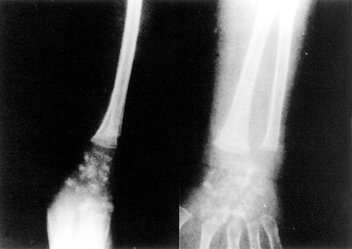
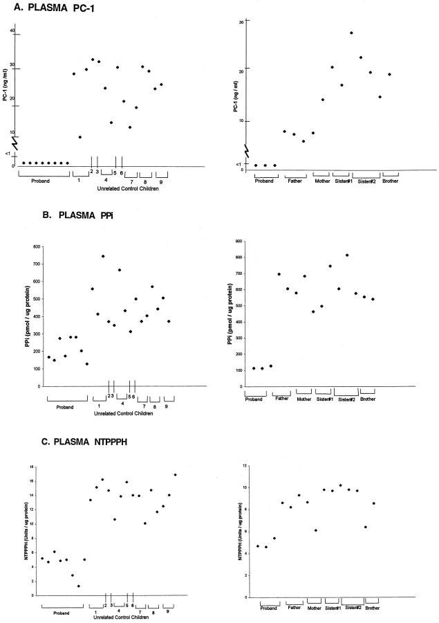
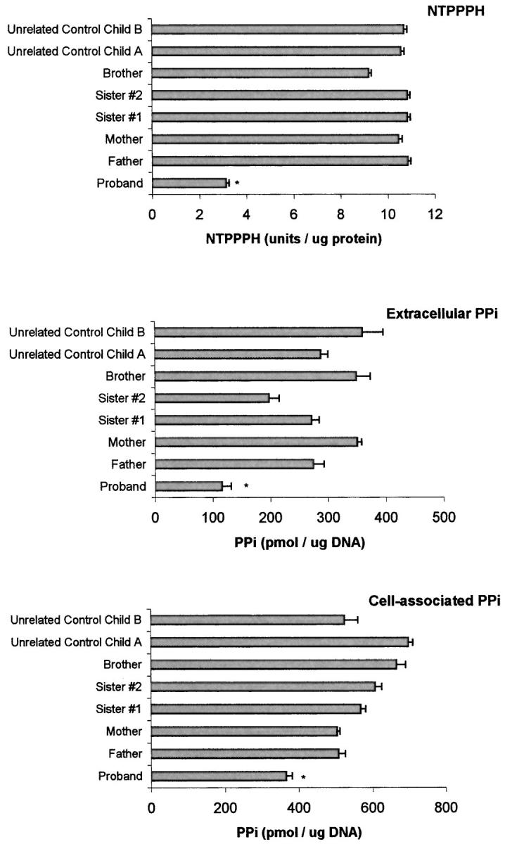
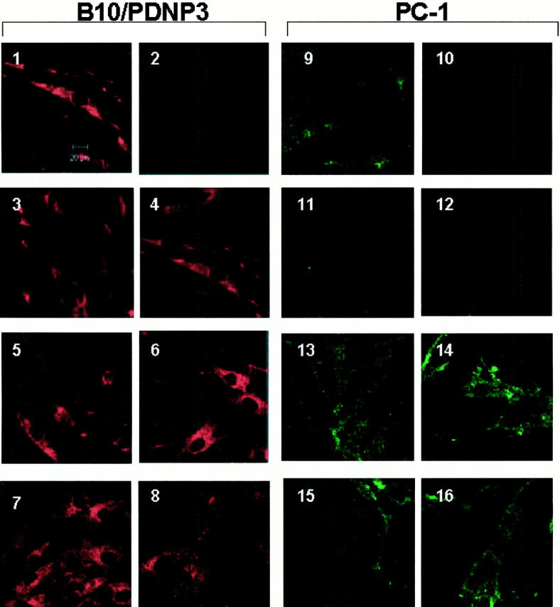
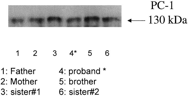
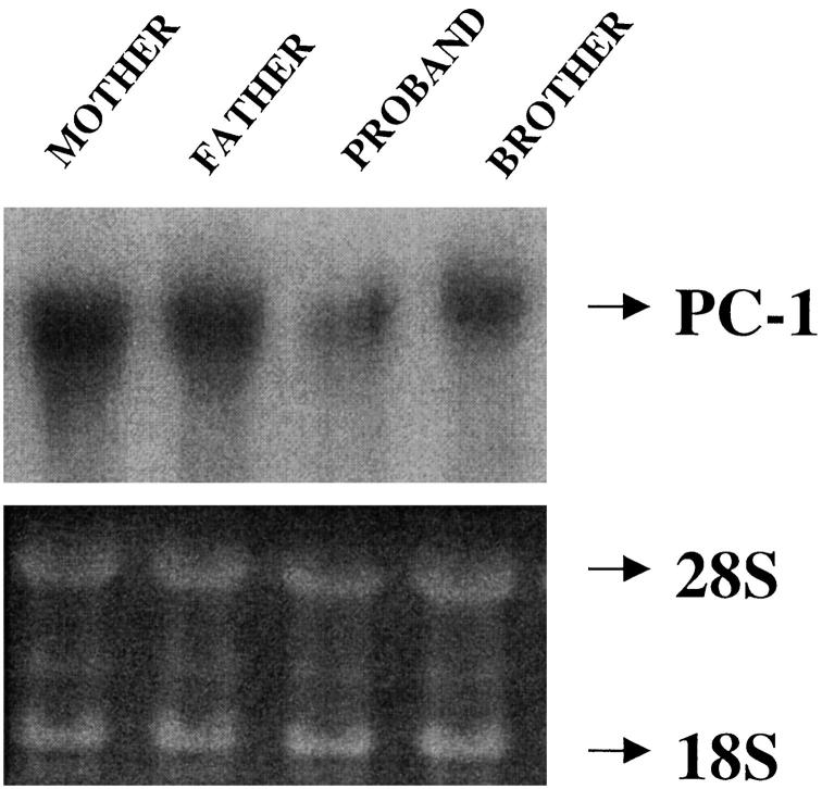
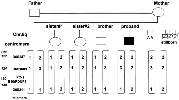
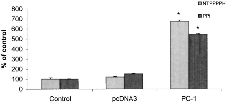
Inogranic pyrophosphate (PPi) inhibits hydroxyapatite deposition, and mice deficient in the PPi-generating nucleoside triphosphate pyrophosphohydrolase (NTPPPH) Plasma cell membrane glycoprotein-1 (PC-1) develop peri-articular and arterial calcification in early life. In idiopathic infantile arterial calcification (IIAC), hydroxyapatite deposition and smooth muscle cell (SMC) proliferation occur, sometimes associated with peri-articular calcification. Thus, we assessed PC-1 expression and PPi metabolism in a 25-month-old boy with IIAC and peri-articular calcifications. Plasma PC-1 was <1 ng/ml by enzyme-linked immunosorbent assay in the proband, but 10 to 30 ng/ml in unaffected family members and controls. PC-1 functioned to raise extracellular PPi in cultured aortic SMCs. However, PC-1 was sparse in temporal artery lesion SMCs in the proband, unlike the case for SMCs in atherosclerotic carotid artery lesions of unrelated adults. Proband plasma and explant-cultured dermal fibroblast NTPPPH and PPi were markedly decreased. The proband was heterozygous at the PC-1 locus, and sizes of PC-1 mRNA and polypeptide, and the PC-1 mRNA-coding region sequence were normal in proband fibroblasts. However, immunoreactive PC-1 protein was relatively sparse in proband fibroblasts. In conclusion, deficient extracellular PPi and a deficiency of PC-1 NTPPPH activity can be associated with human infantile arterial and peri-articular calcification, and may help explain the sharing of certain phenotypic features between some IIAC patients and PC-1-deficient mice.


Figures

Figure 1.
Figure 1.
Radiological evidence for peri-articular calcifications in the IIAC proband. Lateral (left) and PA radiographic (right) views of the right wrist of the proband at 8 months of age are shown, and revealed dense globular calcifications. Similar findings also were seen in both wrists and ankles.
Figure 2.
Figure 2.
ELISA for soluble plasma PC-1 (A), plasma PPi (B) and plasma NTPPPH (C) in the IIAC proband, unrelated control children, and unaffected family members. A: PC-1 ELISA was performed in triplicate, as described in Materials and Methods, in a blinded manner on heparinized plasma samples obtained on multiple dates from the IIAC proband (ME) and unrelated normal control children (left) and the unaffected family members (right). Studies were done on plasmas collected between 1998 to 1999. Each data point for each donor represents a different sampling date, and the dates of sampling for the proband in the left and right panels were completely different. Dates of birth for the IIAC proband and unaffected family members were: proband, January 1997; father, September 1963; mother, February 1959; sister no. 1, October 1987; sister no. 2, March 1990; and brother, December 1994. B: Plasma PPi values are indicated for the proband and unrelated control children (left) and the unaffected family members (right). For the PPi measurements, separate aliquots of heparinized plasma were prepared as described in Materials and Methods and then radiometrically measured for PPi, which was performed as described in Materials and Methods. Dates of collection were as in A. C: Plasma NTPPPH activities are indicated for the proband and unrelated control children (left) and the unaffected family members (right). For assessment of plasma NTPPPH, we studied aliquots of plasmas (collected on multiple dates, as above) that were buffered 1:1 in 0.2 mol/L Tris and 1.6 mmol/L MgCl2, pH 8.1. The figure shows the results of measurements of the specific activity of NTPPPH per μg of protein. Relative differences for results of NTPPPH activity/volume (not shown) were essentially the same.
Figure 3.
Figure 3.
Explant-cultured dermal fibroblast NTPPPH-specific activity, and intracellular and extracellular PPi levels in the IIAC proband, unaffected family members, and additional unrelated control children. Explant-cultured dermal fibroblasts were obtained and cultured from excisional dermal forearm biopsies of all patients under informed consent, as described in Materials and Methods. NTPPPH-specific activity levels and intracellular and extracellular PPi for cultured fibroblasts from each donor were measured as described in Materials and Methods. The results shown are pooled from three determinations in triplicate from each donor. *, P < 0.05 for IIAC proband compared to each unaffected family member and each control.
Figure 4.
Figure 4.
Comparison of B10/PDNP3 and PC-1 expression via laser scanning confocal microscopic images of proliferating dermal fibroblasts in culture. Explant cultured dermal fibroblasts (at 50 to 70% confluence) were analyzed at the same passage number via immunofluorescence for expression of B10/PDNP3 (panels 1–8) and PC-1 (panels 9–16), as described in Materials and Methods. As controls, cells were stained only with the secondary antibodies goat anti-mouse Alexa 488 (panel 2) and goat anti-rabbit Alexa 568 (panel 10) in the absence of the primary antibodies. Micrographs represent fibroblasts of: normal unrelated donor child (1 and 9), proband (3, 4, 11, and 12), father (5 and 13), mother (6 and 14), brother (7 and 15), sister no. 2 (8 and 16). Original magnifications, ×800 for each panel. Representative of four different studies for each individual for both B10/PDNP3 and PC-1.
Figure 5.
Figure 5.
Qualitative Western blot analysis of immunoprecipitated explant-cultured dermal fibroblast PC-1. We immunoprecipitated PC-1 from 50 μg of protein derived from donor fibroblast lysates (each from aliquots of ∼5 × 10 cells), as described in Materials and Methods. We then performed sodium dodecyl sulfate-polyacrylamide gel electrophoresis and Western blotting for PC-1 from each immunoprecipitate, as described in Materials and Methods.
Figure 6.
Figure 6.
Northern blot analysis of PC-1 expression in explant-cultured dermal fibroblasts. We isolated total RNA from explant-cultured dermal fibroblasts from patients in the IIAC kindred as indicated and we transferred RNA samples (20 μg) from formaldehyde-agarose gels to nylon membranes and UV cross-linked the membranes as described in Materials and Methods. Hybridization was performed at 42°C using human PC-1 probe (top) with specific activity >10 cpm/μg, prepared by random primer labeling, as described in Materials and Methods. Loading of RNA is shown at the bottom. Estimated size of PC-1 mRNA was 3.5 kb.
Figure 7.
Figure 7.
Schematized results of polymorphic DNA analysis for PC-1 and B10/PDNP3 linkage in the IIAC proband and surviving unaffected members of the same kindred. Possible genetic linkage of the IIAC disorder to the PC-1 locus and the neighboring B10/PDNP3 on chromosome 6q was tested by analysis of polymorphic variable-number tandem repeats around the region of interest. The markers analyzed were D6S287, D6S1009, and D6S311. The markers were amplified using fluorescence-labeled markers and their length determined using a Perkin-Elmer ABI 310 automated DNA sequence apparatus. In the diagram, the numbers 1, 2, and 3 are used to designate specific differences in lengths when using each marker (A = spontaneous abortion).
Figure 8.
Figure 8.
Immunohistochemical analysis of PC-1, B10/PDNP3, and SMC α-actin expression in the temporal artery of the IIAC proband (A), and in atherosclerotic carotid artery lesions from unrelated adults (B). For immunohistochemical analysis of artery lesion PC-1 and B10/PDNP3, formalin-fixed and paraffin-embedded tissues from the proband’s temporal artery biopsy (A) and from unrelated human adult atherosclerotic carotid endarterectomy samples (B) were sectioned and stained as described in Materials and Methods, using specific antibodies for PC-1, B10/PDNP3, and α-actin antibody for SMC staining. Negative control sections shown in A (C, F, and I) were treated by using appropriate dilutions of the normal sera of the species in which the primary antibodies were made. A: Original magnifications, ×40 (A, C, D, F, G, and I), ×100 (B, E, and H). B: Serial sections from one donor. Original magnifications, ×100 (A, D, G, and J). Serial sections from another donor. Original magnifications, ×100 (B, E, H, and K). C, F, I, and L: Higher magnifications (×250) of the bottom right portion of the corresponding sections of B, E, H, and K. For comparative purposes, trichrome-stained sections are shown in J–L of B.
Figure 9.
Figure 9.
Direct effects of PC-1 expression on extracellular PPi in cultured human aortic SMCs. Human aortic SMCs were transfected with wild-type PC-1 cDNA or empty vector, and then cell-associated NTPPPH and extracellular PPi determined at 72 hours, as described in Materials and Methods. Results are pooled from three separate experiments done in triplicate. Control SMC extracellular PPi was 284.1 ± 17.0 pmol/μg DNA. *, P < 0.05.

Similar articles

Cited by

References

    1. Schinke T, McKee MD, Kiviranta R, Karsenty G: Molecular determinants of arterial calcification. Ann Med 1998, 30:538-541 - PubMed
    1. Schinke T, McKee MD, Karsenty G: Extracellular matrix calcification: where is the action? Nat Genet 1999, 21:150-151 - PubMed
    1. Stary HC: Natural history of calcium deposits in atherosclerosis progression and regression. Zeitschrift fur Kardiologie 2000, 89(Suppl 2):S28-S35 - PubMed
    1. Bini A, Mann KG, Kudryk BJ, Schoen FJ: Noncollagenous bone matrix proteins, calcification, and thrombosis in carotid artery atherosclerosis. Arterioscler Thromb Vasc Biol 1999, 19:1852-1861 - PubMed
    1. Maayan C, Peleg O, Eyal F, Mogle P, Rosenmann E, Bar Ziv J: Idiopathic infantile arterial calcification: a case report and review of the literature. Eur J Pediatr 1984, 142:211-215 - PubMed

Publication types

MeSH terms