Age-dependent induction of congophilic neurofibrillary tau inclusions in tau transgenic mice
- PMID: 11159192
- PMCID: PMC1850303
- DOI: 10.1016/S0002-9440(10)63997-1
Age-dependent induction of congophilic neurofibrillary tau inclusions in tau transgenic mice
Abstract
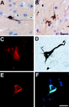
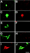
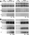
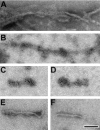
Intraneuronal filamentous tau inclusions such as neurofibrillary tangles (NFTs) are neuropathological hallmarks of Alzheimer's disease (AD) and related sporadic and familial tauopathies. NFTs identical to those found in AD brains have also been detected in the hippocampus and entorhinal cortex of cognitively normal individuals as they age. To recapitulate age-induced NFT formation in a mouse model, we examined 12- to 24-month-old transgenic (Tg) mice overexpressing the smallest human brain tau isoform. These Tg mice develop congophilic tau inclusions in several brain regions including the hippocampus, amygdala, and entorhinal cortex. NFT-like inclusions were first detected in Tg mice at 18 to 20 months of age and they were detected by histochemical dyes that bind specifically to crossed beta-pleated sheet structures (eg, Congo red, Thioflavin S). Moreover, ultrastructurally these lesions contained straight tau filaments comprised of both mouse and human tau proteins but not other cytoskeletal proteins (eg, neurofilaments, microtubules). Isolated tau filaments were also recovered from detergent-insoluble tau fractions and insoluble tau proteins accumulated in brain in an age-dependent manner. Thus, overexpression of the smallest human brain tau isoform resulted in late onset and age-dependent formation of congophilic tau inclusions with properties similar to those in the tangles of human tauopathies, thereby implicating aging in the pathogenesis of fibrous tau inclusions.
Figures


Similar articles
-
Tau filament formation and associative memory deficit in aged mice expressing mutant (R406W) human tau.Proc Natl Acad Sci U S A. 2002 Oct 15;99(21):13896-901. doi: 10.1073/pnas.202205599. Epub 2002 Oct 4. Proc Natl Acad Sci U S A. 2002. PMID: 12368474 Free PMC article.
-
Formation of filamentous tau aggregations in transgenic mice expressing V337M human tau.Neurobiol Dis. 2001 Dec;8(6):1036-45. doi: 10.1006/nbdi.2001.0439. Neurobiol Dis. 2001. PMID: 11741399
-
A novel transgenic mouse expressing double mutant tau driven by its natural promoter exhibits tauopathy characteristics.Exp Neurol. 2008 Jul;212(1):71-84. doi: 10.1016/j.expneurol.2008.03.007. Epub 2008 Mar 21. Exp Neurol. 2008. PMID: 18490011
-
[Analysis of mouse model exhibiting neurofibrillary changes].Rinsho Shinkeigaku. 2001 Dec;41(12):1111-2. Rinsho Shinkeigaku. 2001. PMID: 12235811 Review. Japanese.
-
[Significance of tau in the development of Alzheimer's disease].Brain Nerve. 2010 Jul;62(7):701-8. Brain Nerve. 2010. PMID: 20675874 Review. Japanese.
Cited by
-
Accelerated tau aggregation, apoptosis and neurological dysfunction caused by chronic oral administration of aluminum in a mouse model of tauopathies.Brain Pathol. 2013 Nov;23(6):633-44. doi: 10.1111/bpa.12059. Epub 2013 May 3. Brain Pathol. 2013. PMID: 23574527 Free PMC article.
-
What can rodent models tell us about cognitive decline in Alzheimer's disease?Mol Neurobiol. 2003 Jun;27(3):249-76. doi: 10.1385/MN:27:3:249. Mol Neurobiol. 2003. PMID: 12845151 Review.
-
Microtubule-stabilizing agents as potential therapeutics for neurodegenerative disease.Bioorg Med Chem. 2014 Sep 15;22(18):5040-9. doi: 10.1016/j.bmc.2013.12.046. Epub 2013 Dec 30. Bioorg Med Chem. 2014. PMID: 24433963 Free PMC article. Review.
-
What we can learn from animal models about cerebral multi-morbidity.Alzheimers Res Ther. 2015 Jan 29;7(1):11. doi: 10.1186/s13195-015-0097-2. eCollection 2015. Alzheimers Res Ther. 2015. PMID: 25810783 Free PMC article.
-
The Role of Protein Misfolding and Tau Oligomers (TauOs) in Alzheimer's Disease (AD).Int J Mol Sci. 2019 Sep 20;20(19):4661. doi: 10.3390/ijms20194661. Int J Mol Sci. 2019. PMID: 31547024 Free PMC article.
References
-
- Hong M, Trojanowski JQ, Lee VMY: Tau-based neurofibrillary lesions. Clark CM Trojanowski JQ eds. Neurodegenerative Dementias: Clinical Features and Pathological Mechanisms. 2000, :pp 161-176 McGraw Hill, New York
-
- Lee VMY, Trojanowski JQ: Distinct tau gene mutations induce specific dysfunctions/toxic properties in tau proteins associated with specific FTDP-17 phenotypes. Lee VMY Trojanowski JQ Buee L Christen Y eds. Fatal Attractions: Protein Aggregates in Neurodegenerative Diseases. 2000, :pp 87-104 IPSEN Foundation, Paris
-
- Hof PR, Bouras C, Morrison JH: Cortical Neuropathology In Aging And Dementing Disorders: Neuronal Typology, Connectivity And Selective Vulnerability. Peters A Morrison JH eds. Cerebral Cortex 14. 1999, :pp 175-276 Kluwer Academic/Plenum Publishers, New York
-
- Andreadis A, Brown WM, Kosik KS: Structure and novel exons of the human tau gene. Biochemistry 1992, 31:10626-10633 - PubMed
-
- Goedert M, Spillantini MG, Jakes R, Rutherford D, Crowther RA: Multiple isoforms of human microtubule-associated protein tau: sequences and localization in neurofibrillary tangles of Alzheimer’s disease. Neuron 1989, 3:519-526 - PubMed
Publication types
MeSH terms
Substances
LinkOut - more resources
Full Text Sources
Other Literature Sources
Medical
Molecular Biology Databases
Miscellaneous

