Basement membrane pores in human bronchial epithelium: a conduit for infiltrating cells?
- PMID: 11159204
- PMCID: PMC1850329
- DOI: 10.1016/S0002-9440(10)64009-6
Basement membrane pores in human bronchial epithelium: a conduit for infiltrating cells?
Abstract
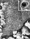
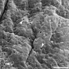
This study reports the presence of oval-shaped pores in the basement membrane of the human bronchial airway that may be used as conduits for immune cells to traffic between the epithelial and mesenchymal compartments. Human bronchial mucosa collected after surgery was stripped of epithelial cells without damaging the basement membrane. Both scanning and transmission electron microscopy showed oval-shaped pores 0.75 to 3.85 microm in diameter in the bronchial basement membrane at a density of 863 pores/mm2. Transmission electron microscopy showed that the pores spanned the full depth of the basement membrane, with a concentration of collagen-like fibers at the lateral edges of the pore. Infiltrating cells apparently moved through the pores, both in the presence and absence of the epithelium. Taken together, these results suggest that immune cells use basement membrane pores as predefined routes to move between the epithelial and mesenchymal compartments without disruption of the basement membrane. As a persistent feature of the basement membrane, pores could facilitate inflammatory cell access to the epithelium and greatly increase the frequency of intercellular contact between trafficking cells.
Figures


Similar articles
-
Distribution of basement membrane pores in bronchus revealed by microscopy following epithelial removal.J Struct Biol. 2002 Sep;139(3):137-45. doi: 10.1016/s1047-8477(02)00589-0. J Struct Biol. 2002. PMID: 12457843
-
[Mechanisms of bronchial self-cleaning. Surface studies on the bronchial mucosa, bronchial basement membrane and subepithelial connective tissue. A scanning electronmicroscopic study].Fortschr Med. 1977 Dec 1;95(45):2691-6. Fortschr Med. 1977. PMID: 924334 German.
-
Prenatal and postnatal changes of the human tonsillar crypt epithelium.Acta Otolaryngol Suppl. 1996;523:28-33. Acta Otolaryngol Suppl. 1996. PMID: 9082803
-
Pathology of asthma.Br Med Bull. 1992 Jan;48(1):23-39. doi: 10.1093/oxfordjournals.bmb.a072537. Br Med Bull. 1992. PMID: 1617392 Review.
-
Airway morphology: epithelium/basement membrane.Am J Respir Crit Care Med. 1994 Nov;150(5 Pt 2):S14-7. doi: 10.1164/ajrccm/150.5_Pt_2.S14. Am J Respir Crit Care Med. 1994. PMID: 7952583 Review.
Cited by
-
Progress in Vocal Fold Regenerative Biomaterials: An Immunological Perspective.Adv Nanobiomed Res. 2022 Feb;2(2):2100119. doi: 10.1002/anbr.202100119. Epub 2021 Dec 18. Adv Nanobiomed Res. 2022. PMID: 35434718 Free PMC article.
-
Dual sources of vitronectin in the human lower urinary tract: synthesis by urothelium vs. extravasation from the bloodstream.Am J Physiol Renal Physiol. 2011 Feb;300(2):F475-87. doi: 10.1152/ajprenal.00407.2010. Epub 2010 Nov 3. Am J Physiol Renal Physiol. 2011. PMID: 21048021 Free PMC article.
-
Mimicking the Natural Basement Membrane for Advanced Tissue Engineering.Biomacromolecules. 2022 Aug 8;23(8):3081-3103. doi: 10.1021/acs.biomac.2c00402. Epub 2022 Jul 15. Biomacromolecules. 2022. PMID: 35839343 Free PMC article. Review.
-
Extracellular matrix mediates moruloid-blastuloid morphodynamics in malignant ovarian spheroids.Life Sci Alliance. 2021 Aug 10;4(10):e202000942. doi: 10.26508/lsa.202000942. Print 2021 Oct. Life Sci Alliance. 2021. PMID: 34376568 Free PMC article.
-
A Fresh Glimpse into Cartilage Immune Privilege.Cartilage. 2022 Dec;13(4):119-132. doi: 10.1177/19476035221126349. Epub 2022 Oct 15. Cartilage. 2022. PMID: 36250484 Free PMC article. Review.
References
-
- Carolan EJ, Mower DA, Casale TB: Cytokine-induced neutrophil transepithelial migration is dependent upon epithelial orientation. Am J Respir Cell Mol Biol 1997, 17:727-732 - PubMed
-
- Liu LX, Zuurbier AEM, Mul FPJ, Verhoeven AJ, Lutter R, Knol EF, Roos D: Triple role of platelet-activating factor in eosinophil migration across monolayers of lung epithelial cells: eosinophil chemoattractant and priming agent and epithelial cell activator. J Immunol 1998, 161:3064-3070 - PubMed
-
- Frew AJ, St Pierre J, Teran LM, Trefilieff A, Madden J, Peroni D, Bodey KM, Walls AF, Howarth PH, Carroll MP, Holgate ST: Cellular and mediator responses twenty-four hours after local endobronchial allergen challenge of asthmatic airways. J Allergy Clin Immunol 1996, 98:133-143 - PubMed
-
- Shaw SK, Hermanowski-Vosatka A, Shibahara T, McCormick BA, Parkos CA, Carlson SL, Ebert EC, Brenner MB, Madara JL: Migration of intestinal intraepithelial lymphocytes into a polarised epithelial monolayer. Am J Physiol 1998, 38:G584-G591 - PubMed
-
- Holt PG, Haining S, Nelson DJ, Sedgwick JD: Origin and steady-state turnover of class II MHC-bearing dendritic cells in the epithelium of conducting airways. J Immunol 1994, 153:256-261 - PubMed
Publication types
MeSH terms
LinkOut - more resources
Full Text Sources
Research Materials

