

Loss of heterozygosity on chromosome 11q22-23 in melanoma is associated with retention of the insertion polymorphism in the matrix metalloproteinase-1 promoter
- PMID: 11159206
- PMCID: PMC1850328
- DOI: 10.1016/S0002-9440(10)64011-4
Loss of heterozygosity on chromosome 11q22-23 in melanoma is associated with retention of the insertion polymorphism in the matrix metalloproteinase-1 promoter
Abstract
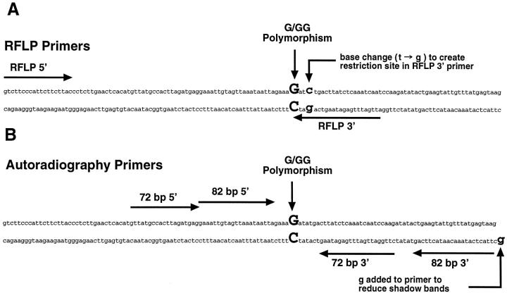
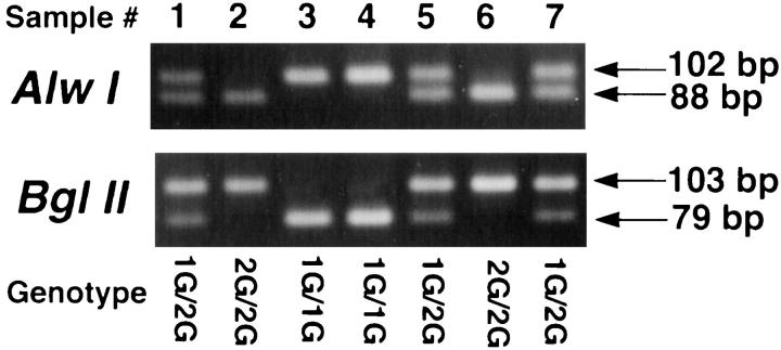
Matrix metalloproteinase-1 (MMP-1, collagenase-1), which degrades interstitial collagen, is expressed at high levels by some tumor cells and is thought to enhance their invasiveness and metastatic potential. We recently described a common single nucleotide insertion polymorphism (2G allele) at -1,607 bp in the promoter of the MMP-1 gene that creates a binding site for the ETS family of transcription factors, and that is associated with enhanced transcription of this gene and increased enzyme activity. Allelic loss at the MMP-1 locus on chromosome 11 occurs in many tumors including melanoma, an invasive and aggressive cancer. We hypothesized that although loss of either the 1G or 2G allele from 1G/2G heterozygotes is random, retention of the transcriptionally more active 2G allele would favor tumor invasion and metastasis. As a result, a higher proportion of metastases would contain the 2G genotype than the 1G genotype. We report here the development of quantitative methods for assessing allelic loss at the MMP-1 locus, and demonstrate that 83% of the metastatic melanomas with loss of heterozygosity at this locus retained the 2G allele. This supports the hypothesis that retention of the 2G allele favors tumor invasion and metastasis in melanoma.
Figures


Similar articles
-
Polymorphisms of the promoter regions of matrix metalloproteinases genes MMP-1 and MMP-9 in breast cancer.Breast Cancer Res Treat. 2006 Jan;95(1):65-72. doi: 10.1007/s10549-005-9042-6. Epub 2005 Nov 3. Breast Cancer Res Treat. 2006. PMID: 16267613
-
The influence of interstitial collagenase-1 genotype polymorphism on colorectal cancer risk in Iranian population.Cancer Invest. 2008 Oct;26(8):836-42. doi: 10.1080/07357900801953204. Cancer Invest. 2008. PMID: 18798060
-
A single nucleotide polymorphism in the matrix metalloproteinase-1 promoter in endometrial carcinomas.Jpn J Cancer Res. 2000 Jun;91(6):612-5. doi: 10.1111/j.1349-7006.2000.tb00989.x. Jpn J Cancer Res. 2000. PMID: 10874213 Free PMC article.
-
Effect of matrix metalloproteinase-1 promoter genotype on interleukin-1beta-induced matrix metalloproteinase-1 production in human periodontal ligament cells.J Periodontal Res. 2010 Feb;45(1):109-15. doi: 10.1111/j.1600-0765.2009.01208.x. Epub 2009 Jul 8. J Periodontal Res. 2010. PMID: 19602108
-
An association between the matrix metalloproteinase 1 promoter gene polymorphism and lymphnode metastasis in breast cancer.J Exp Clin Cancer Res. 2004 Mar;23(1):121-5. J Exp Clin Cancer Res. 2004. PMID: 15149160
Cited by
-
How matrix metalloproteinases regulate cell behavior.Annu Rev Cell Dev Biol. 2001;17:463-516. doi: 10.1146/annurev.cellbio.17.1.463. Annu Rev Cell Dev Biol. 2001. PMID: 11687497 Free PMC article. Review.
-
The BRAF(V600E) inhibitor, PLX4032, increases type I collagen synthesis in melanoma cells.Matrix Biol. 2015 Oct;48:66-77. doi: 10.1016/j.matbio.2015.05.007. Epub 2015 May 16. Matrix Biol. 2015. PMID: 25989506 Free PMC article.
-
The collagenase-1 (MMP-1) gene promoter polymorphism - 1607/2G is associated with favourable prognosis in patients with colorectal cancer.Br J Cancer. 2007 Mar 12;96(5):783-92. doi: 10.1038/sj.bjc.6603630. Epub 2007 Feb 20. Br J Cancer. 2007. PMID: 17311017 Free PMC article.
-
Identification of a cigarette smoke-responsive region in the distal MMP-1 promoter.Am J Respir Cell Mol Biol. 2009 Jan;40(1):4-12. doi: 10.1165/rcmb.2007-0310OC. Epub 2008 Jul 10. Am J Respir Cell Mol Biol. 2009. PMID: 18617682 Free PMC article.
-
Differential mechanisms of tumor progression in clones from a single heterogeneous human melanoma.J Cell Physiol. 2013 Apr;228(4):773-80. doi: 10.1002/jcp.24225. J Cell Physiol. 2013. PMID: 23001823 Free PMC article.
References
-
- Vincenti MP, White LA, Schroen DJ, Benbow U, Brinckerhoff CE: Regulating expression of the gene for matrix metalloproteinase-1 (collagenase): mechanisms that control enzyme activity, transcription and mRNA stability. Crit Rev Eucaryotic Gene Express 1996, 6:391-411 - PubMed
-
- Borden P, Heller RA: Transcriptional control of matrix metalloproteinases and the tissue inhibitors of matrix metalloproteinases. Crit Rev Eukaryotic Gene Express 1997, 7:159-178 - PubMed
-
- Nagase H, Woessner JF, Jr: Matrix metalloproteinases. J Biol Chem 1999, 274:21491-21492 - PubMed
-
- Nomura T, Ishii A, Oishi Y, Kohma H, Hara K: Tissue inhibitor of metalloproteinases level and collagenase activity in gingival crevicular fluid: the relevance to periodontal disease. Oral Dis 1998, 4:231-240 - PubMed
Publication types
MeSH terms
Substances
Grants and funding
LinkOut - more resources
Full Text Sources
Other Literature Sources
Medical
Research Materials
