Activation of potassium conductance by ophiopogonin-D in acutely dissociated rat paratracheal neurones
- PMID: 11159695
- PMCID: PMC1572569
- DOI: 10.1038/sj.bjp.0703818
Activation of potassium conductance by ophiopogonin-D in acutely dissociated rat paratracheal neurones
Abstract
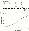
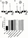
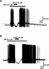
1. The effect of ophiopogonin-D (OP-D), a steroidal glycoside and an active component of Bakumondo-to, a Chinese herbal antitussive, on neurones acutely dissociated from paratracheal ganglia of 2-week-old Wistar rats was investigated using the nystatin-perforated patch recording configuration. 2. Under current-clamp conditions, OP-D (10 microM) hyperpolarized the paratracheal neurones from a resting membrane potential of -65.7 to -73.5 mV. 3. At the concentration of 1 microM and above, OP-D concentration-dependently activated an outward current accompanied by an increase in the membrane conductance under voltage-clamp conditions at a holding potential of -40 mV. 4. The reversal potential of the OP-D-induced current (I(OP-D)) was -79.4 mV, which is close to the K(+) equilibrium potential of -86.4 mV. The changes in the reversal potential for a 10 fold change in extracellular K(+) concentration was 53.1 mV, indicating that the current was carried by K(+). 5. The I(OP-D) was blocked by an extracellular application of 1 mM Ba2+ by 59.0%, but other K(+) channel blockers, including 4-aminopyridine (3 mM), apamin (1 microM), charybdotoxin (0.3 microM), glibenclamide (1 microM), tolbutamide (0.3 mM) and tetraethylammonium (10 mM), did not inhibit the I(OP-D). 6. OP-D also inhibited the ACh- and bradykinin-induced depolarizing responses which were accompanied with firing of action potentials. 7. The results suggest that OP-D may be of benefit in reducing the excitability of airway parasympathetic ganglion neurones and consequently cholinergic control of airway function and further, that the hyperpolarizing effect of OP-D on paratracheal neurones via an activation of K(+) channels might explain a part of mechanisms of the antitussive action of the agent.
Figures


Similar articles
-
Bradykinin activates airway parasympathetic ganglion neurons by inhibiting M-currents.Neuroscience. 2001;105(3):785-91. doi: 10.1016/s0306-4522(01)00211-1. Neuroscience. 2001. PMID: 11516842
-
Nitric oxide modulation of calcium-activated potassium channels in postganglionic neurones of avian cultured ciliary ganglia.Br J Pharmacol. 1993 Nov;110(3):995-1002. doi: 10.1111/j.1476-5381.1993.tb13912.x. Br J Pharmacol. 1993. PMID: 7905346 Free PMC article.
-
Potassium currents operated by thyrotrophin-releasing hormone in dissociated CA1 pyramidal neurones of rat hippocampus.J Physiol. 1993 Dec;472:689-710. doi: 10.1113/jphysiol.1993.sp019967. J Physiol. 1993. PMID: 8145166 Free PMC article.
-
A Kv3-like persistent, outwardly rectifying, Cs+-permeable, K+ current in rat subthalamic nucleus neurones.J Physiol. 2000 Sep 15;527 Pt 3(Pt 3):493-506. doi: 10.1111/j.1469-7793.2000.t01-1-00493.x. J Physiol. 2000. PMID: 10990536 Free PMC article.
-
Resting membrane potential and potassium currents in cultured parasympathetic neurones from rat intracardiac ganglia.J Physiol. 1992 Oct;456:405-24. doi: 10.1113/jphysiol.1992.sp019343. J Physiol. 1992. PMID: 1284080 Free PMC article.
Cited by
-
Quality Evaluation of Ophiopogonis Radix from Two Different Producing Areas.Molecules. 2019 Sep 4;24(18):3220. doi: 10.3390/molecules24183220. Molecules. 2019. PMID: 31487946 Free PMC article.
-
Alpha 1-adrenoceptor-activated cation currents in neurones acutely isolated from rat cardiac parasympathetic ganglia.J Physiol. 2003 Apr 1;548(Pt 1):111-20. doi: 10.1113/jphysiol.2002.033100. Epub 2003 Feb 21. J Physiol. 2003. PMID: 12598585 Free PMC article.
-
Liriopogons (Genera Ophiopogon and Liriope, Asparagaceae): A Critical Review of the Phytochemical and Pharmacological Research.Front Pharmacol. 2021 Dec 3;12:769929. doi: 10.3389/fphar.2021.769929. eCollection 2021. Front Pharmacol. 2021. PMID: 34925027 Free PMC article. Review.
-
A metabonomic study of cardioprotection of ginsenosides, schizandrin, and ophiopogonin D against acute myocardial infarction in rats.BMC Complement Altern Med. 2014 Sep 23;14:350. doi: 10.1186/1472-6882-14-350. BMC Complement Altern Med. 2014. PMID: 25249156 Free PMC article.
-
An electrophysiological study of muscarinic and nicotinic receptors of rat paratracheal ganglion neurons and their inhibition by Z-338.Br J Pharmacol. 2002 Mar;135(6):1403-14. doi: 10.1038/sj.bjp.0704610. Br J Pharmacol. 2002. PMID: 11906953 Free PMC article.
References
-
- AIBARA K., AKAIKE N. Acetylcholine-activated ionic currents in isolated paratracheal ganglion cells of the rat. Brain Res. 1991;558:20–26. - PubMed
-
- ASANO T., MARUYAMA T., HIRAI Y., SHOJI J. Comparative studies on the constituents of ophiopogonins tuber and its congeners. VIII. Studies on the glycosides of the subterranean part of Ophiopogon japonicus Ker-Gawler cv. Nanus. (2) Chem. Pharm. Bull. 1993;41:566–570. - PubMed
-
- BAKER D.G., MCDONALD D.M., BASBAUM C.B., MITCHELL R.A. The architecture of nerves and ganglia of the ferret trachea as revealed by acetylcholinesterase histochemistry. J. Comp Neurol. 1986;246:513–526. - PubMed
MeSH terms
Substances
LinkOut - more resources
Full Text Sources
Miscellaneous

