. 2001 Feb 1;15(3):294-303.
doi: 10.1101/gad.865401.

mRNA 5'-leader trans-splicing in the chordates

Affiliations

mRNA 5'-leader trans-splicing in the chordates

A E Vandenberghe et al. Genes Dev. .

Abstract

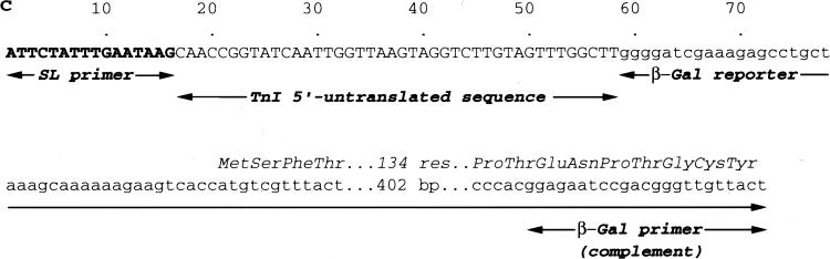
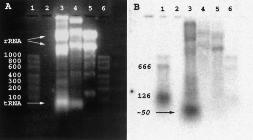
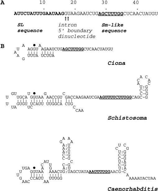
We report the discovery of mRNA 5'-leader trans-splicing (SL trans-splicing) in the chordates. In the ascidian protochordate Ciona intestinalis, the mRNAs of at least seven genes undergo trans-splicing of a 16-nucleotide 5'-leader apparently derived from a 46-nucleotide RNA that shares features with previously characterized splice donor SL RNAs. SL trans-splicing was known previously to occur in several protist and metazoan phyla, however, this is the first report of SL trans-splicing within the deuterostome division of the metazoa. SL trans-splicing is not known to occur in the vertebrates. However, because ascidians are primitive chordates related to vertebrate ancestors, our findings raise the possibility of ancestral SL trans-splicing in the vertebrate lineage.


Figures

Figure 1
Figure 1
A common 5′-sequence in several Ciona mRNAs. (A) A common 16-nucleotide sequence (bold) at the 5′-end of TnI mRNA, determined by 5′-RACE, and three additional Ciona mRNAs found by BLAST (Altschul et al. 1997) search of the GenBank database with the TnI 5′-sequence. The first 35 nucleotides of each mRNA are shown; dots indicate identity with the TnI mRNA sequence. Ci-zeta mRNA (GenBank AJ002142) obtained from ovary, encodes a proteasome subunit (Marino et al. 1999). CiMDFa mRNA (Genbank U80079) is expressed in larval tail muscle and adult body-wall muscle (Meedel et al. 1997). Cs-Endo-1 mRNA (GenBank AB024925) is a maternal mRNA from oocytes of Ciona savignyi (Imai et al. 1999). (B) RT–PCR amplification of TnI mRNAs with TnI-specific leftward priming using the SL primer for rightward priming. (Lane 1) Size markers (pBR322 HaeIII digest; top bands 587–434 bp, middle bands 267–184 bp, bottom bands ≤124 bp). (Lanes 2,4) Heart RNA template; (lanes 3,5) body-wall muscle RNA template; (lane 6) no RNA. Reverse transcriptase was omitted in lanes 4 and 5. Products of 381 bp (heart) and 240 bp (body-wall muscle) are expected; the size difference reflects tissue-specific alternative RNA splicing (MacLean et al. 1997).
Figure 2
Figure 2
Additional mRNAs containing the SL sequence. (A) Amplification of multiple mRNAs by RT–PCR with SL and oligo(dT)-based primers. (Lane 1) Size markers [λ DNA, HindIII and EcoRI digest; in kilobases, from top to bottom, 21, 5.1/5.0, 4.2, 3.5, 2.0/1.9, 1.6, and (not visible) 1.4, 0.95, 0.83, and 0.5]. (Lanes 2,3) Body-wall muscle RNA template; (lanes 4,5) heart RNA template; (lane 6) no RNA. Reverse transcriptase was omitted in lanes 3 and 5. (B) 5′-untranslated sequences of three apparently complete mRNAs (GenBank AF237689–-AF237691) recovered by cloning DNA from the 1.0 and 0.8-kb bands in A, lane 2. The SL sequence is bolded; not shown is the BamHI site engineered at the 5′-end of the SL primer. The overlined ATG codons initiate ORFs of the lengths indicated, each terminated by a TAA stop codon and followed by an apparently complete 3′-untranslated sequence. In-frame stop codons within the 5′-untranslated sequences are underlined. The mRNA 1 ORF encodes a protein resembling HR-29 (Takagi et al. 1993), a myofibrillar protein from body wall muscle of the ascidian Halocynthia roretzi (46% identity over 207 aligned residues). The mRNA 2 ORF encodes a novel protein containing 17 PTDAVTL repeats resembling the mucin heptad [PTE(E/V)(P/T)TV] repeats of mammalian zonadhesins (Gao and Garbers 1998). The mRNA 3 ORF encodes a protein resembling vertebrate 27-kd heat-shock protein, hsp27 (Cooper and Uoshima 1994) (42% identity over 177 aligned residues).
Figure 3
Figure 3
Comparison of TnI mRNA 5′-sequence and corresponding genomic DNA. The first 35 nucleotides of the mRNA are shown; the SL sequence is bolded and a vertical line marks its junction with the rest of the 5′-untranslated sequence. The sequences of two genomic DNA alleles, originating from Atlantic (A allele) or Pacific (P allele) coast animals, are shown. (The mRNA sequence derives from Atlantic coast animals.) Dots in the genomic DNA sequence show identity with the mRNA sequence (except at the left ends, where they signify additional upstream DNA). Differences between the A and P alleles are shown in lower case; a 4-base deletion in the P allele is shown by dashes. The AG dinucleotide present in the genomic DNA at the point of mRNA/genomic DNA sequence divergence is indicated by asterisks adjacent to the vertical line. A third asterisk marks the A residue in the branch point consensus sequence YRCTRAY.
Figure 4
Figure 4
In vivo expression and SL trans-splicing of a chimeric TnI/β-gal mRNA from a TnI/β-gal gene construct lacking the SL sequence. (A) Expression of β-gal, revealed by X-Gal staining (blue), in tail muscle of embryos 12 h following introduction of CiTnILacZ(−1.5) DNA into zygotes by electroporation. (B) RT–PCR amplification of β-gal mRNA with β-gal-specific leftward priming, and rightward priming with the SL primer. (Lane 1) Size markers (MBI Fermentas ladder mix, 10–0.1 kb; top visible band, 3 kb, bottom visible band 500 bp). (Lanes 2,3) RT–PCR products from two different batches of transfected embryos. The template in lane 4 was tRNA, used as a carrier in embryo RNA isolations. A 550-bp product, the size predicted for SL-ended β-gal mRNA, was produced from both embryo batches. [Production of this product required the presence of both primers and did not occur when CiTnILacZ(−1.5) plasmid DNA was used as the amplification template. An additional product of ∼400 bp seen in lanes 2 and 3 required only the β-gal-specific primer and was produced in control amplifications of CiTnILacZ(−1.5) plasmid DNA; it apparently results from rightward mis-priming by the β-gal-specific primer upstream of its normal leftward priming site.] (C) DNA sequence of 550-bp RT–PCR product. The 550-bp product (as in B) was recovered and sequenced using the SL primer (right) and β-gal-specific primer (left). The leftward sequence confirmed the presence of the SL primer (bold) immediately upstream of TnI mRNA nucleotide 17. Not shown is the BamHI site engineered at the 5′-end of the SL primer. Sequences deriving from the β-gal reporter gene are shown in lower case.
Figure 4
Figure 4
In vivo expression and SL trans-splicing of a chimeric TnI/β-gal mRNA from a TnI/β-gal gene construct lacking the SL sequence. (A) Expression of β-gal, revealed by X-Gal staining (blue), in tail muscle of embryos 12 h following introduction of CiTnILacZ(−1.5) DNA into zygotes by electroporation. (B) RT–PCR amplification of β-gal mRNA with β-gal-specific leftward priming, and rightward priming with the SL primer. (Lane 1) Size markers (MBI Fermentas ladder mix, 10–0.1 kb; top visible band, 3 kb, bottom visible band 500 bp). (Lanes 2,3) RT–PCR products from two different batches of transfected embryos. The template in lane 4 was tRNA, used as a carrier in embryo RNA isolations. A 550-bp product, the size predicted for SL-ended β-gal mRNA, was produced from both embryo batches. [Production of this product required the presence of both primers and did not occur when CiTnILacZ(−1.5) plasmid DNA was used as the amplification template. An additional product of ∼400 bp seen in lanes 2 and 3 required only the β-gal-specific primer and was produced in control amplifications of CiTnILacZ(−1.5) plasmid DNA; it apparently results from rightward mis-priming by the β-gal-specific primer upstream of its normal leftward priming site.] (C) DNA sequence of 550-bp RT–PCR product. The 550-bp product (as in B) was recovered and sequenced using the SL primer (right) and β-gal-specific primer (left). The leftward sequence confirmed the presence of the SL primer (bold) immediately upstream of TnI mRNA nucleotide 17. Not shown is the BamHI site engineered at the 5′-end of the SL primer. Sequences deriving from the β-gal reporter gene are shown in lower case.
Figure 5
Figure 5
Northern blot detection of Ciona SL RNA. (A) Fluorescence of ethidium bromide stained gel. (B) Autoradiography following transfer to nylon membrane and hybridization with a 5′-32P-labeled oligonucleotide complementary to the SL sequence. (Lanes 1,6) An RNA marker set (100–1000 nucleotide sizes indicated), to which has been added either 50 ng (lane 1) or 5 ng (lane 6) each of 126-nucleotide, and 666-nucleotide SL-containing in vitro transcripts of a plasmid encoding mRNA 3 (see Fig 2). (Lane 2) Blank; (lane 3) Ciona body-wall muscle RNA (not salt precipitated); (lane 4) quail muscle RNA (not salt precipitated); (lane 5) Ciona body-wall muscle RNA (salt precipitated). Large and small subunit rRNA and tRNA bands are indicated in A. Because the samples had not been salt precipitated, lanes 3 and 4 also contain genomic DNA (near sample wells).
Figure 6
Figure 6
Sequence of Ciona SL RNA and predicted secondary structure comparison with SL RNAs of the flatworm Schistosoma and nematode Caenorhabditis. (A) Sequence of the 46-nucleotide Ciona SL RNA. 3′-poly(A), added before amplification and cloning, has been removed from the sequence; the original RNA may have contained one or more A residues at the 3′-end. The SL sequence is bolded and the Sm-like sequence is underlined and bolded [the canonical Sm consensus is RA(U)nGR]. Arrows mark the intron 5′-boundary GU dinucleotide. (B) Predicted secondary structures. The Ciona SL RNA structure was generated by mfold 3.0 (Mathews et al. 1999) and the Schistosoma and Caenorhabditis structures are from Davis (1996); in all structures the Sm-like sequence (bold, underlined) was constrained to be single stranded. In terms of ΔG, the Ciona structure shown was among the top two or three generated by calculations based on 22°C or 37°C and was within 1 kcal/mole of the optimal solution. Three-H-bond base pairs (i.e., G–C) are indicated by lines, and 2-H-bond base pairs (i.e., A–U and G–U) are indicated by paired dots. A single large dot marks the first G of the intron moiety.

References

    1. Agabian N. Trans splicing of nuclear pre-mRNAs. Cell. 1990;61:1157–1160. - PubMed
    1. Akopian AN, Okuse K, Souslova V, England S, Ogata N, Wood JN. Trans-splicing of a voltage-gated sodium channel is regulated by nerve growth factor. FEBS Lett. 1999;445:177–182. - PubMed
    1. Altschul SF, Madden TL, Schaffer AA, Zhang J, Zhang Z, Miller W, Lipman DJ. Gapped BLAST and PSI-BLAST: A new generation of protein database search programs. Nucleic Acids Res. 1997;25:3389–3402. - PMC - PubMed
    1. Ausoni S, Campione M, Picard A, Moretti P, Vitadello M, De Nardi C, Schiaffino S. Structure and regulation of the mouse cardiac troponin I gene. J Biol Chem. 1994;269:339–346. - PubMed
    1. Baldwin AS, Jr, Kittler EL, Emerson CP., Jr Structure, evolution, and regulation of a fast skeletal muscle troponin I gene. Proc Natl Acad Sci. 1985;82:8080–8084. - PMC - PubMed

Publication types

LinkOut - more resources