. 2001 Feb 1;15(3):340-51.
doi: 10.1101/gad.861401.

Transcriptional regulation of the Drosophila gene zen by competing Smad and Brinker inputs

Affiliations

Transcriptional regulation of the Drosophila gene zen by competing Smad and Brinker inputs

C Rushlow et al. Genes Dev. .

Abstract

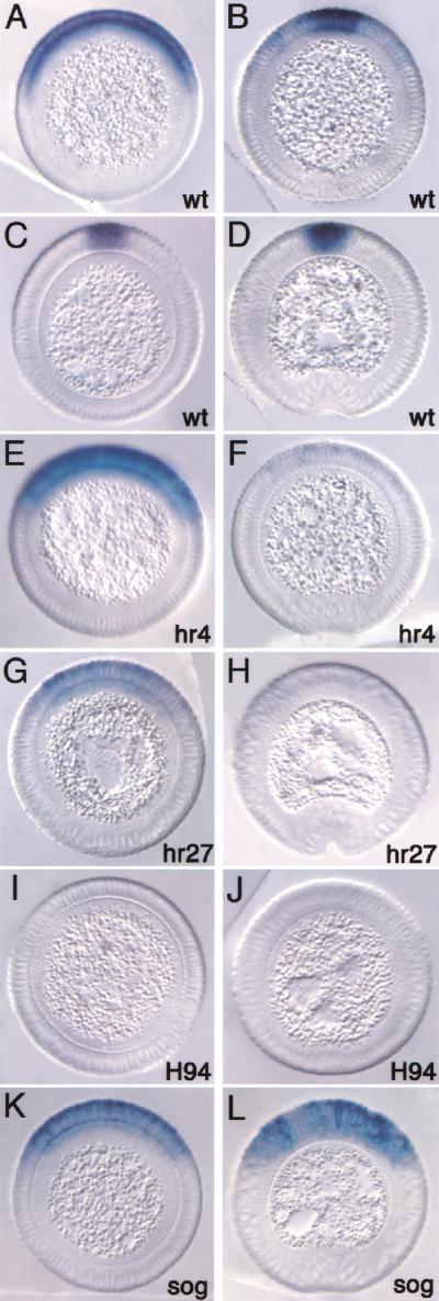
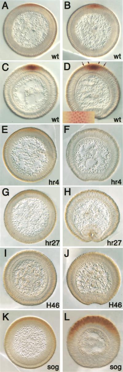
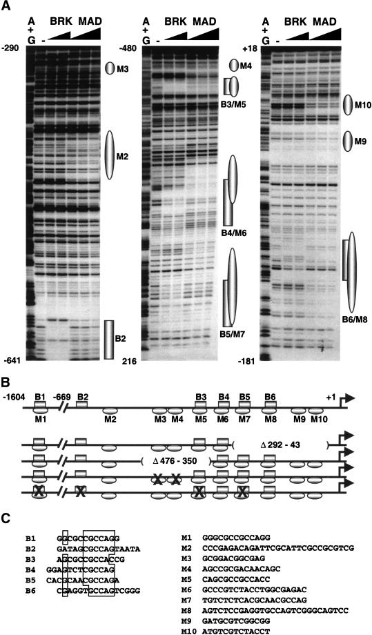
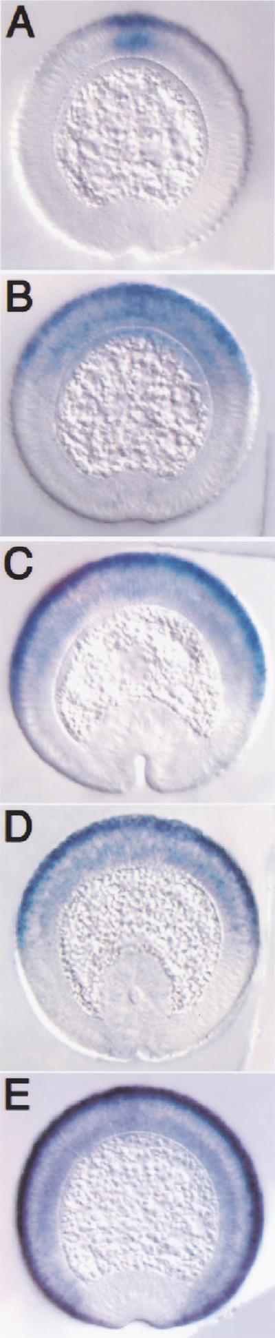
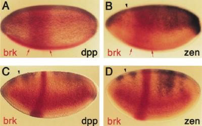
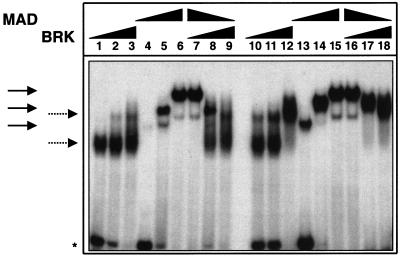
The establishment of expression domains of developmentally regulated genes depends on cues provided by different concentrations of transcriptional activators and repressors. Here we analyze the regulation of the Drosophila gene zen, which is a target of the Decapentaplegic (Dpp) signaling pathway during cellular blastoderm formation. We show that low levels of the Dpp signal transducer p-Mad (phosphorylated Mad), together with the recently discovered negative regulator Brinker (Brk), define the spatial limits of zen transcription in a broad dorsal-on/ventral-off domain. The subsequent refinement of this pattern to the dorsal-most cells, however, correlates with high levels of p-Mad that accumulate in the same region during late blastoderm. Examination of the zen regulatory sequences revealed the presence of multiple Mad and Brk binding sites, and our results indicate that a full occupancy of the Mad sites due to high concentrations of nuclear Mad is the primary mechanism for refinement of zen. Interestingly, several Mad and Brk binding sites overlap, and we show that Mad and Brk cannot bind simultaneously to such sites. We propose a model whereby competition between Mad and Brk determines spatially restricted domains of expression of Dpp target genes.


Figures

Figure 1
Figure 1
Dpp-dependent regulation of zen expression. Cross sections of embryos hybridized with zen antisense RNA probes. Dorsal is up. (A–D) Wild type, (E,F) dpphr4/dpphr4, (G,H) dpphr27/dpphr27, (I,J) dppH94/dppH94, (K,L) sogYS06/Y. (A) Embryo in early to mid-cellularization showing the broad dorsal-on/ventral-off zen pattern. (B) Embryo in mid-cellularization undergoing zen refinement. (C) Embryo in mid- to late cellularization showing the refined zen pattern restricted to the presumptive amnioserosa. (D) Embryo undergoing gastrulation with continued strong zen expression. Remaining embryos on the left are in mid- to late cellularization and can be compared with (C), and those on the right are beginning gastrulation and can be compared with (D). Note the decrease in the level of zen transcripts during mid- to late cellularization with decreasing dpp activity, and the loss of expression by gastrulation. Although a reduction in the level of transcripts was observed in dpphr27 embryos, the ventral limit of zen expression did not change significantly. In the absence of sog, zen expression is maintained to gastrulation, but there is no refinement.
Figure 2
Figure 2
High levels of activated Mad correlate with refined zen expression during late cellularization. Cross sections of embryos stained with anti-phospho-Smad antibodies. Dorsal is up; cellularization stages are as in Fig. 1. (A–D) Wild type, (E,F) dpphr4/dpphr4, (G,H) dpphr27/dpphr27, (I,J) dppH94/dppH46, (K,L) sogYS06/Y. (A–D) p-Mad accumulates as cellularization proceeds, reaching highest levels in the presumptive amnioserosa. The inset in D shows a surface view of a region of the dorsal side of a whole-mount staining, the same region delimited by the outer arrows on the section. Note that staining gradually decreases laterally. (E–H) p-Mad accumulation to high levels is not observed in dpp mutants. (I,J) In dppH46 embryos, no p-Mad staining is observed, indicating that the antibody is specific for Mad. (K,L) In sog mutants, p-Mad accumulates in a broad domain but may not reach the peak levels seen in the wild type.
Figure 3
Figure 3
Mad and Brk bind to the zen promoter. (A) DNase I footprinting analysis of Mad and Brk GST fusion proteins bound to the proximal 669 bp of the zen promoter. Increasing amounts of Brk (50 ng and 150 ng) and Mad (500 ng, 1500 ng, and 4500 ng) were incubated with zen-promoter DNA fragments: (left panel) EcoRI–AccI fragment labeled at the EcoRI site (−669 to −290), (middle panel) XbaI–AvaI fragment labeled at the XbaI site (−198 to −480), (right panel) XbaI–BamHI fragment labeled at the XbaI site (−198 to +18). The numbers correspond to the positions of the nucleotides relative to the transcription start site (+1). The (−) lanes show DNase I digestion of the DNA probes. The G+A lanes show the chemical degradation of the probes on G+As. Regions protected by Mad and Brk proteins are depicted as ovals and rectangles, respectively. (B) Schematic representation of the Smad (Mad/Medea) and Brk binding sites on the 1604-bp zen promoter, and the deletion and point mutations used in the transgenic analysis. The drawing is in scale only for the proximal 669 bp of the promoter. The locations of Brk binding sites (B2–B6) and Mad sites (M2–M10) are based on the footprinting data. The location of B1/M1 and all of the Medea sites were found by gel-shift analysis (not shown). The deletions are designated as absent lines inside parentheses, and the binding sites with point mutations are designated by X. (C) Alignment of the sequences of the BRK binding sites (B1–B6) (left). A list of Mad/Medea sites that cannot be aligned because of their degeneracy (right). Note that none of the Mad/Medea sites contains the inverted repeat 5′-GTC TAGAC-3′, which was determined as an optimal site for Smad3 and Smad4 proteins (Zawel et al. 1998).
Figure 4
Figure 4
Mutation of Smad and Brk binding sites results in altered expression of a zen–lacZ reporter. Cross sections of transgenic embryos undergoing gastrulation carrying the zen–lacZ fusion constructs (described in Fig. 3B) hybridized with lacZ antisense RNA probes. Dorsal is up. (A) Embryo carrying the full-length 1.6-kb zen promoter-lacZ shows refinement. (B) Embryo carrying the deletion Δ292–43. (C) Embryo carrying the deletion Δ476–350. (D) Embryo carrying a mutation that eliminates Smad binding sites M3 and M4. (E) Embryo carrying a mutation that eliminates four Brk binding sites (see Fig. 3B). Proper refinement is not observed in embryos carrying any of the mutant constructs.
Figure 5
Figure 5
Brk represses dpp and zen directly. Transgenic embryos carrying the FLP-out construct eve–stripe 2-brk were hybridized with either brk and dpp (A,C) or brk and zen (B,D) antisense RNA probes. dpp and zen are repressed in the region of eve–stripe 2 (parasegment 3) as Brk protein accumulates during mid- to late cellularization (C,D), but not earlier in precellular stages (A,B). Red arrows delimit the early broad stripe 2 expression. Arrowheads point to the site where normal loss of dpp and zen expression during cellularization occurs.
Figure 6
Figure 6
Mad and Brk compete in vitro for binding to DNA. Electrophoretic mobility shift assay (EMSA) of complexes formed by 2 ng, 10 ng, and 50 ng (lanes 1,2,3 and 10,11,12) of Brk; 20 ng, 100 ng, and 500 ng (lanes 4,5,6 and 13,14,15) of Mad; and 2 ng + 500 ng, 10 ng + 100 ng, and 100 ng + 20 ng of Brk and Mad, respectively (lanes 7,8,9 and 16,17,18) with 0.2 ng of labeled oligonucleotides spanning B5/M7 (lanes 1–9) and B4/M6 (lanes 10–18) sites. Solid and dotted arrows indicate the positions of the Mad and Brk complexes, respectively. The asterisk indicates the position of free DNA.

References

    1. Arnosti DN, Gray S, Barolo S, Zhou J, Levine M. The gap protein Knirps mediates both quenching and direct repression in the Drosophila embryo. EMBO J. 1996;15:3659–3666. - PMC - PubMed
    1. Ashe HL, Levine M. Local inhibition and long-range enhancement of Dpp signal transduction by Sog. Nature. 1999;398:427–431. - PubMed
    1. Ashe HL, Mannervik M, Levine M. Dpp signaling thresholds in the dorsal ectoderm of the Drosophila embryo. Development. 2000;127:3305–3312. - PubMed
    1. Biehs B, François V, Bier E. The Drosophila short gastrulation gene prevents Dpp from autoactivating and suppressing neurogenesis in the neuroectoderm. Genes & Dev. 1996;10:2922–2934. - PubMed
    1. Campbell G, Tomlinson A. Transducing the Dpp morphogen gradient in the wing of Drosophila: Regulation of Dpp target genes by brinker. Cell. 1999;96:553–562. - PubMed

Publication types

MeSH terms