

Interleukin-6 deficiency increases inflammatory bone destruction
- PMID: 11159963
- PMCID: PMC97947
- DOI: 10.1128/IAI.69.2.744-750.2001
Interleukin-6 deficiency increases inflammatory bone destruction
Abstract
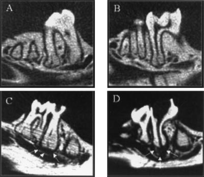
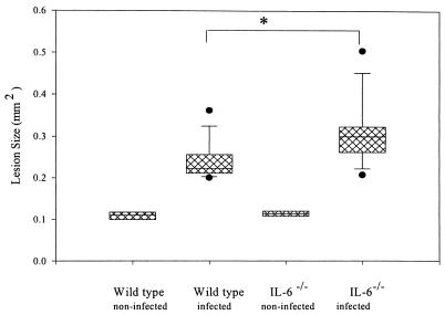
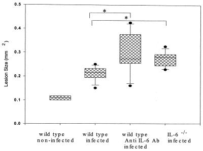
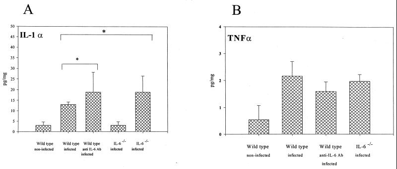
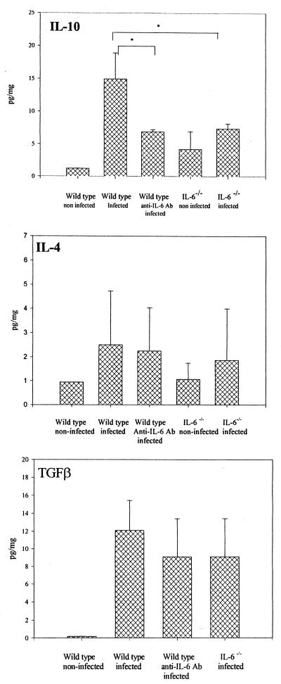
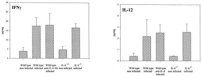
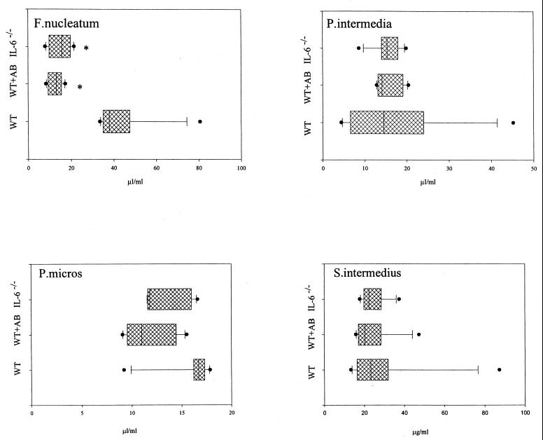
Periapical bone destruction occurs as a consequence of pulpal infection. In previous studies, we showed that interleukin-1 (IL-1) is the primary stimulator of bone destruction in this model. IL-6 is a pleiotropic cytokine that is induced in these infections and has both pro- and anti-inflammatory activities. In the present study, we determined the role of IL-6 in regulating IL-1 expression and bone resorption. The first molars of IL-6 knockouts (IL-6(-/-)) and wild-type mice were subjected to surgical pulp exposure and infection with a mixture of four common pulpal pathogens, including Prevotella intermedia, Fusobacterium nucleatum, Peptostreptococcus micros, and Streptococcus intermedius. Mice were killed after 21 days, and bone destruction and cytokine expression were determined. Surprisingly, bone destruction was significantly increased in IL-6(-/-) mice versus that in wild-type mice (by 30%; P < 0.001). In a second experiment, the effects of chronic (IL-6(-/-)) IL-6 deficiency and short-term IL-6 deficiency induced by in vivo antibody neutralization were determined. Both IL-6(-/-) (30%; P < 0.001) and anti-IL-6 antibody-treated mice (40%; P < 0.05) exhibited increased periapical bone resorption, compared to wild-type controls. The increased bone resorption in IL-6-deficient animals correlated with increases in osteoclast numbers, as well as with elevated expression of bone-resorptive cytokines IL-1alpha and IL-1beta, in periapical lesions and with decreased expression of the anti-inflammatory cytokine IL-10. These data demonstrate that endogenous IL-6 expression has significant anti-inflammatory effects in modulating infection-stimulated bone destruction in vivo.
Figures
References
-
- Aderka D, Le J, Wallach D. IL-6 inhibits lipopolysaccharide-induced tumor necrosis factor production in cultured human monocytes, U937 cells, and in mice. J Immunol. 1989;143:3517–3523. - PubMed
-
- Ahmed N, Thorley R, Xia D, Samols D, Webster R O. Transgenic mice expressing rabbit C-reactive protein exhibit diminished chemotactic factor-induced alveolitis. Am J Respir Crit Care Med. 1996;153:1141–1147. - PubMed
-
- Akira S, Taga T, Kishimoto T. Interleukin-6 in biology and medicine. Adv Immunol. 1993;54:1–78. - PubMed
-
- Balto K, Muller R, Carrington D, Dobeck J, Stashenko P. Quantification of periapical bone destruction in mice by micro-computed tomograhy. J Dent Res. 2000;79:35–40. - PubMed
-
- Barkhodar R, Hayashi C, Hussain M. Detection of interleukin-6 in human dental pulp and periapical lesions. Endod Dent Traumatol. 1999;15:26–27. - PubMed
Publication types
MeSH terms
Substances
Grants and funding
LinkOut - more resources
Full Text Sources
Other Literature Sources
Medical
Molecular Biology Databases
