. 2001 Feb;69(2):912-6.
doi: 10.1128/IAI.69.2.912-916.2001.

Initial characterization of the hemolysin stachylysin from Stachybotrys chartarum

Affiliations

Initial characterization of the hemolysin stachylysin from Stachybotrys chartarum

S J Vesper et al. Infect Immun. 2001 Feb.

Abstract

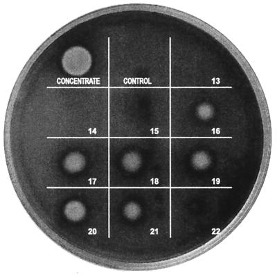
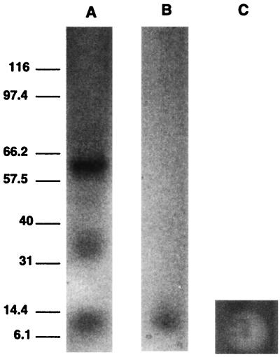
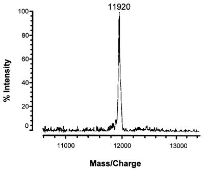
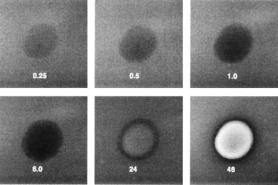
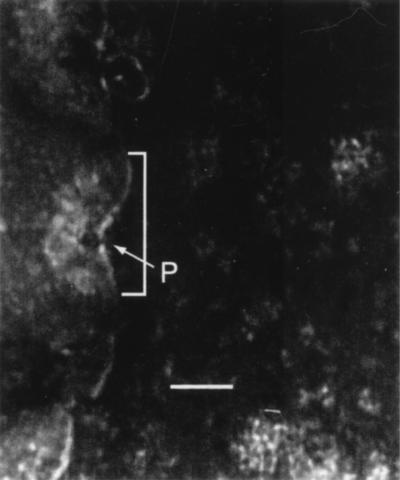
Stachybotrys chartarum is a toxigenic fungus that has been associated with human health concerns, including pulmonary hemorrhage and hemosiderosis. This fungus produces a hemolysin, stachylysin, which in its apparent monomeric form has a molecular mass of 11,920 Da as determined by matrix-assisted laser desorption ionization-time of flight mass spectrometry. However, it appears to form polydispersed aggregates, which confounds understanding of the actual hemolytically active form. Exhaustive dialysis or heat treatment at 60 degrees C for 30 min inactivated stachylysin. Stachylysin is composed of about 40% nonpolar amino acids and contains two cysteine residues. Purified stachylysin required more than 6 h to begin lysing sheep erythrocytes, but by 48 h, lysis was complete. Stachylysin also formed pores in sheep erythrocyte membranes.


Figures

FIG. 1
FIG. 1
Test for hemolytic activity in gel filtration fractions. Strain 58-06 of S. chartarum was grown in TSB. The proteins greater than 50- kDa were concentrated as described in Materials and Methods and then subjected to gel filtration in 0.2 M sodium azide. Fractions (0.25 ml) were collected, and 10 μl of each fraction was plated on SBA (Becton Dickinson) and incubated at 37°C for 48 h. The spot labeled as “concentrate” was applied before gel filtration. Ten microliters of the 0.2 M sodium azide solution was used as the control.
FIG. 2
FIG. 2
Gel electrophoresis (after lyophilization) of combined hemolytically active fractions in 0.2 M sodium azide (A). Gel electrophoresis (with SDS) after double desalting and lyophilization (B). Molecular mass standards in kilodaltons are shown on the left. (C) Lysis response to 10 μl of doubly-desalted stachylysin solution applied to SBA and then incubated at 37°C for 48 h.
FIG. 3
FIG. 3
MALDI-TOF mass spectrum of the doubly-desalted stachylysin fractions. Mass spectra were acquired in positive linear high-power mode, and pulsed extraction was used to improve resolution. Calibration was done with apomyoglobin and angiotensin II. The MALDI target was spotted with 0.25 μl of the sample solution. Before the sample dried, 0.25 μl of matrix solution was applied on top of the sample. The resultant spot was allowed to air dry. The matrix solution was 10 mg of 3,5-dimethoxy-4-hydroxy-cinnamic acid per ml in 50% acetonitrile in water containing 0.05% trifluoroacetic acid.
FIG. 4
FIG. 4
MALDI-TOF mass spectrum of the exhaustively dialyzed stachylysin fractions. Mass spectra were acquired in the positive linear high-power mode, and pulsed extraction was used to improve resolution. Calibration was done with apomyoglobin and angiotensin II. The MALDI target was spotted with 0.25 μl of the sample solution. Before the sample dried, 0.25 μl of matrix solution was applied on top of the sample. The resultant spot was allowed to air dry. The matrix solution was 10 mg of 3,5-dimethoxy-4-hydroxy-cinnamic acid per ml in 50% acetonitrile in water containing 0.05% trifluoroacetic acid.
FIG. 5
FIG. 5
Rate of stachylysin hemolysis of sheep RBCs. Approximately 0.2 μg of purified stachylysin in 10 μl of sterile, deioinzed water was spotted onto an SBA plate. The plate was incubated at 37°C and photographed at intervals of 0.25, 0.5, 1.0, 6.0, 24, and 48 h during the incubation.
FIG. 6
FIG. 6
TEM observations of stachylysin-treated RBCs. Approximately 0.2 μg of purified stachylysin was added to a 50-μl suspension of sheep RBCs and incubated statically at 37°C. After 48 h, the stachylysin-treated cells were placed on carbon-coated grids, stained with 1% phosphotungstic acid, and then examined by TEM (JEOL 1200EX). The bracketed area shows the membrane depression resulting from the stachylysin pore-forming process, and P is the actual pore. The scale bar is 100 nm.

References

    1. Baker C J, Edwards M S. Group B streptococcal infections. In: Remington J, Klein J O, editors. Infectious diseases of the fetus and newborn infant. 4th ed. Philadelphia, Pa: W. B. Saunders; 1995. pp. 980–1054.
    1. Bhakdi S, Greulich S, Muhly M, Korom S, Schmidt G. Effects of E. coli hemolysin on human monocytes: cytocidal action and stimulation of the interleukin-1 release. J Clin Investig. 1990;85:1746–1753. - PMC - PubMed
    1. Bhakdi S, Tranum-Jensen J. Alpha-toxin of Staphylococcus aureus. Microbiol Rev. 1991;55:733–751. - PMC - PubMed
    1. Bhakdi S, Bayley H, Valeva A, Walev I, Walker B, Weller U, Kehoe M, Palmer M. Staphylococcal alpha-toxin, streptolysin-O, and Escherichia coli hemolysin prototypes of pore-forming bacterial cytolysins. Arch Microbiol. 1996;165:73–79. - PubMed
    1. Cavalieri S J, Bohach G A, Snyder I S. Escherichia coli α-hemolysin characteristics and probable role in pathogenicity. Microbiol Rev. 1984;48:326–343. - PMC - PubMed

Publication types

MeSH terms

Substances

LinkOut - more resources