. 2001 Feb;69(2):1002-8.
doi: 10.1128/IAI.69.2.1002-1008.2001.

Regulation of expression of major histocompatibility antigens by bovine macrophages infected with Mycobacterium avium subsp. paratuberculosis or Mycobacterium avium subsp. avium

Affiliations

Regulation of expression of major histocompatibility antigens by bovine macrophages infected with Mycobacterium avium subsp. paratuberculosis or Mycobacterium avium subsp. avium

D J Weiss et al. Infect Immun. 2001 Feb.

Abstract

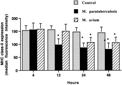
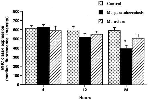
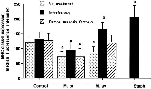
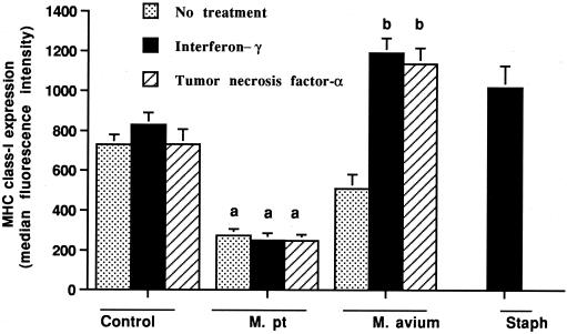
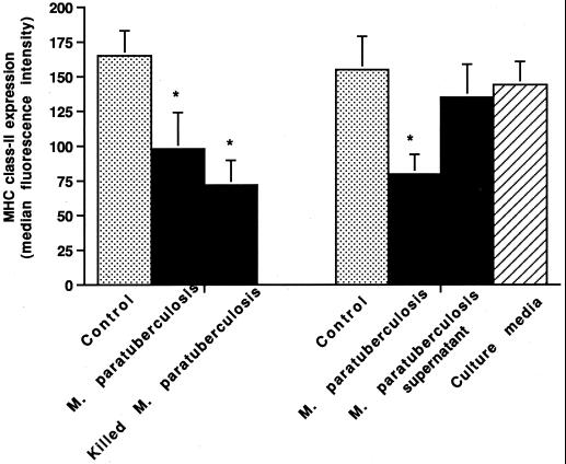
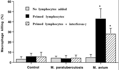
Mycobacterium avium subsp. paratuberculosis and Mycobacterium avium subsp. avium are antigenically and genetically very similar organisms; however, they differ markedly in their virulence for cattle. We evaluated the capacity of bovine macrophages infected with M. avium subsp. paratuberculosis or M. avium subsp. avium to express major histocompatibility complex (MHC) class I and class II antigens on their surface and to interact with primed autologous lymphocytes. Our results indicate that infection of bovine macrophages with M. avium subsp. paratuberculosis promoted the downregulation of MHC class I and class II molecules on the macrophage surface within 24 and 12 h, respectively. Alternatively, MHC class II expression by M. avium subsp. avium-infected macrophages was not detected until 24 h after infection, and the magnitude of the decrease was smaller. Decreased MHC class I expression by M. avium subsp. avium-infected macrophages was not detected. Unlike M. avium subsp. paratuberculosis-infected macrophages, M. avium subsp. avium-infected macrophages upregulated MHC class I and class II expression after activation by gamma interferon or tumor necrosis factor alpha. Further, M. avium subsp. avium-infected macrophages were lysed by primed autologous lymphocytes, whereas M. avium subsp. paratuberculosis-infected macrophages were not. Overall, the results support the hypothesis that the difference in the virulence of M. avium subsp. paratuberculosis and M. avium subsp. avium for cattle is dependent on a difference in the capacity of the organisms to suppress mycobacterial antigen presentation to T lymphocytes.


Figures

FIG. 1
FIG. 1
Incubation of bovine macrophages with M. avium subsp. paratuberculosis (M. paratuberculosis) or M. avium subsp. avium (M. avium) decreases surface expression of MHC class II antigens, as detected by flow cytometry. Bovine monocyte-derived macrophages (106) were cultured for various times with or without M. avium subsp. paratuberculosis or M. avium subsp. avium added. The mean and SE for four separate experiments are shown. Asterisks indicate statistically significant differences relative to control values (P < 0.05).
FIG. 2
FIG. 2
Flow cytometric scatter plots of MHC class II expression by bovine monocyte-derived macrophages. Just before analysis, 100 μl of propidium iodide was added to identify dead cells. Cells were displayed as log green fluorescence intensity (FL1-H; i.e. a measure of surface MHC class II expression) versus log red fluorescence intensity (FL2-H; a measure of propidium iodide uptake). Quadrant gates were set to eliminate dead cells (upper right and left quadrants) and to identify cells with decreased MHC class II expression compared to that of uninfected macrophages (lower left quadrant). (A) Uninfected macrophages incubated for 24 h. (B) Macrophages infected with M. avium subsp. paratuberculosis organisms for 24 h. (C) Macrophages infected with M. avium subsp. avium organisms for 24 h.
FIG. 3
FIG. 3
Incubation of bovine macrophages with M. avium subsp. paratuberculosis (M. paratuberculosis) but not M. avium subsp. avium (M. avium) decreases surface expression of MHC class I antigens, as detected by flow cytometry. Bovine monocyte-derived macrophages (106) were cultured for various times with or without M. avium subsp. paratuberculosis or M. avium subsp. avium added. The mean and SE for three separate experiments are shown. The asterisk indicates a statistically significant difference relative to the control value (P < 0.05).
FIG. 4
FIG. 4
Effects of gamma interferon and TNF-α on MHC class II expression by bovine macrophages incubated for 24 h with or without M. avium subsp. paratuberculosis (M. pt) or M. avium subsp. avium (M. av). Unlike M. avium subsp. paratuberculosis-infected macrophages, M. avium subsp. avium-infected macrophages upregulated MHC class II molecules in response to gamma interferon. As a positive control, uninfected macrophages were incubated with gamma interferon and killed S. intermedius (Staph) organisms. The mean and SE for three separate experiments are shown. a, statistically significant differences relative to control values (P < 0.05); b, statistically significant differences relative to samples incubated with M. avium subsp. avium organisms alone (P < 0.05).
FIG. 5
FIG. 5
Effects of gamma interferon and TNF-α on MHC class I expression by bovine macrophages incubated for 24 h with or without M. avium subsp. paratuberculosis (M. pt) or M. avium subsp. avium. Unlike M. avium subsp. paratuberculosis-infected macrophages, M. avium subsp. avium-infected macrophages upregulated MHC class I molecules in response to gamma interferon and TNF-α. As a positive control, uninfected macrophages were incubated with gamma interferon and killed S. intermedius (Staph) organisms. The mean and SE for three separate experiments are shown. a, statistically significant differences relative to control values (P < 0.05); b, statistically significant differences relative to samples incubated with M. avium subsp. avium organisms alone (P < 0.05).
FIG. 6
FIG. 6
Effects of live versus killed M. avium subsp. paratuberculosis (M. paratuberculosis) organisms and culture supernatants on MHC class II expression by bovine macrophages. Bovine monocyte-derived macrophages (106) were incubated for 24 h with or without the addition of live or heat-killed M. avium subsp. paratuberculosis organisms or culture supernatant. The mean and SE for two separate experiments are shown. Asterisks indicate statistically significant differences relative to control values (P < 0.05).
FIG. 7
FIG. 7
Effects of autologous primed lymphocytes on killing of macrophages incubated with or without M. avium subsp. paratuberculosis (M. paratuberculosis) or M. avium subsp. avium (M. avium) added. Bovine monocyte-derived macrophages (106) were incubated with autologous primed lymphocytes (107) and with or without gamma interferon. The mean and SE for three separate experiments are shown. Asterisks indicate statistically significant differences relative to control values and M. avium subsp. paratuberculosis values (P < 0.05).

References

    1. Antoine J C, Prima E, Lang T, Courret N. The biogenesis and properties of the parasitophorous vacuoles that harbor Leishmania in murine macrophages. Trends Microbiol. 1998;6:392–401. - PubMed
    1. Antoine J C, Lang T, Prima E, Courret N, Hellio R. H-2M molecules, like MHC class II molecules, are targeted to parasitophorous vacuoles of Leishmania-infected macrophages and internalized by amastigotes of L. amazonensis and L. mexicana. J Cell Sci. 1999;112:2559–2570. - PubMed
    1. Bassey E O, Collins M T. Study of T-lymphocyte subsets of healthy and Mycobacterium avium subsp. paratuberculosis-infected cattle. Infect Immun. 1997;65:4869–4872. - PMC - PubMed
    1. Bonecini-Almeida M G, Chitale S, Boutsikakis I, Geng J, Doo H, He S, Ho J L. Induction of in vitro human macrophage anti-Mycobacterium tuberculosis activity: requirement for INF-γ and primed lymphocytes. J Immunol. 1998;160:4490–4499. - PubMed
    1. Brennan P L, Nikaido H. The envelope of mycobacteria. Annu Rev Biochem. 1995;64:29–63. - PubMed

Publication types

Substances