. 2001 Feb;69(2):1016-24.
doi: 10.1128/IAI.69.2.1016-1024.2001.

Microsporidian invasion apparatus: identification of a novel polar tube protein and evidence for clustering of ptp1 and ptp2 genes in three Encephalitozoon species

Affiliations

Microsporidian invasion apparatus: identification of a novel polar tube protein and evidence for clustering of ptp1 and ptp2 genes in three Encephalitozoon species

F Delbac et al. Infect Immun. 2001 Feb.

Abstract

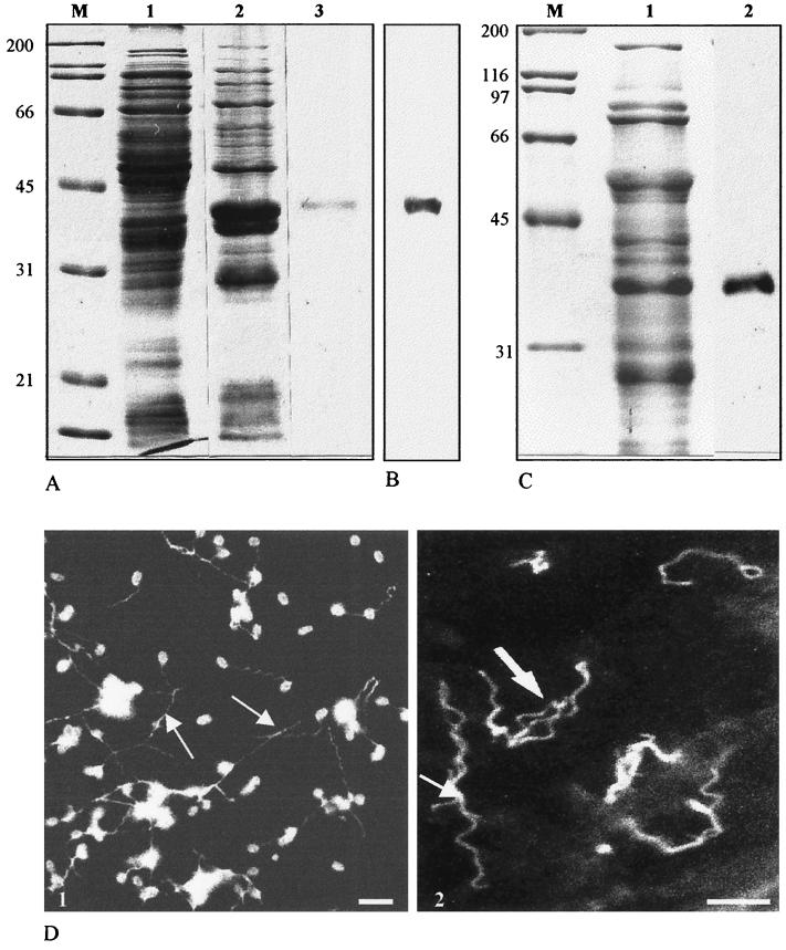
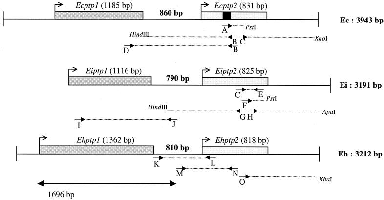
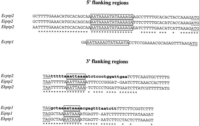
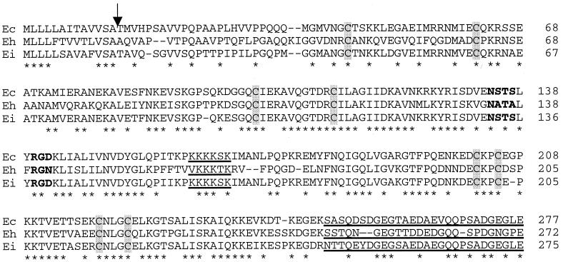
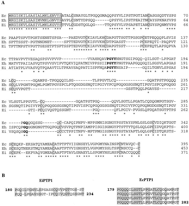
Microsporidia are unicellular eukaryotes occurring as obligate intracellular parasites which produce resistant spores. A unique motile process is represented by the sudden extrusion of the sporal polar tube for initiating entry of the parasite into a new host cell. The complete sequence of an acidic proline-rich polar tube protein (renamed PTP1) has been previously reported for Encephalitozoon cuniculi and E. hellem. Our immunological investigations provided evidence for an additional PTP in E. cuniculi, termed PTP2. The corresponding gene was sequenced and then expressed in Escherichia coli. As expected, mouse antibodies raised against the recombinant protein reacted specifically with the polar tube. The single copy ptp1 and ptp2 genes of E. cuniculi were tandemly arranged on chromosome VI. Polyadenylation of the mRNAs was demonstrated. Identification and sequencing of homologous genes in the two other human-infecting Encephalitozoon species (ptp2 in E. hellem and ptp1 and ptp2 in E. intestinalis) were facilitated by conserved gene clustering. PTP2 appears as a novel structural protein (30 kDa) with a basic lysine-rich core and an acidic tail. Unlike PTP1, this protein is devoid of large tandem repeats. The interspecies conservation of cysteine residues supports a major role of disulfide bridges in polar tube assembly. The two PTPs should serve as both molecular markers of spore differentiation and diagnostic tools.


Figures

FIG. 1
FIG. 1
(A) SDS-PAGE analysis of E. coli-expressed recombinant EcPTP2 (Coomassie blue staining). Lanes 1 and 2, total E. coli lysate before (lane 1) and after (lane 2) IPTG induction; lane 3, recombinant EcPTP2 after purification on an Ni-NTA column; lane M, molecular mass standards in kilodaltons. (B) Immunoblotting reactivity of MAb Ec102 with E. coli proteins 4 h after IPTG induction. The blot was probed with a 1:10,000 dilution of MAb Ec102 and developed using ECL (Amersham). (C) Lane 2, immunoblot of E. cuniculi whole-cell homogenates probed with anti-recombinant EcPTP2 antiserum. Lane 1, total E. cuniculi proteins stained with Coomassie blue. (D) Indirect immunofluorescence of E. cuniculi spores with extruded polar tubes (arrows). (1) Labeling with an antiserum against total proteins; (2) specific labeling of polar tubes with anti-recombinant PTP2. Bars, 5 μm.
FIG. 2
FIG. 2
Schematic representation of ptp1 and ptp2 gene clusters with positions of PCR primers (A to O) in the three Encephalitozoon species, E. cuniculi (Ec), E. intestinalis (Ei), and E. hellem (Eh). The peptide obtained by microsequencing of EcPTP2 (AVQGTDRCILAGIID) is indicated by a black square. Sense primer D, designed 186 bp upstream from the stop codon of Ecptp1, combined with antisense primer B, designed 380 bp downstream from the initiation codon of Ecptp2, were used to amplify a 1.4-kbp DNA fragment. Two primers deduced from the Ecptp2 sequence (C and E, positions 419 to 699 in the Ecptp2 ORF) were used to amplify a 280-bp DNA fragment in E. intestinalis. The corresponding sequence shared about 90% identity with that of Ecptp2 but showed some differences that were useful to determine specific oligonucleotides. Downstream and upstream regions of the 280-bp known sequence were amplified with PstI and HindIII ligation products, respectively, using two specific primers (F and G) and the reverse vector primer. Thus, SSP-PCR experiments led to 120-bp (3′ with PstI) and 1,150-bp (5′ with HindIII) amplicons, respectively. The 3′ end of Eiptp2 was completed through another SSP-PCR step with primer H, resulting in a 570-bp amplification from the ApaI ligation product. The final sequence is 1,968 bp in length with a predicted 825-bp ORF coding for EiPTP2. The Eiptp1 gene was amplified using a combination of an antisense primer in the 5′ flanking region of Eiptp2 (J) and a sense primer (I) determined from the alignment of conserved regions encoding signal peptides of EcPTP1 and EhPTP1. For E. hellem, primer K, determined in the 3′ known flanking region of Ehptp1, was combined with the reverse primer L, determined by the alignment of the highly conserved N-terminal sequence encoding the PTP2 signal peptide in E. cuniculi and E. intestinalis. The whole coding sequence of Ehptp2 and its 3′ UTR were completed by SSP-PCR amplification with primer O. The 1,696-bp sequence is from reference .
FIG. 3
FIG. 3
Alignments of ptp1 and ptp2 gene 5′ and 3′ flanking regions showing conserved signals. Start and stop codons are underlined. Identical nucleotides are indicated by asterisks. The AT-rich consensus sequence in the 5′ region and potential site of polyadenylation are boxed. Partial E. cuniculi cDNAs with short 3′ UTRs (less than 30 nucleotides) are in boldface lowercase letters.
FIG. 4
FIG. 4
Alignment of the three complete PTP2 amino acid sequences. Amino acids are numbered on the right. Identical residues are indicated by asterisks. The common putative cleavage site for the signal peptide is shown by an arrow. The eight cysteine residues are shaded. The common putative N-glycosylation site and the RGD sequence are indicated in boldface. The lysine-rich central region and the acidic tail are underlined.
FIG. 5
FIG. 5
(A) Alignment of the PTP1 amino acid sequences from E. cuniculi (Ec), E. hellem (Eh), and E. intestinalis (Ei). Amino acids are numbered on the right. Identical amino acids are indicated by asterisks. The highly conserved 22-aa signal peptide is boxed. The three PTP1s share common N- and C-terminal parts but diverge in their tandemly repeated sequences, forming a central core delimited by common boundaries (in boldface). Conserved cysteine residues are shaded. (B) Alignment of the tandemly repeated sequences of EiPTP1 (two repeats) and EcPTP1 (four repeats). Identical amino acids are shaded.

References

    1. Altschul S F, Madden T L, Schäffer A A, Zhang J, Zhang Z, Miller W, Lipman D J. Gapped BLAST and PSI-BLAST: a new generation of protein database search programs. Nucleic Acids Res. 1997;25:3389–3402. - PMC - PubMed
    1. Beauvais B, Sarfati C, Challier S, Derouin F. In vitro model to assess effect of antimicrobial agents on Encephalitozoon cuniculi. Antimicrob Agents Chemother. 1994;38:2440–2448. - PMC - PubMed
    1. Beckers P J A, Derks G J M M, Van Gool T, Rietveld F J R, Sauerwein R W. Encephalitozoon intestinalis-specific monoclonal antibodies for laboratory diagnosis of microsporidiosis. J Clin Microbiol. 1996;34:282–285. - PMC - PubMed
    1. Biderre C, Pagès M, Méténier G, Canning E U, Vivarès C P. Evidence for the smallest nuclear genome (2, 9 Mb) in the microsporidium Encephalitozoon cuniculi. Mol Biochem Parasitol. 1995;74:229–231. - PubMed
    1. Biderre C, Canning E U, Méténier P G, Vivarès C P. Comparison of two isolates of Encephalitozoon hellem and E. intestinalis (Microspora) by pulsed field gel electrophoresis. Eur J Protistol. 1999;35:194–196.

Publication types

Substances